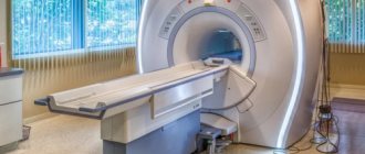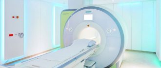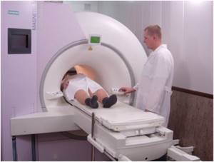
The photo shows a traditional closed-circuit MRI scanner. Magnetic resonance imaging is a high-tech diagnostic method that allows you to visualize bone structures and surrounding soft tissues of the body. The advantage of the procedure is the safety and information content of the images. There are several types of devices: open and closed MRI. The devices have their differences, pros and cons. A closed-type MRI device is considered classic, but the principle of operation of tomographs is identical.
The research is based on the use of magnetic induction. The field generated by the device interacts with hydrogen atoms, exciting them. Depending on the saturation of tissues with water, the intensity of the return signal will change. The device's sensors record the response, and the computer processes the information. The minimum imaging step is 1 mm, so pathological areas of even small size can be identified, for example, when examining the brain or oncology. Radiologists receive images, interpret the information and issue a report. The picture can be combined into a 3D model and video.
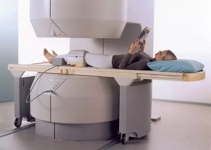
The difference between open and closed MRI is their design. A traditional tomograph is represented by a tube into which the subject is placed. The open-loop device looks like two “plates” between which the patient is positioned. The device is suitable for people with a fear of confined spaces, but the results are less informative. Some authors highlight the semi-open appearance of the devices, but this is incorrect.
The most important classification of tomographs is based on the strength of the magnetic field generated by the device. There are low-field devices (0.15-1 T), high-field (up to 3 T) and ultra-high-field (more than 3 T). High-field devices with a closed loop are used more often in clinical practice due to the reliability of the results, the speed of image acquisition and the absence of negative effects on the human body. The terms “half-open” and “half-closed” are not used in relation to MRI scanners, being a marketing ploy.
Features of open MRI
Carrying out magnetic resonance imaging using an open-type device provides the same opportunities as a conventional procedure. But there are also some peculiarities. For example, in this case, the study takes a little longer, and the images are characterized by lower resolution. This is due to the lower magnetic field strength.
Our clinic offers open MRI in Moscow at a competitive price. The address of the medical center is listed on the website; additional information can be obtained by phone. We use MRI machines from General Electric and other well-known manufacturers, and the highly qualified doctors allow us to accurately interpret the results.
What is the difference
If you look at what a semi-open type MRI looks like, it becomes clear that it is easier to place a person with impressive body dimensions on it, whereas a closed device has limitations in terms of parameters. But this also depends on the specific type of device. At the same time, semi-open equipment combines all the advantages:
- there is a round camera for volumetric scanning;
- this compartment is shortened, and the patient is placed in it only with the area of the body being examined;
- there is no effect of a long narrow pipe, not dangerous for people with claustrophobia.
It is difficult to answer the question which MRI is better, closed or open. It's always individual. For more detailed scanning, closed or semi-open equipment is traditionally offered.
However, there are cases when their use is impossible, and the patient undergoes diagnostics using an open device.
Advantages of open tomographs
Open type MRI machines have certain advantages:
- Suitable for examining people suffering from claustrophobia - finding such patients in a confined space can cause panic attacks and other negative manifestations;
- They can be used when performing MRI on large people with significant body weight - in open-circuit devices there are no restrictions on these indicators. For the same reason, it becomes possible to perform the procedure on women at a significant stage of pregnancy;
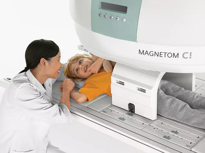
- They are used to diagnose diseases in young children - with a closed MRI, the child may be frightened, and a mother or father may be next to the open tomograph to calm the baby;
- Used to examine people with mental illness who also need monitoring;
- Allows MRI to be done for people in serious condition while simultaneously performing medical procedures;
- They give accurate results even if the patient makes minor movements.
People with serious injuries that prevent them from taking a lying position cannot be in a closed-type apparatus. This happens, for example, when the spine is damaged. In such cases, examinations are carried out using an open-type tomograph. Another plus is the absence of strong noise, which is typical for closed devices.
Closed-type MRI
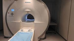
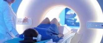
Photo of an MRI scanner tunnel
MRI on a closed-type tomograph is a classic imaging method. The device looks like a short and wide pipe in which magnetic field sources are located. The latter affects the body from all sides in a circle. The structural features provide greater power compared to other equipment - up to 3 Tesla in clinical practice. The described difference makes diagnosis faster, and the images are of better quality and detail.
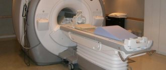
The subject is placed on a mobile table, fastened with belts and placed inside the tunnel. The work process is accompanied by loud sounds, crackling, knocking. It is important to remain still during the procedure. If the patient moves, the reliability of the image will decrease.
Doctors leave the room and monitor the process from a separate room. You can maintain constant communication using a radio device. During the diagnostic test, one of the parents is allowed to stay with the child during the MRI to calm the baby.
The limitation for using the apparatus of this design option is the presence of claustrophobia. Prolonged stay in a confined space accompanied by loud noises can trigger an attack. However, if you notify the specialists in advance, they will select a sedative for you, put on headphones with relaxing music, and the examination will take place without unnecessary stress. Another limitation is the patient’s weight is more than 120 kg. The structure may not withstand a large mass and break.
Advantages of closed-type MRI
There are a few differences that may make it better to choose the traditional option. These include:
- high accuracy of the result, regardless of the area of the body (suitable for examining the brain, determining the malignant potential of tumors, imaging the heart, etc.);
- difference in scanning duration - the average duration of native MRI is 15 minutes.
Disadvantages of closed-type MRI
There are the following disadvantages of using the device:
- limiting the weight of the subject to 120 kg;
- claustrophobia may be a contraindication to tomography;
- high noise level (you can use earplugs or headphones with pleasant music);
- complete stillness is required to obtain high-quality photographs.
Indications for the procedure
Open tomography is used to study joints, spine, soft tissues, blood vessels, including the brain. To identify pathologies of the pelvic or abdominal organs, a closed device with a higher power is often used, but if necessary, an MRI can be done on an open type tomograph.
Open type devices make it possible to identify the causes:
- Frequent dizziness and headaches;
- Impaired visual acuity;
- Numbness of the limbs and stiffness of movement;
- Painful sensations in the joints and spine;
- Infertility.
Tomography allows you to determine the condition of internal organs, identify the presence of tumors, their location, extent and type (malignant or benign). To control the tumor process, a procedure using a contrast agent can be performed. This technique helps to determine the structure and boundaries of the tumor and identify metastases after surgery.
Advantages of Sofia Cancer Center
The development of open devices has brought MR diagnostics to a new level - now this examination method is available to the widest group of patients. The Sofia Cancer Center uses some of the best equipment, which makes our capabilities even more valuable. Thanks to high technologies, it is possible to detect oncological, pathological and other processes before clinical manifestations and before they are noticeable in other studies.
Advantages of the Sofia Cancer Center:
- experienced doctors;
- everything in one place: in Sofia there are two large buildings standing next to each other with a total area of 35,000 sq.m;
- 30 years of unique experience;
- individual support around the clinic;
- increased comfort: cafe with panoramic views, modern interior, design, like in a five-star hotel;
- conducting research strictly according to standards (including JCI);
- large parking for patients.
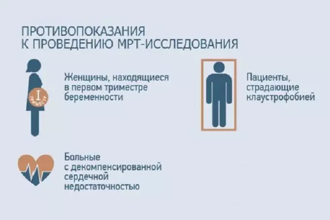
Main contraindications
Contraindications to MRI using an open tomograph are:
- The presence of a pacemaker or fixed prostheses made of metal (exceptions are elements made of titanium);
- Installed insulin pump;
- The presence of hemostatic clips on the cerebral vessels;
- Performed stenting of coronary heart vessels.
In the first trimester of pregnancy, the need for examination is determined by the doctor. MRI with contrast is not performed if you are allergic to the components of the injected substance.
Features of the resulting image
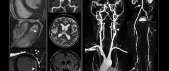
The main advantage of images created using MRI is that they are obtained layer by layer. Just imagine that you are cutting some product into small slices with a very sharp knife. This makes it possible to get a detailed, clear image of everything that is inside.
The design of the device uses special gradient coils. Modern methods help create magnetic resonance on a specific selected layer. Doctors can move between layers.
The result is a tomogram. It can be called a map of the organ - many small sections, each of which is made in three directions. Thanks to this, even the smallest changes at an early stage of development will not be hidden from the physician’s gaze.
How is MRI performed on an open machine?
During the procedure, the patient is in a special room where a tomograph is installed. The doctor remains in the adjacent room and monitors the process through a transparent partition, while maintaining voice contact. If necessary, accompanying persons may be present in the premises.
First you need to remove metal jewelry. Open tomographs usually have side loading. The patient is positioned on the platform in a supine position. The area of the body being examined should be approximately in the middle of the table. As a rule, the procedure takes about half an hour, but in some cases it can last longer, for example, when examining the pelvic organs. It is important to remain still throughout the examination.
Based on the results of the study, the doctor describes the results obtained and issues a conclusion to the patient. The document is accompanied by graphic images (photographs) and a recording of the study on electronic media (disk or memory card).
Open MRI is offered in many diagnostic centers in Moscow.
Our advantages are an individual approach to each patient, a detailed interpretation of the results obtained and diagnostic accuracy due to the decent professional level of specialists and high quality equipment. Rate this article: (1 rated 5 out of 5)
Much experience has been accumulated over time
in conducting MRI examinations of patients with limited mobility, as well as patients suffering from claustrophobia (fear of closed spaces).
The center’s specialists are always patient, polite, understanding of this problem, and always result-oriented. Our medical center has a high percentage of successful examinations for patients even with severe claustrophobia
, and the data obtained is always of high quality.
Open type MRI scanners are absolutely safe for humans. A constant magnetic field and a huge selection of scanning programs allow you to obtain images of the highest quality.
With the advent of an open-type tomograph, the procedure has become more comfortable
compared to a closed-type device (tunnel). For people suffering from claustrophobia, the elderly, children, and patients who cannot assume a completely horizontal position due to injury or physiological characteristics, MRI on an open-type machine will allow them to undergo an examination.
An open-type tomograph can also be used to examine overweight people. The tables of open devices are quite wide and comfortable, you can sit and lie on them comfortably. The open circuit device table can support up to 140kg
, however, there are restrictions on the volume of the patient’s torso. These restrictions are always individual and depend on body composition (height, weight, coverage of the abdomen and chest in a lying position) and the type of examination.
How is an MRI examination performed?
During the procedure, the patient is in a special diagnostic room, where his relatives or accompanying persons can be present if desired.
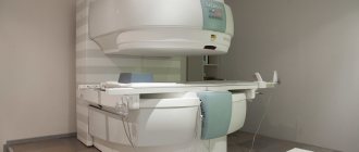
In open-type tomographs, the magnetic field is constant and is located between the upper and lower parts of the device. The patient is comfortably positioned on the tomograph table in a supine position so that the part of the body being examined is located in the center of the tomograph. A radiofrequency coil is placed over the area of interest. At the same time, the device produces monotonous sounds, which are an order of magnitude quieter than in closed-type devices. Depending on the type of examination, you will be offered headphones with soothing music or earplugs.
To make an emergency call to personnel during an examination, a signal “bulb” is placed in the patient’s hand. The duration of the diagnostic procedure is on average 20-30 minutes, but in some cases this time can be extended. When conducting an examination with intravenous contrast enhancement, the examination time will be 40-50 minutes.
Before the procedure, the patient must remove jewelry and clothing with metal elements, shoes, and also leave personal belongings in the locker room (there is also a safe), including phones, magnetic cards, watches and other electronics, as they may stop working under the influence of magnetic fields.
It is important that the patient lies still during the examination to avoid motion artifacts (image distortions).
The images are described by an experienced doctor
by a radiologist, and the conclusion and the study itself in the form of an archive are sent by email to the patient, so there is no need to wait for the conclusion.
Differences between open and closed MRI equipment
The modern open-type tomograph OPENMARK 4000 is not only not inferior to closed-type equipment in terms of the quality of the tomograms obtained, but also has a number of advantages.
The main advantage of open MRI is the ability to obtain high-quality images of any part of the human body without experiencing discomfort in a confined space. Open circuit device
allows you to position the patient in a way that is convenient for tomography. In addition, the device configuration allows the physician to approach the site at any time and adjust the equipment, change the coil, move the patient, or administer contrast enhancement.
Patients undergoing closed-type MRI often complain of noise. Open type devices are much quieter
, which reduces anxiety and stress levels.
An important advantage of open MRI scanners is the ability to conduct examinations of young children under the supervision of their parents. In a closed device, the child does not see what is happening around him, gets scared and does not want to lie down. With open magnetic resonance imaging, a small patient can see and hear his mother. A loved one can calm the baby, hold his hand, read his favorite story, providing the patient with the stillness necessary to obtain high-quality images.
On the technical side, the devices differ in the following:
Open type devices have a constant magnetic field created by a large mass of magnets (the weight of the device is 16 tons), due to which the magnetic field power is 0.44 Tesla, which is absolutely safe for the human body. These devices are successfully used and perform an excellent job in 95% of all diagnostic cases, thanks to special programs and highly sensitive magnetic coils, the quality of the resulting tomograms is very high, images of the brain and spine are comparable to 1 Tesla devices, and when examining joints and pelvic organs, the image quality superior to 1 Tesla devices.
In closed, that is, superconducting magnets, the magnetic field is raised and created by applying an electric current to a magnetic coil, and the output power of the magnetic field reaches 1-3 Tesla. Visualization on a three-tesla device is naturally higher, but how safe and advisable is it to place a person in a magnetic field of such high power? Unfortunately, this question remains open. It is well known that in Europe and already in Russia, specialists with low levels of hemoglobin in the blood are not allowed to work with powerful MRI machines.
It is important to understand that the conclusion about the examination result is written not by an MRI machine, but by a radiologist trained to work with various types of tomographs.
Our clinic also uses a method called aromatherapy
during MRI. Special equipment and natural oils with a calming effect are used.
The doctors of our center are highly qualified and have extensive experience.
conducting an open MRI, which guarantees an error-free conclusion based on the examination results.
What diseases can be detected using this method?
Since the list of diseases depending on the patient is very extensive. In the table below you can see detailed information.
| Systems and organs | Detectable diseases |
| Brain | It is with the help of MRI that doctors successfully determine the signs of ischemic stroke. This is done early, which allows action to be taken. Thanks to the possibility of examining pregnant women, it is possible to determine the presence of pathologies of brain development in the child. In the early stages of development, magnetic resonance reveals both malignant and benign tumors, aneurysms, encephalitis, and multiple sclerosis. In some cases, it is even possible to establish the cause of mental disorders. An examination for epilepsy is also prescribed - it helps to understand where the focus is located. |
| Spine | Using MRI, you can check the spine, both in a calm and loaded state. In the first case, standard horizontal scanners are used, in the second - vertical ones. The technique helps to check the condition of the bone marrow, vertebrae, nerves, adjacent tissues, and intervertebral discs. MRI detects protrusions and hernias, inflammation of the spinal cord, compression fractures. A complete picture of neoplasms and the metastases they produce is given. |
| Bones and joints | Magnetic resonance allows you to check whether a person has injuries, where foci of degenerative and inflammatory lesions are located. It is an effective method for monitoring femoral head necrosis, dislocations, traumatic joint injuries, and a variety of other problems. |
| Large vessels and heart | MRI as a diagnostic approach is excellent in early detection of problems leading to heart attack and stroke. It shows aneurysms, tumors, areas affected by necrosis or ischemia. You can check the pulmonary and coronary arteries, the myocardium, and identify pericarditis, myocarditis and epicarditis. |
| Other organs | The examination can be carried out on almost all internal organs. This is how diseases of the pelvic and abdominal organs, lymph nodes, adrenal glands, liver, kidneys and spleen are checked. |
Main purposes of MRI
There are several purposes that MRI testing serves. Among them the main ones are the following:
- It is necessary to clarify where the pathology is located, how intensively it spreads, and what danger it poses. This allows you to choose an effective further treatment method.
- Check whether there are neoplasms of a benign or malignant type in the human body. At the same time, all problems can be identified at an early start - a proven contrast enhancement system works for this.
- Monitoring the success of treatment or rehabilitation period. MRI is often performed after injuries, treatment of oncology and other pathologies of the body.
- Analysis of the presence of problems in the body. These may include bleeding, foci of inflammation, necrosis, the current state of cysts and other types of neoplasms in the patient’s body.
Among the goals is the need to find foci of ischemic brain damage.
What can you see from MRI results?
Many patients are interested in the question of what an MRI shows. To date, this method is the most informative. With its help, you can examine the entire body, internal organs, tissues, and vascular system. At the same time, not only the current state is shown, but also the structure and its minor changes, which is also very important for patients.
Unlike most alternative diagnostic tools, this one helps doctors obtain not only static, but also dynamic images. This is the only way to check foci of brain activity, check the state of blood flow in organs, and the movement of cerebrospinal fluid.
When examining the cardiovascular system and identifying its pathologies, diagnostics also shows good results - it is possible to check parameters such as the degree of perfusion, myocardial contractility and other important parameters.
There are also serious opportunities for testing tissues - from cartilage and blood vessels to ligaments, muscles, gray matter, and nerves.
Is it possible to undergo an MRI procedure under a medical insurance policy?
In Russia, you can undergo an MRI under two types of health insurance policies.
These include:
- Compulsory medical insurance. This is a compulsory health insurance policy, the procedures for which are paid by the organizers.
- VHI. A voluntary health insurance policy can be paid for either by the person himself or by the company that employs him. In this case, the composition of services may be different.
Let's look at the features of MRI for each type of policy
MRI according to compulsory medical insurance
For the owner of a state insurance policy, it is possible to undergo an MRI for free. In this case, the examination can only be carried out if the medical institutions included in the system have magnetic tomographs available.
The procedure itself will be as follows:
- Consult a specialist based on your symptoms. In this case, the doctor will give you an official referral, which must be signed by the head of such institution.
- Get detailed information about whether it is possible to go with such a referral under compulsory medical insurance to a specific clinic.
- Sign up for an examination. Please note that, in accordance with regulations, the waiting period for your turn cannot be more than 30 days. There are also cases when the waiting process can be reduced to 14 days. This is permissible in cases where there is a suspicion of cancer or the patient’s condition is rapidly deteriorating.
- Come for examination. You must have with you not only a referral, but also a compulsory medical insurance policy and SNILS. Please note that when the procedure is performed on children, they must be accompanied by an adult. You must have your birth certificate with you.
Not all doctors decide to issue such a referral. Refusal can be expected when you do not have symptoms for an MRI and there are contraindications for examination.
But since the procedure is quite common and there are long queues for it, free treatment is often denied under the pretext of the availability of similar, less expensive diagnostic methods.
Attempts to replace MRI with X-rays or ultrasound can have negative consequences - incorrect diagnosis or significant delay in the process.
MRI according to VHI
A voluntary health insurance policy helps you undergo a free tomography examination if such an option is included in the insurance package and the clinic with which the insurance company has contracts has the necessary equipment.
When using this policy option, there are several basic steps:
- A specialized doctor writes a referral. It indicates which symptoms led to the appointment of an MRI.
- The patient contacts the insurance company with which he has a contract. You need to clarify whether an MRI procedure is included in your insurance package. It is also worth deciding on a clinic in advance and checking whether it provides services for your VHI.
- The insurance company itself contacts the medical center. You must obtain a letter of guarantee written specifically in your name.
- The patient is given an appointment date. At this time, you will need to come to the session and have a set of documents with you - a VHI policy, a referral from a doctor, a passport.
As you can see, you can conduct an examination using different means. The main thing is to get a referral in advance, choose a quality clinic and an experienced doctor who is well versed in diagnostics.
Features of the examination
The entire process of conducting an MRI can be divided into three main stages - preparation, actual execution and writing of the report. Let's look at each of them in more detail.
Stage 1: Preparation
Before undergoing an MRI examination, you need to make sure of several aspects:
- Complete the documents. To undergo an MRI, you need to have a referral in hand. It will tell you what type of diagnosis you will need to get, as well as additional details, such as when contrast enhancement and other add-ons will need to be used.
- Make sure that the particular clinic provides services according to your type of policy. If the work is supposed to be carried out under voluntary health insurance, you will need to clarify whether the contract has been concluded with your insurance company. The same applies to compulsory medical insurance, because not all hospitals operate under such a policy.
- If you want to pay for the service from your budget, enter into an agreement with the clinic. This is a special agreement for the provision of paid medical services. An agreement on the protection of personal data is also concluded.
Usually, when searching for a suitable clinic, you should also immediately check with the doctor in what form the results will need to be provided. Thus, some only ask for a snapshot, while others require recording on a special medium.
The next step will be a meeting with a doctor. It is very important to determine whether you have contraindications for a certain type of examination.
Doctors pay great attention to preparation because it can affect the quality of the resulting image.
It is also worth paying attention to the correct selection of contrast that will be introduced into the circulatory system.
It is selected at the preparation stage; you need to make sure that there are no personal incompatibility, allergic reactions and other potential problems.
To undergo the procedure you will need to choose the right clothes. You should not be wearing chains, things with metal buttons, and much more.
Also, before the process, you will have to remove rings, bracelets, and watches.
Stage 2: conducting research
After preparation, the patient enters a room with an installed scanner. There he is placed on a special medical table.
The nurse talks about how the procedure will be performed, what you will feel inside the device, and what you should be prepared for.
It is important to understand that although the CT scanner is usually comfortable, many people may be concerned about the loud noise. The doctor will also tell you that you should not move during the process.
We advise you to remember that the process is completely safe. Although the doctors go into another room during the procedure, they monitor your condition - you don’t have to worry about anything.
If you are concerned that loud noise will disturb you during the procedure, you can wear earplugs or special headphones. They are issued at the medical center.
It is especially worth considering preparation for contrast-enhanced MRI. The patient is given an injection with a previously selected medical solution.
Remember that you will need to remain still so that the finished image does not appear distorted, making it difficult to read and make a diagnosis.
Stage 3: preparation of results
The standard package for transmitting results is photographs and a conclusion signed by a specialist.
Previously, doctors were given only ordinary images, but today the equipment helps record data on various media, including flash drives.
The images go not only to the patient, but also to the doctor. He studies the information received and prepares a conclusion. Preparation takes from 30 to 60 minutes. Sometimes the process can take longer – up to two days.
All patients receive information based on the results of the conclusion. Based on such a document, it will be possible to prescribe further treatment with the correct choice of drugs.