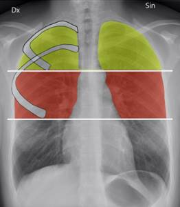
When radiography is performed in direct projection, the right and left pulmonary fields , which represent the projection of the corresponding lungs onto the plane of the X-ray film. The right pulmonary field is short and wide, the left one is narrow and long due to the peculiarities of the location of the mediastinal organs and the domes of the diaphragm. Surrounding the organs of the mediastinum, the lungs seem to envelop them and therefore are partially projected onto the median shadow. These parts of the lung, as well as areas of the lung covered by the diaphragm, are not visible on a direct radiograph. They are best seen in lateral and oblique projections.
For convenience, the pulmonary fields are usually divided into 3 belts and 3 zones . Horizontal lines drawn at the level of the lower edges of the II and IV ribs divide the pulmonary field into 3 zones - upper, middle and lower. The supraclavicular region or the apices of the lungs do not belong to any of the belts. Vertical lines drawn through the intersection point of the clavicle with the outer costal contour and through the middle of the clavicle segment projected against the background of the pulmonary field divide the pulmonary field into 3 zones - internal, middle and external.
The main characteristic of the pulmonary fields is their transparency, which is determined by three main factors: air filling, blood filling of the vessels, and the amount of lung parenchyma. The ratio of these factors determines the degree of transparency of the pulmonary fields. Obviously, transparency is directly proportional to the amount of air contained in the lungs, and inversely proportional to the number of blood vessels and lung tissue per unit volume.
In addition to intrapulmonary factors, the transparency of the pulmonary fields is also influenced by the condition of the chest wall. Thus, the transparency of zones and belts under normal conditions is not the same due to the projection overlay of the soft tissues of the chest on them. Therefore, for men, the lower belts are most transparent, then the upper ones, and the middle belts are the least transparent; in women, the lower belts are the least transparent due to the overlap of shadows of the mammary glands, the upper belts are the most transparent. The transparency of the zones in both men and women decreases from the middle to the lateral and inner zones.
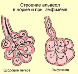
With emphysema, the lungs expand. Moreover, this process is associated with stretching of the pulmonary alveoli with air. During emphysema, acute and chronic processes are distinguished.
Chronic pulmonary emphysema develops as a result of a gradual loss of elasticity of the alveoli. Loss of elasticity is associated with constant stretching in chronic respiratory diseases. What is important in this process is the persistent irreversible expansion of the air spaces.
This pathological process is accompanied by increased swelling of the lung tissue of the terminal bronchioles. A complication of this disease is the development of cardiopulmonary pathology. Moreover, there is a high risk of mortality with emphysema, and the ability to work is significantly reduced.
What it is?
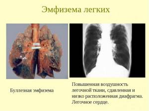
Pulmonary emphysema is a pathological change in lung tissue, which is accompanied by irreversible phenomena and persistent clinical manifestations. Lung tissue undergoes changes. Its increased airiness is noted.
With emphysema, there is a significant increase in lung size. Destruction of alveolar septa is often observed. Which also indicates a pathological process.
Pulmonary emphysema is also divided into primary and secondary pathology. Primary is associated with the direct predominance of congenital factors. Secondary pulmonary emphysema is associated with various diseases. That is, it is a consequence of various chronic diseases.
In the development of pulmonary emphysema, emphasis is placed on the unclear etiology of the disease. Since in some cases emphysema is not associated with any disease. In particular, congenital or primary pulmonary emphysema is distinguished. In this case, one lobe of the lung is affected.
Selecting the right tools to increase transparency
You won't achieve greater transparency by simply declaring your desire to know how things work. To do this you need the right tool with the right capabilities. When choosing such a tool, pay attention to the following functions:
- Taskbars . They will allow you to cover all important data at a glance.
- Reports . Regular automated reports on team performance, status and other aspects of project execution.
- Workload schedule . Makes it possible to check the workload level of an individual participant or the entire team.
- Resource management . Helps allocate team resources (both physical and financial).
“Detailed, customizable reports on projects, people and team progress helped us achieve excellent results when launching projects,” says Glineke from Only-apartments.
Causes
The causes of pulmonary emphysema are some pathological processes. In particular, chronic diseases are important. The etiology of the disease is associated with the following diseases:
Also important in the etiology of the disease are the processes that provoke the development of emphysema. These provoking factors are:
- congenital antitrypsin deficiency;
- tobacco smoke;
- toxic substances;
- employment in hazardous production.
Spontaneous pneumothorax is of particular importance in the pathological process. It is spontaneous pneumothorax that can cause rupture of air cysts. That is, in this case, air cysts are a consequence of swelling and overstretching of the lung tissue.
Causes and types of emphysema
Based on the cause and mechanism of development, primary and secondary pulmonary emphysema are distinguished. Primary pulmonary emphysema is an independent nosological form. It develops without previous bronchopulmonary pathology. In its development, great importance is attached to genetic factors (a1-antitrypsin deficiency). Secondary pulmonary emphysema occurs against the background of other respiratory diseases, primarily chronic obstructive bronchitis.
Based on the prevalence of the lesion, pulmonologists distinguish diffuse and local pulmonary emphysema. The diffuse form of the disease includes primary and secondary pulmonary emphysema, which developed against the background of chronic obstructive bronchitis. Local forms of pulmonary emphysema (bullous, irregular, peri-scar) develop for the following reasons:
- Cicatricial changes in the pleura and pulmonary parenchyma;
- Congenital changes in lung tissue;
- Local disorders of bronchial obstruction;
- Overextension of part of the lung due to shrinkage or surgical removal of other parts.
The following external factors play a great role in the development of pulmonary emphysema: air pollution, smoking, pulmonary infections. Pollutants (chemicals or compounds that are in the air in quantities that exceed background values) and occupational hazards, which lead to the formation of chronic processes in the respiratory tract and an imbalance in the proteolysis-antiproteolysis system, have a damaging effect on lung tissue. When air pollutants penetrate through inhalation, the membranes of the apical part of epithelial cells are damaged, inflammatory mediators, leukotrienes are released, and the balance in the oxidant-antioxidant system is disrupted. When the antioxidant system is depleted, inflammation of the mucous membrane of the respiratory tract continues. The development of emphysema is caused by exposure to particulate matter, suspended dust, and hydrocarbons. Fossil coal dust is especially dangerous.
The severity of the disease depends on the intensity and duration of smoking. The development of pulmonary emphysema is promoted by components of tobacco smoke - cadmium, sulfur, nitric oxide. They activate the work of alveolar macrophages and neutrophils. This helps to increase the level of metalloproteinases and neutrophil elastase. With prolonged smoking, the activity of proteolysis inhibitors decreases, and proteases, under conditions of the slightest deficiency of antienzyme function, cause significant damage to elastic fibers and destruction of the fine structures of the respiratory part of the lung. Tobacco smoke contains oxidants. They suppress the activity of anti-elastase inhibitors and inhibit recovery processes in the damaged elastic framework of the lung. Under the influence of smoking, the content of antioxidants in the blood plasma decreases, which together leads to the development of pulmonary emphysema.
One of the reasons for the development of pulmonary emphysema is a pulmonary infection. In the presence of infectious inflammation, the proteolytic activity of macrophages and neutrophils is stimulated. Bacteria and respiratory viruses can act as an additional source of proteolytic agents. Viruses themselves do not stimulate the production of neutrophils or macrophages, but due to their ability to suppress the immune system, they contribute to the exacerbation of inflammatory processes and the development of bacterial infection.
Symptoms
What are the main clinical signs of the disease? The main symptoms of emphysema include shortness of breath. Moreover, shortness of breath is associated with difficulty in exhaling. In terms of symptoms, this symptom resembles bronchial asthma. It is not without reason that bronchial asthma is the cause of the development of emphysema.
Shortness of breath has a progressive course. This is due to the fact that in the initial period shortness of breath is mainly associated with physical exertion. Subsequently, shortness of breath manifests itself at rest.
Cough is also observed with pulmonary emphysema. The cough is characterized by the production of scanty mucous sputum. In cases of respiratory failure, the following signs of the disease are significant:
- cyanosis;
- puffiness;
- swelling of the veins of the neck.
Patients with emphysema begin to lose weight. Even so to speak, they have a cachectic appearance. That is, symptoms of cachexia often predominate.
What is the cause of cachexia in emphysema? Cachexia in pulmonary emphysema is associated with high energy costs. These costs are calculated for intensive work of the respiratory muscles. The most dangerous variant of pulmonary emphysema is a repeated episode of spontaneous pneumothorax.
A complication of pulmonary emphysema is the process of irreversible phenomena in the cardiopulmonary system. Often the main consequence of this process is respiratory failure. Patients also experience swelling.
Read also: Classification of liver cirrhosis according to Child
Swelling is predominantly in the lower extremities. Ascites is also characteristic. This includes hepatomegaly, that is, enlargement of the liver. Spontaneous pneumothorax requires urgent measures, namely drainage and aspiration of air.
You can get more detailed information on the website: bolit.info
This site is for informational purposes only!
Increased transparency of lung fields
Definition of the concept
Here we consider an increase in the transparency of the lungs, not limited to clearly defined boundaries.
The exception is pneumothorax. The following diseases are considered: acquired pulmonary emphysema, congenital pulmonary emphysema, agenesis and hypoplasia of the branches of the pulmonary artery, progressive pulmonary dystrophy.
Research methods When diagnosing and differentially diagnosing these diseases, the main research methods are:
- X-ray examination, including examination in lateroposition.
- Radiography using samples from Yu. N. Sokolov and I. S. Amosov.
- Tomography of the root and parenchyma of the lung.
- Angiopulmonography.
- Radionuclide research.
- The transparency of the lung fields is normal.
- When analyzing the transparency of the pulmonary fields, it should be borne in mind that normally it is not the same throughout.
If you compare transparency along the horizontal belts, you will notice that in men it is highest in the lower sections, where the volume of lung tissue is greatest. On the contrary, in women, the lower belts are the least transparent due to the overlap of shadows of the mammary glands.
In men engaged in physical labor, the transparency of the middle belts is slightly reduced, which is associated with the development of the pectoral muscles, especially on the right. For women, these belts are more transparent than others.
Normally, the difference in the transparency of different parts of the lungs is expressed insignificantly and is subject to certain patterns. In pathological conditions it can be quite significant.
“Differential X-ray diagnosis of diseases of the respiratory organs and mediastinum”, L.S.Rozenshtraukh, M.G.Winner
Bloating of the lung due to partial blockage of the bronchial tube
Acute swelling of the lung due to obstructions located in the bronchial tree is of great practical importance. This primarily applies to intrabronchial non-contrast foreign bodies and the initial phases of development of both malignant and benign intrabronchial tumors. Foreign bodies have to be dealt with more often in children, and intrabronchial tumors in adults. Particularly great difficulties arise in...
Bloating of the lung due to partial blockage of the bronchus (Partial blockage of the bronchi)
With partial blockage of the bronchus, a heterogeneous decrease in the transparency of a certain area or the entire lung is visible. When you inhale deeply, a healthy lung becomes more transparent; when you exhale, its transparency decreases. In the affected lung, there is no noticeable difference in transparency between inhalation and exhalation. All this should be recorded in photographs. It is advisable to perform Sokolov or Amosov tests or use modifications of these...
Congenital emphysema
The disease is a malformation of the parenchyma and small bronchi of the lung. It occurs frequently, although it is not always diagnosed. This causes death in newborns due to respiratory failure. The most common synonyms of the disease: lobar emphysema of newborns, hypertrophic emphysema, progressive lobar emphysema, infantile lobar emphysema. The disease is detected in the first days or weeks of life. Usually one of the lobes of the lung is affected, more often...
Congenital pulmonary emphysema (Clinical picture)
The clinical picture of lobar emphysema depends on the degree of overextension of the affected area of the lung and the body’s ability to compensate for it. Respiratory disorders in newborns may appear immediately after birth or 2 to 3 weeks later. Characteristic symptoms include shortness of breath, cyanosis, worsening during feeding or crying, and attacks of asphyxia. With minor respiratory failure, all these symptoms are mildly expressed and progress relatively slowly….
Congenital emphysema and bloating with partial bronchial obstruction, spontaneous pneumothorax, giant air cyst
The main disease from which lobar emphysema should be differentiated is emphysema of the lobe or lung as a result of blockage of the bronchial tube by an accidentally ingested foreign body. The clinical and radiological pictures for both diseases may be identical. The issue is resolved with the help of subanesthetic bronchoscopy. One should also keep in mind spontaneous pneumothorax due to rupture of one of the bullae. However, on structural survey radiographs with...
Hypoplasia of the branches of the pulmonary artery
This anomaly may occur without clinically apparent symptoms. Patients are identified during a routine examination due to the conspicuous increased transparency of one lung. At the same time, the pulmonary veins are absent or underdeveloped, and the blood supply to the lung is carried out through the development of bronchial and other arteries arising from the aorta. X-ray, in addition to increased transparency of the pulmonary field on one side (unipulmonary emphysema, hypertransparent...
Progressive pulmonary dystrophy
L. Heilmeyer and A. Schmid (1956) described a picture of the disease, which was characterized by the disappearance of lung tissue, including bronchi, vessels in part of the lung (segment, lobe) or in the entire lung. The disease tends to progress. The etiology and pathogenesis are unclear. The disease is referred to differently: vanishing lung, idiopathic pulmonary atrophy, bullous pulmonary emphysema. Having examined lung preparations for progressive dystrophy, it was found that small and...
Progressive pulmonary dystrophy (X-ray picture)
The X-ray picture is very similar to that described for obstructive emphysema and differs in the following features: the lesion can be symmetrical or asymmetrical, but never completely involves both lungs. More often the upper lobes are affected on one or both sides; there is a sharper rarefaction of the pulmonary pattern in a limited area, a condensed and deformed pulmonary pattern is visible next to the affected area...
Progressive dystrophy and other lung conditions
Progressive pulmonary dystrophy and the system of bronchial cysts, hypoplasia of the pulmonary artery, giant cysts, spontaneous pneumothorax Progressive pulmonary dystrophy should be distinguished from several diseases that give a similar radiological picture: from the system of bronchial cysts, forming a picture of a “honeycomb” lung. With this disease, sharp walls of the cavities are clearly visible on radiographs and tomograms, which is not observed with progressive lung dystrophy. In addition, bronchial...
Progressive dystrophy and other lung conditions (Janus syndrome)
With a diffuse unilateral increase in the transparency of one lung (Janus syndrome), it is necessary to keep in mind the following main options: increased transparency of circulatory origin - congenital vascular pathology (absence of one pulmonary artery, in 50% of cases combined with tetralogy of Fallot) and acquired (local thrombosis and embolism of the branch pulmonary artery); increased transparency of ventilation origin - congenital (congenital emphysema, congenital cystic...
1 Next
Diagnostics
Anamnesis plays an important role in the diagnosis of pulmonary emphysema. At the same time, a certain trend can be traced in the anamnesis. Patients with pulmonary emphysema are usually employed in hazardous work. They also have a long history of smoking.
A history of chronic lung diseases is significant. Hereditary history also plays a role. Patients are noted to have lung diseases along a hereditary line. Diagnosis is also based on examination of patients.
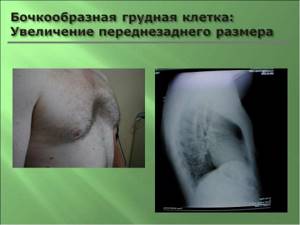
Patients have an enlarged chest. This includes protrusion of the supraclavicular fossa. On auscultation, shallow breathing is noted. In particular, there is a tendency for the presence of muffled heart sounds.
In laboratory diagnostics for pulmonary emphysema, the blood value is important. Erythrocytosis is observed in the blood. This includes an increased level of hemoglobin. Diagnosis is also based on the use of radiography.
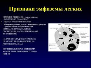
X-ray of the lungs indicates transparency of the lung fields. There is also a limitation in the mobility of the diaphragm dome. CT scan of the lungs allows you to clarify the presence of pathological changes. This is mainly associated with the presence and location of bullae.
An additional diagnostic method for pulmonary emphysema is spirometry. It is designed to identify the pathology of the respiratory reflex. Blood gas analysis can detect hypoxemia. This includes detecting hypercapnia.
Diagnostics includes consultation with a specialist. This specialist is a pulmonologist. A pulmonologist is not only able to make an accurate diagnosis, but also to identify complications or an increased risk of complications in a particular pathological process.
Fluorography results
On the left in the lower lobe of the lung in C9-10 there is infiltration of the lung tissue with a reaction of the costal pleura. The rest of the lung fields are without features. What does it mean? And is this dangerous for our unborn child? If this is something new and he knows nothing about it in principle, then he simply urgently needs to be further examined by a phthisiatrician at an anti-tuberculosis dispensary.
If you feel normal, there are no other changes in the lungs, and there are no complaints from the respiratory system, then there is nothing to worry about. Bronchospasm occurs in strong winds and frost. Sometimes it is difficult to breathe. I am 54 years old, how dangerous is this? Pneumosclerosis as a morphological phenomenon and radiological characteristics always indicates a previous disease of the bronchopulmonary system or the presence of a chronic disease now.
The essence of this phenomenon is this: when lung tissue is damaged, the affected areas are replaced with connective tissue. All the best! Hello, Lena! Increased transparency of the lung fields means that the amount of active lung tissue has decreased. This phenomenon accompanies emphysema.
But equally, all this can be both a defect in the fluorographic film and the characteristics of the radiologist’s description. I decided to do fluorography, the conclusion was that the pulmonary pattern was enriched. The enrichment of the pulmonary pattern is the body’s reaction to an acute respiratory viral disease. In this case, you need to exclude these factors and (or) smoking. An enrichment of the pulmonary pattern is observed with increased blood supply to the lungs.
Now she is ill with a temperature of 37.5-39.0 ARVI with an unproductive cough. But first, and the doctor is right, a posterior radiograph, which will either confirm or reject the suspected pathological changes. I am very concerned about receiving such a conclusion. There were always conclusions “the norm”! I never suffered from pneumonia or bronchitis.
A decrease in the transparency of the lung tissue is also accompanied by an increase or decrease in the volume of the affected area. Under various pathological conditions, the transparency of the lung tissue changes; first it is necessary to determine whether it is increased or decreased.
Prevention
Emphysema can also be prevented. Prevention is aimed at eliminating occupational hazards. Let’s say that people with a history of hereditary predisposition should protect themselves from harmful production.
Prevention of pulmonary emphysema is aimed at maintaining a healthy lifestyle. A healthy lifestyle not only involves giving up bad habits, but also proper nutrition. In particular, it is necessary to observe the rest and work regime.
Prevention of pulmonary emphysema should also be aimed at timely treatment of various lung diseases. And to eliminate possible complications of pulmonary emphysema it is necessary:
- strictly follow the doctor’s recommendations;
- take certain medications.
Prevention is based not only on preventing the development of emphysema, but also on preventing complications of this process. Active smoking is often a provoking factor. Therefore, active smokers have a responsibility to think about this.
Drinking alcoholic beverages in excessive quantities is also undesirable. Since pulmonary emphysema also affects the liver. And if alcohol affects liver cells, the risk of disease increases.
To prevent emphysema, it is also necessary to eliminate cardiac pathologies. Namely, to cure diseases of the cardiovascular system. Often, pulmonary emphysema affects the cardiac system.
Clinical examination plays a special role in the prevention of this disease. Since pulmonary emphysema is best detected at the initial stage. Subsequent symptoms can be quite severe. And the disease will smoothly move into the chronic stage.
Treatment
An important element of the treatment process is the elimination of factors predisposing to the disease. These include active smoking and inhalation of harmful substances. A major role in the treatment of pulmonary emphysema is given to the treatment of chronic lung diseases.
Drug treatment is aimed at eliminating pronounced symptoms. The following medications are indicated:
- salbutamol;
- fenoterol;
- theophylline;
- glucocorticoids.
These drugs are also used in the form of inhalations and tablets. Glucocorticoids include budesonide and prednisolone. If there is respiratory and heart failure, oxygen treatment is used. Namely, oxygen therapy.
Diuretics are prescribed for pulmonary emphysema. Preference is also given to breathing exercises. Breathing exercises can improve the course of the disease. Including improving the functioning of the respiratory system.
The mandatory method of treatment for pulmonary emphysema is surgery. However, this method is used according to indications. Surgery for pulmonary emphysema is aimed at reducing their volume.
It should also be noted that resection of areas of lung tissue is used. This helps to significantly improve lung function. In severe cases of pulmonary emphysema, lung transplantation is indicated.
In adults
Emphysema in adults develops mainly due to the harmful effects of environmental factors. And also as a result of the harmful effects of tobacco smoke. Smoking greatly complicates the disease process.
Emphysema is more common in men. The age category is most often from sixty years. It is in old age that all diseases worsen and this is due to the same influence of unfavorable factors.
In adults, as a result of chronic lung diseases, a severe obstructive process occurs in the pulmonary alveoli. What are the main signs of emphysema in adults? The main symptoms of the disease in adults include:
- cough:
- sputum production;
- body temperature may increase;
- swelling of the lower extremities;
- weight loss;
- weakness.
In adults, in the absence of proper treatment, the acute process of the disease passes into the chronic stage. The chronic stage of the disease leads to a long course and the development of complications. Respiratory and cardiac failure are often noted.
Diagnosis in adults contributes to early detection of the disease. Treatment with drug therapy can achieve good results. Surgical intervention helps to improve the disease process and even leads to recovery.
Read also: Preparing a patient for an ECG
In children
Emphysema in children is most often a congenital pathology. To prevent this disease in children, a comprehensive examination of the fetus is performed. This reduces the risk of developing intrauterine lesions.
Emphysema in children is associated with underdevelopment of lung tissue. And also with underdevelopment of the lung. What are the main symptoms of emphysema? The main signs of emphysema in children include:
- dyspnea;
- cyanosis;
- whistling breathing;
- asphyxia;
- convulsions;
- loss of consciousness.
These phenomena are considered the most severe symptoms of emphysema. If certain therapeutic measures are not carried out, complications will arise. These complications are cardiac and respiratory failure.
In newborns, shortness of breath increases, especially during breastfeeding. Symptoms of emphysema in school-age children are the following:
Significant signs of pulmonary emphysema in school-age children are also deformation of the chest and curvature of the spine. In some cases, pulmonary emphysema in children is accompanied by the presence of heart defects. This includes deviations in bone formation.
From these symptoms it follows that pulmonary emphysema leads to irreversible phenomena. The child may become disabled due to developmental defects. Diagnosis is primarily based on the use of radiography.
Diagnostic and therapeutic tactics for congenital lung malformations
Increased transparency of one or both pulmonary fields can be explained by emphysematous changes in the lungs, or changes in vascularization (hypoplasia of pulmonary vessels). A decrease in the size of the lung is possible with its hypoplasia, or pulmonary fibrosis, or a combination of these factors.
3. Due to the fact that when deciding on treatment tactics, surgery may be the method of choice, bronchography should provide information about the condition of the bronchi and the opposite lung. Good day, Elena. There are pathological changes in the lungs and heart. Pulmonary symptoms most likely indicate a previous illness, while cardiac ones should be addressed to a cardiologist - there are signs of pathology.
Forecast
With emphysema, the prognosis is most often unfavorable. This is due to the presence of complications. But when using inhalations, the prognosis improves significantly. Surgical intervention also has an impact on the formation of a favorable prognosis.
In some cases, surgical intervention leads to favorable prognosis. This is due to lung transplantation. However, it is carried out only when indicated.
The prognosis is also influenced by the course of the disease. The chronic course of the disease is the most unfavorable phenomenon. Since the chronic stage lasts for a long time and is difficult to treat.
Exodus
Death is possible due to respiratory and heart failure. But, if the development of these complications is prevented in time, the outcome improves. However, in case of pulmonary emphysema, supportive therapy plays an important role.
Maintenance therapy includes the use of inhalations. Even with persistent severe development of the disease, inhalations smooth out the symptoms of emphysema. The outcome of pulmonary emphysema is often disability.
Disability reduces quality of life. And the combination of pulmonary emphysema with heart defects in children leads to irreversible heart failure. The outcome in this case is death.
Lifespan
Life expectancy decreases with severe complications of the disease. And the presence of disability, ascites and edema leads to a decrease in its quality. Patients often require maintenance drug therapy.
Life expectancy is higher if the underlying pathology is eliminated in a timely manner. Mostly chronic diseases. For example, bronchial asthma.
The chronic course of the disease leads to the duration of the pathological process. The use of surgical methods is often required. However, only according to indications. Surgery not only promotes recovery, but also improves the quality of life.
What it is?
The term “pulmonary emphysema” refers to pathological processes in the lungs, characterized by an increased content of air in the lung tissue; it is a chronic pulmonary disease characterized by impaired breathing and gas exchange in the lungs. The name of the disease comes from the Greek. emphysao - “to blow in”, “to inflate”.
In recent years, the incidence of emphysema has been increasing, especially among older people.
The significant prevalence of this disease, its progressive course, temporary disability and early disability of patients due to the development of respiratory failure and cor pulmonale cause significant economic damage. Emphysema, along with chronic obstructive bronchitis and bronchial asthma, belongs to the group of chronic obstructive pulmonary diseases (COPD). All these diseases are accompanied by impaired bronchial obstruction, which is why their clinical picture is somewhat similar. However, each form of COPD has its own specific characteristics, and correct, timely diagnosis of these diseases allows for targeted prevention and rational therapy.
Dance of life
In the absence of pneumosclerosis, the pulmonary pattern is sparse and depleted.
In the diagnosis and differential diagnosis of PP and lung cancer, the radiological method is of leading importance. L. Heilmeyer and A. Schmid (1956) described a picture of the disease, which was characterized by the disappearance of lung tissue, including bronchi, vessels in part of the lung (segment, lobe) or in the entire lung.
The “ground glass” symptom is a nonspecific radiological symptom that reflects various pathological changes in the lung tissue at the level of the alveoli. Since this symptom is nonspecific, it is necessary to keep in mind the anamnestic data, clinical picture and concomitant pathology. The “ground glass” symptom may be observed around areas of compaction of the lung tissue (“halo symptom”).
CT examination in the expiratory phase helps to differentiate the presence of expiratory swelling of the lung tissue, helping to identify “air traps”. Strengthening the pulmonary pattern means a numerical increase in the elements of the pulmonary pattern per unit area of the pulmonary space (i.e., in the costal rhomboids).
The proliferation of peribronchial and perivascular compacted tissue, the alternation of pulmonary parenchyma with areas of emphysema leads to deformation of the bronchial tree and vascular bundles of this area.
It may also be called depletion (rarefaction) or a decrease in the pulmonary pattern. In women, these belts are more transparent than others. Normally, the difference in the transparency of different parts of the lungs is insignificant and is subject to certain patterns. Sometimes subpleurally located bullae are visible on tomograms. The roots of the lungs are expanded; Their characteristic shape is in the form of commas due to the expansion of the main trunks of the pulmonary artery.
The diaphragm domes are flattened and can be deformed into a tent shape. The excursion of the domes of the diaphragm is sharply reduced. Changes in the heart and blood vessels. The typical configuration of the developing cor pulmonale is caused by hypertrophy of the right ventricle, low standing of the diaphragm and the associated rotation of the heart to the right.
As a rule, emphysema is accompanied by chronic bronchitis and pneumosclerosis. On the other hand, these diseases, like bronchial asthma, are accompanied by emphysema. Acute swelling of the lung due to obstructions located in the bronchial tree is of great practical importance.
Causes of emphysema
The main cause of the disease is chronic bronchitis, which implies a chronic infection. Chronic bronchitis usually develops between the ages of 30 and 60 and occurs in men much more often than in women. In fact, the result of chronic bronchitis is the formation of pulmonary emphysema.
In the development of bullous emphysema, hereditary factors play an important role, as well as previous lung diseases (tuberculosis, etc.).
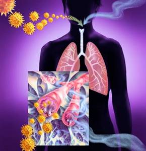
Smoking, air pollution with various dust particles and certain working conditions associated, for example, with constant inhalation of coal dust or asbestos and silicon particles, also contribute to the development of the disease.
At the same time, emphysema, leading to severe respiratory failure, can develop without a previous disease of the respiratory tract, that is, it can be primary.
Classification and causes of emphysema
Primary emphysema, which occurs due to congenital abnormalities in the body, is the most difficult to treat. In newborns it may appear due to obstruction of one of the bronchi. Secondary emphysema is a milder form, and at first it can even occur without obvious symptoms; the patient seeks treatment only when the disease has become chronic.
Tuberculosis can cause emphysema, its focal variety, which causes changes in the parenchyma around the foci of tuberculosis. Senile emphysema occurs as a result of age-related changes in blood vessels and deterioration in the elasticity of the walls of the alveoli.
What happens in the lungs?
The development of emphysema is associated with irreversible changes in the wall of the bronchi and lungs under the influence of prolonged inflammation and prolonged narrowing of the airways. The elastic properties of the lungs are disrupted: after exhalation, a larger amount of air begins to remain in them than should be normal, which causes overextension (inflation) of the lungs. Such excess air does not participate in breathing and the overstretched lung tissue does not function fully. Which, in turn, is accompanied by a loss of the ability to sufficiently contract and difficulty exhaling, as a result of which the supply of oxygen to the blood and the removal of carbon dioxide from it is disrupted. Compensatory, in order to improve the removal of carbon dioxide, shortness of breath occurs.
Read also: Is liver cirrhosis curable?
Also in the bronchi and lungs, the amount of connective tissue begins to progressively increase, which, as it were, “replaces” the air areas of the lung tissue, and also contributes to a long-term narrowing of the bronchi, regardless of the existing inflammation.
As a result of these changes, numerous air sacs of different sizes are formed in the lungs, which can be scattered throughout the lung (diffuse form of emphysema). Sometimes swollen areas of the lungs are combined with normal lung tissue (local form of emphysema). Bullous emphysema is also distinguished separately (a bulla is an emphysematous (swollen) area more than 1 cm in size).
Normal metabolism
What does this mean and how serious is it? Could reduced transparency have formed due to the fact that I somehow stood incorrectly during the flg, or is this still a process in the lung?
Then in December, against the background of ARVI, it reappeared, and also disappeared after 3 weeks. This may be as a result of a defect in the processing of the X-ray film, errors in the examination, and a number of other technical reasons. If we talk about possible pathology, this may be a manifestation of emphysema and a number of other problems, often associated with a long history of smoking.
An increase in the transparency of the lung tissue can be unilateral or bilateral; in some cases there is an increase in the volume of the lung tissue, in others it remains normal. When analyzing the X-ray picture, it is necessary to determine whether the volume of the area of lung tissue in which airiness is reduced is increased or decreased and what its structure is.
Noteworthy is the decrease in the volume of the right lung and the decrease in transparency of its upper sections. Frosted glass" is a term used to characterize processes accompanied by a decrease in the density of lung tissue - a sign of the interstitial type of infiltration. Frosted glass is an area of moderately reduced airiness of the lung tissue; the key sign of this condition is the visibility of the pulmonary vessels and bronchial walls.
Symptoms of emphysema
The “classic” manifestations of diffuse pulmonary emphysema include:
- severe shortness of breath;
- cyanosis;
- an increase in the volume (barrel shape) of the chest and a decrease in its respiratory movements;
- expansion and sometimes bulging of intercostal spaces;
- widening or bulging of the supraclavicular areas.
In the early stages of emphysema, the main symptom is shortness of breath on exertion. At first it is unstable and appears more often in winter, then at any time of the year. Subsequently, shortness of breath occurs at the slightest physical effort and, finally, can occur at rest. Patients experience a short, “sharp”, “grasping” inhalation and a prolonged exhalation. They exhale with their lips closed, puffing out their cheeks (“puffing”). The respiratory movements of the chest are reduced; additional muscles are involved in breathing: the chest and neck.
Shortness of breath that for many years, without noticeably manifesting itself and gradually progressing, it turns into a condition that threatens the patient’s life.
Patients with pulmonary emphysema in the initial stages of the disease take a forced position on their stomach with their head and shoulder girdle down, which brings them relief. However, with severe emphysema with pronounced changes in the chest and fatigue of the respiratory muscles, the horizontal position causes intense work of the diaphragm, so patients are even forced to sleep in a sitting position. Patients with pulmonary emphysema often take a sitting position with their torso slightly bent forward, resting their hands on their knees or the edge of the bed, which allows them to fix the shoulder girdle and include additional muscles in the act of breathing.
In advanced cases, cyanosis appears: the tongue appears blue; lips and nails become bluish, especially after physical activity.
Questions and answers
How is pulmonary emphysema treated?
Patients with emphysema are prescribed a conservative course of treatment, which allows them to cope with symptoms, ensure normal functioning of the respiratory tract and eliminate possible complications. The duration of treatment depends on the severity of the patient's condition and the symptomatic picture. It is possible to adjust the previously chosen tactics in the direction of adding or eliminating some of the medications taken.
Is there a cure for emphysema?
We can talk about the chances of curing the disease only on the basis of research results and timely seeking medical help. In the early stages, it is possible to almost completely eliminate the signs of pathology and return the patient to the ability to breathe fully. In advanced cases with numerous complications, it will not be possible to cope with the disease without consequences.
What is the main symptom of emphysema?
The disease is indicated by shortness of breath after physical exertion or without it (in the later stages), as well as a cough with a large volume of sputum with purulent or mucous contents. Other symptoms - wheezing and whistling, protrusion of the chest, puffiness and swelling of the face - may vary depending on the nature of the pathology.
What can you do?
Treatment should begin at the stage of bronchitis, even before the development of emphysema. Since most often, due to the patient’s late visit to the doctor, by the time of the first visit, irreversible changes have usually already occurred in the lungs, which significantly complicates subsequent treatment.
It is necessary for the sick person to be directly involved in the treatment. He must understand and realize the seriousness of the disease and possible complications.
It is necessary to categorically exclude smoking and other harmful substances, incl. occupational, effects on lung tissue, limitation of physical activity, rational employment.
Quitting smoking is an extremely important undertaking. It should take first place in the treatment of this pathology. It is necessary to keep in mind the following: stopping smoking immediately has a greater effect than gradually reducing the number of cigarettes smoked; high motivation to quit smoking is the main factor determining success; Chewing gum and skin patches containing nicotine can help reduce smoking urges, especially when used as part of a smoking cessation program.
What can your doctor do?
Your doctor (pulmonologist or therapist) will conduct the necessary examinations:
- examination, auscultation (listening), percussion (tapping) of the chest;
- X-ray examination of the lungs (characteristic is swelling of the lung tissue and an increase in its airiness, downward displacement of the diaphragm);
- computed tomography of the lungs is often used to diagnose and determine the exact location of bullae;
- examination of the function of external respiration: allows you to identify the degree of impairment of lung function (to reduce the amount of air that the patient is able to exhale).
The main methods of treating emphysema:
- quitting smoking: as already mentioned, the main method of preventing and treating emphysema;
- oxygen therapy (inhalation of air with a high oxygen content, possibly at home);
- special breathing exercises;
- adequate and thorough treatment of the disease that led to emphysema (chronic bronchitis, bronchial asthma): antibiotics should be used for infectious processes and for their prevention. They also use drugs that reduce the amount of sputum and thin it, which makes expectoration easier; Substances that dilate the bronchi and relieve spasm of the bronchial muscles are also administered.
For bullous emphysema, surgical treatment is recommended. The essence of treatment is removal of bullae. Such operations can be performed either using a classic approach with opening the chest, or endoscopically (using special instruments through punctures of the chest). Timely removal of bullae prevents the development of such a serious complication as pneumothorax.
In any case, you should not self-medicate. If you suspect that you or your relative have emphysema, you should immediately contact a specialist for timely diagnosis and initiation of treatment. In case of severe forms of the disease, your doctor may suggest registration of a disability group. But in order for the disease not to lead to complications and disability of the patient, you need to contact a specialist and be observed by him if you suffer from chronic bronchitis, have bad habits or occupational hazards associated with inhaling coal dust or asbestos and silicon particles.