Medical X-ray equipment is a set of equipment that produces X-ray radiation and uses it for diagnostic and therapeutic purposes.
This radiation was discovered in 1895 by the German scientist Wilhelm Roentgen. To better study his discovery, the scientist had to invent a special tube. Later, other devices were needed that made it possible not only to study, but also to use this radiation. So gradually an X-ray machine appeared.
X-ray units for all directions
On our company’s website you can buy any X-ray machines.
In order for you to be able to easily decide on the choice of installation, we have collected in our catalog the best models of devices in this area from the world's most famous manufacturers of medical equipment. At your discretion, we offer both particularly popular devices and X-ray systems that have proven themselves to be affordable, budget options suitable for equipping small private clinics. Any X-ray systems in our company can be purchased at a discount, which we will gladly provide to our partners as part of the program for the development of Russian medicine, with the transition to new generation technologies for the diagnosis, prevention and treatment of various diseases. We present to your attention a wide range of devices for radiography, fluorography and fluoroscopy, both classical and digital. Some of them belong to modern remote-controlled complexes, others are equipped with a C-arm module, which will allow the operator to carry out exposures directly in the hospital room, with higher quality and accuracy of the image. Foreign X-ray machines include leading devices from companies such as Philips, Canon, Samsung, Siemens, SG HealthCare, Medonica, POSKOM, Porta, GIERTH, ItalRay, LISTEM, EUROCOLUMBUS, Dixion and EcoRay. Many of them are equipped with advanced Toshiba X-ray tubes and powerful microprocessor-controlled high-frequency radiation generators.
In our company, Russian equipment remains available, which is often distinguished by a lower price and equally efficient and reliable operation. X-ray systems “RIM”, “S.P. Gelpik", "ADANI", "PROTON", "AMICO", "ELECTRON" are the product of domestic and joint developments. They are successfully used in hundreds of hospitals and medical centers, performing the work of diagnosing complex clinical cases every day, helping doctors make the correct diagnosis and perform world-class operations. On par with foreign products, Russian X-ray devices are often equipped with sets of digital detectors and support the latest technologies for obtaining and processing images, including in automatic mode. You can study the main characteristics and get an idea of the specifics of a particular device on our website.
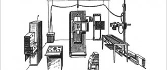
With the discovery of the so-called “X-rays” by physicist Wilhelm Conrad Roentgen in 1895, a new era in medicine began. X-rays literally revolutionized medical practice. After all, it became possible to study organs and tissues in a living person without causing him any harm. In those years, scientists did not yet know about the dangers of excessive exposure to X-rays on the body - both the person being examined and the researcher. And the designs of the X-ray machines themselves were imperfect and cumbersome.
By the way, the first successful X-ray photograph was the hand of Wilhelm Roentgen’s wife, and he received his Nobel Prize in 1901 for the invention of “X-rays” 2 years later, since he was busy with research and could not find time to come for the prize. In general, this scientist was distinguished by selflessness, modesty and nobility.
In Russia, the first X-ray machine was designed in 1896 by physicist and electrical engineer Alexander Stepanovich Popov, known throughout the world as the inventor of radio.
X-ray machines were constantly improved, and the science of radiology moved forward by leaps and bounds. Today this is a high-tech, rapidly developing and at the same time very subtle and elegant area of medicine, which allows solving a wide variety of diagnostic problems. If we consider radiology in a simplified and schematic way, we can distinguish the following sections:
- Radiography is the production of images on film or in digitized form using the reverse negative principle. The positive aspect of the method is minimal radiation exposure and the possibility of detailing. The negative point is the duration of image processing and the lack of dynamics of the process.
- Fluoroscopy is the process of obtaining an image on a screen using the direct negative principle. This method makes it possible to examine in real time and allows you to change the patient’s position during the examination. Which, ultimately, allows you to quickly and accurately identify the localization of the pathological process.
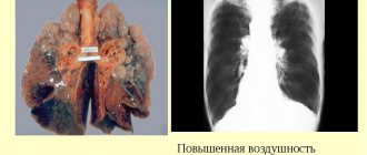
- Fluorography is a reduced image of an x-ray object, with minimal radiation exposure. The fluorographic method mainly examines the mammary glands, organs of the chest and the skeletal system. Currently, film fluorography is gradually being replaced by digital fluorography. And fluorographic devices can be both stationary and mobile.
- Contrast radiography is based on the introduction into the body of radiopaque substances, which are divided into X-ray positive and X-ray negative. The method is based on the fact that these drugs absorb x-rays less or more than soft tissues, thereby creating contrast in relation to the examined organ.
- X-ray television: teleradiography and telefluoroscopy are studies in which an X-ray image is transmitted as a television signal using an electron-optical converter and reproduced as a positive X-ray image on a television monitor. The main advantage of the technique is that research data can be transmitted over any distance.
The modern medical industry produces a wide variety of X-ray machines. According to the method of application, X-ray machines can be system-wide and special, digital and analog. Depending on the area of application, the equipment can be neurodiagnostic, urological, mammographic, cardiological, dental, etc.
As an example, let’s look at the premium dental high-frequency intraoral X-ray machine Carestream CS 2200.
Specifications:
- Digital remote control
- Generator voltage 60/70 kW
- Focal spot - 0.7 mm
- Voltage stabilization system on the generator
- Indication of the radiation dose received by the patient after exposure
- Anti-vibration bracket
- Possibility of connecting an additional (remote) release button
Advantages:
- Full generator voltage control: 60 kV for high contrast or 70 kV for wide grayscale studies
- Convenient and intuitive interface
- Highest image quality with minimal exposure time
- Optimized for use with CS RVG radiovisiographs and CS film
- High-frequency generator technology reduces radiation doses by 25%
- Various wall mounting options (vertical, right/left) and mobile stand options
Conducting X-ray examinations and their interpretation, of course, require knowledge and skills, constant clinical practice, and the ability to interact with colleagues. X-ray equipment is constantly being improved, new methods and research programs, and new methods of image analysis are emerging. After all, a properly selected study can serve as a decisive factor in establishing the correct diagnosis and saving the patient’s life.
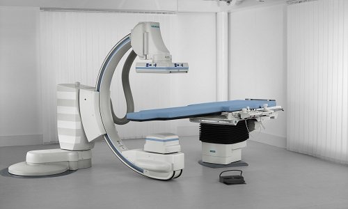
Stationary X-ray machines
Despite the growing popularity of mobile radiography and fluoroscopy, a stationary X-ray unit remains one of the most popular tools when staffing public and large private clinics. Such a device copes with the most complex tasks, especially if it is equipped with digital detectors and powerful automated workstations for the doctor and assistant. As a rule, they easily replace two or three older systems, saving money and providing a universal approach to all operations. Digital X-ray photography dramatically reduces the time usually spent on laboratory tasks such as developing film. The picture can be printed immediately by simply connecting a modern thermal printer to the device.
Thanks to the presence of an automated workstation with the latest software, you can not only quickly process visuals, but also maintain a database. At the same time, many systems are compatible with wireless communication protocols, which will allow the operator to immediately send the image to a single information database of the medical institution. Some devices are mounted on a ceiling mount, which reduces room congestion and adds extra shine to the shooting. However, most devices are mounted on the floor, using rails for motorized smooth movement of the emitter column, a stand for vertical images and other moving parts of the system. As an alternative, our company offers stationary X-ray machines that work with cassette holders, including phosphor and standard cassette formats.
Types of X-ray machines
Many modern clinics have high-quality X-ray equipment. With its help, the doctor can identify pathology in a patient at an early stage of development.
X-ray equipment comes in 3 types:
- permanently installed;
- used in mobile installations;
- portable.
The equipment that is installed in special vehicles is called mobile. Specialized cars, wagons and even water vessels are used as transport. There is also a darkroom for obtaining x-ray photographs. Such equipment must be protected as much as possible from dust and dirt. Exceptional tightness and reliable fastening are required.
Portable X-ray equipment can be transported by any vehicle on any road. But it must be securely fastened, since even minor damage to the device can lead to bad consequences.
Stationary X-ray equipment is installed in special rooms at medical institutions.
Science does not stand still. Every year the technology improves. X-ray machines were not spared this either. They are changing more and more, new functions are appearing. As a result, new research methods are coming to the aid of doctors. Among them:
- A fluorograph designed for examining the chest. This is the most frequently used and probably familiar device to everyone. With its help, the skull is also examined. This device allows you to see a crack just a few microns in size.
- Dental equipment used by dentists to monitor pathologies of dense tissues of the oral cavity. This device is narrow in its specialization, but nevertheless, very important.
- An angiograph designed to help cardiologists. He is able to detect problems in cardiac activity at very early stages of disease development. The device is highly informative and sensitive.
- A computed tomograph is a universal device. It is used to determine various pathologies located in any part of the human body, including the brain. It looks like a large capsule, where the person being examined can fit completely.
Simple X-ray machines began to be replaced by more advanced ones - digital ones. They are more convenient to use and less secure. With their help, images are obtained of higher quality. Thanks to them, it is possible to identify the pathological process much earlier, which contributes to early treatment.
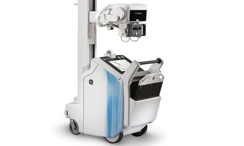
Ward X-ray systems
It often happens that it is not possible to take an x-ray using a stationary device. What to do if the patient is in a coma or needs urgent surgery? The answer to these and many other questions was the emergence of mobile X-ray devices. They are mounted on movable stands with a wheel base, so they can be moved to the desired compartment in a matter of minutes. The technology is on par with programs and algorithms, so such devices include the best software and special automated applications that will allow the doctor to instantly select preset shooting parameters. Ward X-ray devices support more than ten areas of medicine, including cardiology.
It is worth considering that the capabilities of a particular mobile device directly depend on the plug-ins. Choosing between GE Healthcare, LISTEM, Dixion, S.P. Gelpik" and other companies, pay attention to the price, equipment and compatibility with various additional units. Expert-level installations with wireless digital detectors, as well as mobile devices supporting C-arc modules, look especially attractive. These include X-ray systems from EUROCOLUMBUS and some other manufacturers.
Portable X-ray devices
Some areas of modern medicine require a portable X-ray unit that can easily fit in the trunk of a car. An example would be disaster medicine. In the event of an avalanche, mudflow or rockfall, forest fire or man-made accident, it is very often not possible to deliver the victim to a hospital with a stationary X-ray or at least a ward X-ray system. But before evacuation, the patient must be given first aid and factors such as possible spinal injury or head injury must be identified. The outcome of the diagnosis will directly affect the further development of the situation, because even the method of transporting the patient will depend on the examination. Such situations are also typical in sports medicine, traumatology and orthopedics.
EcoRay, PORTA, GIERTH and Poskom portable X-ray machines are the best tools in the hands of a specialist when it comes to portable radiography. The low power of the generators guarantees a low radiation dose, which will allow you to work with minimal or no protection. Such systems do not require a specially prepared office; they can be deployed in a military hospital tent or a mobile community care center. As a rule, they are used to diagnose the condition of the extremities and identify injuries to large internal organs. Such X-ray devices remain just as popular in veterinary medicine, where stationary X-rays are not in demand at all for most operations, and even a mobile unit is an unnecessary luxury due to its excess power.