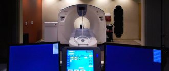
PET/CT (positron emission tomography - computed tomography) is a method for diagnosing diseases that combines the study of the structure and functional characteristics of tissues. This technology is most in demand in oncology for diagnosing and determining the extent of spread of malignant tumors.
- Description of PET/CT technology
- Advantages and disadvantages of PET
- Types of drugs for PET
- When is PET/CT indicated?
- Contraindications to PET/CT
- Preparing for PET
- Passing PET/CT diagnostics
- Complications and side effects of PET/CT
- Radiation exposure to the patient during PET scanning
Description of PET/CT technology
PET technology is based on the study of both structural and functional characteristics of tissues. Tissue function is assessed through metabolism. For example, a universal substance is selected that is necessary for all cells of the body. It is labeled with a radioactive label, injected into the body, and the places of its maximum accumulation are observed.
One of the most versatile substances in the human body is glucose. It is necessary to nourish almost all cells and tissues. But its greatest consumption is in malignant neoplasms, since a lot of energy is spent on the growth and reproduction of the tumor.
In PET/CT studies, glucose is labeled with radioactive atoms with a short half-life, such as fluorine-18. And after introduction into the body, it accumulates in large quantities in tissues with the most intense metabolism, i.e. in malignant neoplasms.
The radioactive tag decays, emitting energy in the form of gamma rays. The radiation is recorded by the device, and based on the data obtained, a visual model is built that shows the location of the tumor, its size and metastases.
Radioactive tracers accumulate only in pathological cells, while healthy tissues are not visualized. When the doctor wants to see both healthy and changed structures, a computed tomography scan is performed. It allows you to obtain a detailed image with millimeter accuracy.
After receiving data from both scanning systems, the software overlays them to create an image that gives the doctor a clear picture of where the pathological lesions are located.
Book a consultation 24 hours a day
+7+7+78
CT: what is it?
Computed tomography is a method of layer-by-layer examination of the body. Any area from the head to the pelvis can be examined. The method is based on the attenuation of X-ray radiation by tissues of different densities.
The X-ray tube rotates around the object of study, irradiating it. The weakened radiation is perceived by detectors on the opposite side. Computed tomography can be stepwise or spiral. With step-by-step CT, the tube rotates around the object while the table is stationary.
After completing a full revolution of the tube, the table moves a few millimeters and scanning starts again. With spiral tomography, the table moves sequentially without stopping. The difference between the original and attenuated radiation is estimated. Cross-sectional images are generated and displayed on the screen for analysis.
Advantages and disadvantages of PET
Advantages:
- The ability to combine several types of diagnostics in one study, which allows you to obtain highly accurate data.
- The examination is painless and does not cause discomfort.
- Possibility of diagnosing cancer at an early, pre-symptomatic stage.
- Possibility of studying all organs at once.
- The procedure is performed on an outpatient basis and does not require hospitalization.
The disadvantages of the method include the need to use radioactive drugs. To reduce risks, the study is carried out in strict accordance with radiation safety standards. In general, the side effects of PET scans are not commensurate with the valuable information this research method provides. In this case, three rules apply:
- The research must be justified. The benefits of diagnostics must outweigh the harm from exposure.
- Optimization. Medical staff are obliged to take all measures that will minimize radiation exposure to the body.
- Rationing. All radiation safety standards and regulations must be strictly observed.
Is positron emission tomography harmful?
Due to the use of two types of ionizing radiation at once (gamma radiation from PET and x-ray radiation from CT), this method has a very significant radiation exposure to the body. The estimated effective radiation dose to the whole body during intravenous administration of diagnostic doses of an isotope drug (150-555 MBq) ranges from 3.7 to 13.9 mSv, and this is only the radiation dose of the gamma camera; to it you need to add the radiation dose from the CT X-ray tube, which can double and sometimes triple. At the same time, the recommended radiation dose for the population does not exceed 5 mSv.
As you can see, when added, the figure turns out to be very impressive, so the method is not used in pregnant women and is not recommended for children. However, the diagnostic value of the technique in most cases exceeds the risk of the consequences of ionization.
Types of drugs for PET
The potential of PET studies is determined by the arsenal of radiopharmaceuticals used—pharmaceuticals that are labeled with unstable isotopes that make the substances radioactive.
Currently, isotopes of the following elements are widely used in PET studies:
- carbon-11;
- nitrogen-13;
- oxygen-15;
- fluorine-18.
Fluorine is most widely used in oncological practice because it has the longest half-life and the lowest radiation energy. This, firstly, allows you to obtain images of high quality and spatial resolution. And secondly, the relatively long half-life (109.8 min) makes it possible to transport the drug from the production site to scanning centers.
The most common radiopharmaceutical in oncology is fluorodeoxyglucose FDG. This is an analogue of glucose, which is a universal substance absorbed by all cells of the body. Cancer cells absorb it faster, so they accumulate the drug in a higher dosage, which is clearly visible during scanning. The disadvantage of this substance is that it accumulates in slightly larger quantities in brain tissue and nephrons, which can cause illumination of these organs even in normal conditions. Therefore, the question arises of searching for other, more specific radiopharmaceuticals.
For the brain, such a drug is 18F-FET. It contains the amino acid tyrosine, labeled with the fluorine-18 isotope. Tyrosine has a very high selective accumulation in brain tissue, which is used for imaging neurotumors. In addition, 18F-FET is used for the diagnosis of oropharyngeal tumors, in the search for metastases and for the diagnosis of lesions of the lymph nodes of the neck.
In addition, there are a number of specific radiopharmaceuticals for the diagnosis of individual tumors:
- 68GaPSMA - used in the diagnosis of prostate cancer.
- 68Ga-DOTA-TATE, -NOC, TOC, -LAN, etc. are used for the diagnosis of neuroendocrine tumors.
- 18 FCH - used to diagnose hepatocellular carcinoma.
The choice of radiopharmaceutical is made by a specialist doctor at the PET center based on the histological type of cancer, sensitivity and specificity of the tracer.
Indications:
- Identification and study of the prevalence of oncological process. SPECT allows you to detect the primary tumor and metastases, including distant ones; In addition, it allows you to study the biological processes of cancer cells (levels of apoptosis, pathological hypervascularization, changes in cell replication, involvement of surrounding cells, etc.) The advantage of the method over PET is the high spatial resolution (less than 1 mm), which allows you to obtain detailed structural information about the pathological hearth.
- Skeletal SPECT is the most widely used, mainly for searching for bone metastases. Bone SPECT is performed using a diphosphonate labeled with technetium 99m.
- Diseases of the cardiovascular system. Allows you to study the functional state of the heart (study of ejection fraction, myocardial perfusion, its viability, oxygen uptake, glucose metabolism). In terms of information content, SPECT is comparable to and superior to echocardiography.
- Study of blood circulation in the brain. Using SPECT, it is possible to perform a local assessment of the state of vascular perfusion capacity, regional blood flow and functional reserve of the cerebrovascular bed. It is also widely used in the evaluation of tissues in acute and chronic cerebrovascular accidents, including to evaluate the effectiveness of treatment. In case of cerebral ischemia, this method is often preferable to others and competes well with PET.
- It is possible to study the serotonergic, dopaminergic and cholinergic systems to detect Alzheimer's and Parkinson's diseases, however, the diagnostic accuracy is significantly inferior to PET.
When is PET/CT indicated?
- Search for the primary tumor site when metastases are detected.
- Staging of an already identified malignant process.
- Differential diagnosis of relapse and post-therapeutic or post-surgical changes.
- Monitoring the course of the disease, detecting relapse (for example, with an increase in the level of tumor markers).
- Planning for radiotherapy and surgery.
- Planning a biopsy of the tumor and determining its most “aggressive” area.
Contraindications to PET/CT
There are absolute and relative contraindications to PET/CT. The following states are considered absolute:
- Pregnancy. The study is not carried out when a diagnosis is established, and in the case when a woman suspects its presence.
- Lactation. It is recommended to stop breastfeeding 6 hours before the intended examination. The timing of resumption of feeding is determined by the tracer used.
A relative contraindication to the study is decompensated diabetes mellitus when planning to use fluorodeoxyglucose (FDG) as a radiopharmaceutical. In this case, consultation with an endocrinologist is required and stabilization of blood glucose levels below 7.7 mmol/l.
The study is carried out with caution in cases of impaired renal function, since the data obtained may be distorted due to retention of the radiopharmaceutical in the tissues.
Book a consultation 24 hours a day
+7+7+78
Preparing for PET
The day before the scheduled date of the study, it is necessary to avoid physical activity. If you plan to use FDG, you should also avoid foods rich in carbohydrates. This includes not only sweets and sugary drinks, but also fruits, vegetables, baked goods, legumes, etc. On the day of the procedure, eating is strictly prohibited. You can only drink clean water.
The patient should arrive at the clinic wearing comfortable clothing. It is advisable that it does not contain metal fittings and decorations (zippers, buttons, buttons, fasteners, etc.). But if necessary, staff can provide a disposable gown.
If the patient has diabetes, glucose levels should be measured before the test. If it is above 7 mmol/l, you need to take half the dose of insulin. The minimum time between insulin injection and radiopharmaceutical administration is 5 hours. If the patient is taking tablets to lower glucose levels, it is necessary to discuss with the endocrinologist the possibility of discontinuing them on the day of the study.
Passing PET/CT diagnostics
The patient must arrive at the clinic exactly at the appointed time. In this case, it is necessary to take all medical documents that are of interest within the framework of the disease - data from previous CT scans, histology data, laboratory tests, a statement of treatment, as well as data on concomitant pathology that may affect the diagnostic results (diabetes mellitus, kidney disease) .
After completing the paperwork, the patient is taken to the treatment room. There they take a blood test for glucose and measure anthropometric data. If everything is in order, the patient is taken to a relaxation room. There he is comfortably positioned, and a peripheral intravenous catheter is installed, through which the radiopharmaceutical is administered. To ensure it is evenly distributed, you need to drink a glass of water (or a contrast agent that the nurse will suggest) every 15 minutes. After administration of the radiopharmaceutical, you must be in a relaxed state and not talk. Any physical activity can lead to uneven distribution of the tracer and distort the results.
After 60–90 minutes, the patient is invited for a scan. First, a CT scan is performed, and then a PET scan. A CT scan takes a matter of minutes, while a PET scan takes up to half an hour. In this case, it is important to lie still so as not to provoke the formation of artifacts in the photographs that can distort the image. Both studies are carried out in the same tomograph and do not cause pain. After the procedure is completed, the patient must remain in the clinic for another hour, as a repeat scan may be necessary to clarify the data.
After a PET examination, the patient should not be in crowded places or have contact with children and pregnant women for 24 hours. In a family circle, you should keep a distance of at least 1 m from each other. Such restrictions apply for 24 hours after administration of the radiopharmaceutical.
In addition, during this period it is necessary to take a sufficient amount of liquid, at least 2.5 liters. This will help remove the contrast and radiopharmaceuticals faster and safer. You can drink more than just water. You can consume any caffeine-free products - juices, sparkling water, fruit drinks, mineral water, etc.
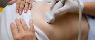
How is the SPECT-CT procedure performed?
- Initially, the patient is intravenously injected with a radiopharmaceutical, after which he waits for its uniform distribution throughout the body (when examining the skeleton - 2.5-3 hours, brain or heart - 30 minutes);
- Before the procedure, it is necessary to empty the bladder to avoid artifacts in this area during scanning;
- The scanning procedure itself takes place in two stages: first, for 15-20 minutes (when examining a certain area), or 40 minutes (when examining the whole body), two emission chambers are scanned, rotating around the patient; then the second part of the examination is scanning with a spiral X-ray tube of a CT machine, which lasts up to 5 minutes;
- To quickly eliminate the radionuclide, it is necessary to increase fluid intake; the half-life (the time during which the concentration in the blood is halved) of technetium is 6 hours.
Complications and side effects of PET/CT
Complications during PET/CT are mainly related to the administration of CT contrast. As a rule, these are allergic reactions and complications associated with vein puncture - vascular damage, hematoma, pain, neuritis, etc. Renal dysfunction occurs extremely rarely. If contrast is taken orally, nausea, vomiting and diarrhea may occur.
Dizziness and weakness may occur after a PET scan due to the need for the test on an empty stomach. Therefore, it is recommended to bring a snack with you that can be eaten after all procedures related to the study are completed.
Difference between SPECT and CT
CT is a structural imaging technique for tissue. It allows you to clearly determine the localization of the pathological process, the size of the affected area, its shape, the degree of involvement of surrounding tissues, etc. SPECT, in turn, is based on the ability of tissues to accumulate special radionuclide preparations to varying degrees due to different blood supply in them. Thus, foci of inflammation, tumors and metastases accumulate intravenously administered radiopharmaceuticals to a greater extent in their tissues, which is recorded by a gamma camera and allows obtaining layer-by-layer images of drug accumulation. However, the SPECT method alone did not have a high diagnostic value due to the poor spatial resolution, which did not allow one to accurately indicate the location of the detected accumulation focus. Therefore, SPECT-CT devices have been created that combine the advantages of both methods and make it possible to establish clear boundaries of organs while simultaneously studying the degree of drug accumulation in them.
Radiation exposure to the patient during PET scanning
Radiation exposure during PET/CT depends on the radiopharmaceutical used and the scope of the study. For example, scanning the head will expose you to less radiation than scanning the whole body. To minimize harm from PET/CT, the following standards are used:
- The research is carried out using ultra-short-lived isotopes, the half-life of which is several minutes or hours.
- An individual dose calculation is used for each patient, taking into account weight, height and age.
- The scanners use special filters and programs that reduce X-ray doses.
- Applying the required amount of liquid not only improves the quality of the resulting image, but also helps to remove the radiopharmaceutical faster.
In general, PET/CT is a fairly safe procedure, but it is performed only for strict indications when other studies do not provide the necessary information.
Book a consultation 24 hours a day
+7+7+78
Difference between SPECT and PET
PET differs from SPECT in that it requires radiopharmaceuticals containing radioactive isotopes that are capable of emitting gamma rays with an energy of more than 1024 keV. The possibility of using different isotopes makes it possible to determine a process that is tropic specifically for the tissue being studied or for a specific oncological process. When interacting with the tissues of the body, each primary quantum creates two particles: an electron and a positron. In the future, this leads to the simultaneous formation of two gamma rays emitted in opposite directions. This physicochemical process provides the unique diagnostic capabilities of PET.
In turn, with SPECT after injection of a radionuclide, a standard gamma camera captures only one quantum leaving the body. The absence of the need to use isotope radiopharmaceuticals makes the price of SPECT much more affordable than that of PET, and the technique more widespread.