Description
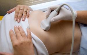
In the case of ultrasound, the machine creates vibrations, or waves;
When an ultrasonic wave reaches tissues with a certain acoustic resistance, it is refracted. That part of the wave that acts on tissues with less resistance will be absorbed by them and will travel further, and the other part, in front of which the resistance of the tissue is stronger, will be reflected. Roughly speaking, the more ultrasonic waves are reflected, the brighter and clearer the picture on the device screen will be. With MRI, the story is a little different - but the main role here also belongs to waves, only electromagnetic ones .
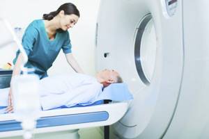
They create a strong magnetic field and record the response to it from some particles (the nuclei of hydrogen atoms are responsible for this). Essentially, the device registers the body’s response electromagnetic radiation and displays an image. This is not a "photograph" of the organ being examined, but rather a map of its electromagnetic signals .
Harmfulness
Such methods are safe for the patient’s health because they propagate sound or electromagnetic waves that are not capable of changing the structure of cells.
Important: Ultrasound and MRI can be done safely during pregnancy - ultrasound is used to determine not only the sex of the child, but also the risk of developing Down syndrome or congenital anomalies. The dangerous effect of ultrasound and MRI on the fetus is no more than a harmful myth , because there is no ionizing radiation from such studies.
Application
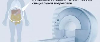
MRI is better suited for soft tissue , allowing tumors and studying, for example, the brain and spinal cord , although again this method can be used for other parts of the body.
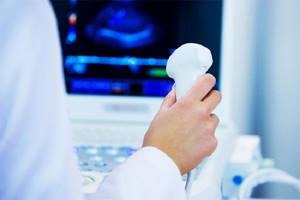
Ultrasound , on the contrary, has a limited spectrum of action . It is believed that it does not see organs that are hidden behind the bones (the ultrasound wave simply does not reach them). It is also not yet amenable to automation, meaning a specialist is needed to interpret the ultrasound results. However, the device can be easily installed right at the patient’s bedside, which is not possible with, for example, a massive MRI tunnel.
To understand what to choose, ultrasound or MRI with contrast, you need to consider their differences:
- Ultrasound allows you to confirm or refute the preliminary diagnosis made according to the clinical picture; if it is blurred, MRI is used;
- tomography of the abdominal cavity helps to examine the organ layer by layer in 3D, ultrasound gives two-dimensional results;
- Tomography better identifies diseases in the early stages.
Indications for ultrasound of the stomach:
- heartburn, nausea, vomiting, belching, pain, gas, bloating, feeling of heaviness in the stomach, coating on the tongue, unpleasant odor and bitterness in the mouth
- detection of ulcers, gastritis and polyps
- pathologies in the abdominal cavity
- intestinal obstruction
- determining the size and contours of the digestive organ
- detection of inflammation of the stomach walls
- assessment of reflexivity, peristalsis and gastric functions
- suspicion of cancer
- tracking the disease over time
Indications for MRI of the stomach:
- pain, nausea, vomiting, decreased and loss of appetite, change in stool, accumulation of gases
- suspicion of hernia, gastritis, peptic ulcer, its perforation and malignancy
- abnormal structure of the stomach
- monitoring the condition of the stomach walls
- suspicion of the development of a tumor and metastases in the gastrointestinal tract
- tracking the localization of the disease and the dynamics of its development
- quality control of treatment
Comparison of possible contraindications and restrictions
Ultrasound of the abdominal cavity is contraindicated in the following pathologies:
- infectious diseases (period of their exacerbation);
- purulent rashes over the dermis;
- heat;
- damage to the skin;
- acute cerebrovascular accidents;
- wounds in the abdomen.
During pregnancy, it is not prohibited to perform an ultrasound of the peritoneum. This diagnostic method, on the contrary, is used to assess the condition of the fetus, its development, and the condition of the woman’s reproductive organs.
Contraindications to MRI in the peritoneal area are:
- the presence of metal elements inside the body (vascular clips, pacemaker, insulin pump);
- allergy to contrast agent;
- weight more than 120 kg;
- claustrophobia (after all, the patient is in a tomograph tunnel. If necessary, he is given a sedative).
In what cases and why is it better to do an MRI?

Undoubtedly, magnetic resonance imaging allows us to find out exactly what is happening in the human body. The main advantage and advantage, compared to other research methods, is that tomography allows you to detect the disease at the initial stage of development , which greatly simplifies treatment and gives the patient a better chance of a speedy recovery in case of serious serious illnesses.
Help Of course, magnetic resonance imaging as a diagnostic method must be chosen in cases where it is extremely important to establish the most
accurate diagnosis , completely eliminating the slightest mistakes and mistakes.
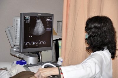
MRI examinations are carried out with subsequent image processing by a computer, so they are reliable and very accurate. Unlike MRI, the ultrasound procedure is directly related to the work of a specialist and the accuracy of the diagnosis may depend on the qualifications of the doctor , as well as on the human factor.
Typically, indications for MRI are: confirmation or exclusion of a diagnosis, prevention of cancer , a complete examination of the functioning of the human body, accurate diagnosis, identification of pathological processes, inflammatory processes, the development of tumors, monitoring after surgical interventions.
Important diagnostic differences
For the initial examination of the human body, none of the described methods are usually used. Ultrasound is more in demand in diagnostics because it is much cheaper and more convenient.
Doctors use CT scans when they urgently need to examine a patient and quickly assess the condition of organs in both the abdominal cavity and the retroperitoneal space. Without contrast, such a study can be performed in just 20 seconds.
This allows you to quickly navigate and perform the operation when minutes count.
At the same time, computed tomography usually gives a more complete picture of the state of the human stomach and intestines.
The reason is that the dependence on pulsation is much less than that of MRI. This way it is possible to track even minor changes in the condition of these organs.
MRI is much more often used when it is necessary to obtain the most detailed and informative conclusion to make a diagnosis.
It should be borne in mind that MRI of the abdominal cavity and retroperitoneal space are two different procedures. A detailed check of one organ can take up to 20 minutes.
An important advantage of using MRI is that the device makes it possible to conduct diffusion-weighted diagnostics of the bile ducts. In this way, the patient’s lymph nodes begin to be very clearly visible.
Even changes up to 2 mm will be noticeable very clearly - this gives an idea of the early stage of cancer.
Also on the list of advantages of MRI is the availability of color contrast. In this way, doctors can superimpose several images on top of each other at once and obtain complete data on the condition of organs and the presence of tumors.
How is MRI fundamentally different from ultrasound?
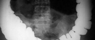
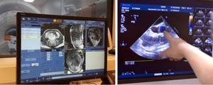
Ultrasound is a traditional method of studying internal organs, which makes it possible to evaluate the structure, anatomical features, already sufficiently developed disorders and progressive pathologies, and tissue structure. Ultrasound diagnostic devices provide an insufficiently detailed image , mainly two-dimensional (in two planes, measuring width and length). Therefore, in a black and white photograph, only some blurry outlines are visible, to understand which you need to be able to “read” them.
The MRI machine takes layer-by-layer sections of internal organs and visualizes the information in the form of a three-dimensional image. The study, with a high degree of accuracy, shows in detail not only the condition of each internal organ, but also its function over time . Therefore, if a tumor or functional impairment is suspected, it is recommended to do an MRI of the abdominal cavity for a more accurate examination and confirmation of the diagnosis.
How to prepare?
To increase the information content, it is necessary to prepare well for the procedure. This includes a number of rules:
- abstaining from food for 5-6 hours before the analysis;
- for 2-3 days it is good to give up all types of cabbage, beans, sour milk, raw fruits and vegetables, carbonated water, and do not drink alcohol;
- if an MRI is planned under anesthesia (for example, for patients with claustrophobia), preparation will include the avoidance of cosmetics that may interfere with the contact of the sensors;
- as prescribed by the doctor, take antispasmodic medications 30 minutes before the procedure, if required;
- Empty your bowels and bladder before the procedure.
Immediately before an MRI of the abdominal cavity, preparation is simple - remove all metal objects. If necessary, contrast is injected into the vein before the procedure begins.
In our clinic, cavity diagnostics are performed daily. To make an appointment, simply call us or fill out the form on the website. The specialist will advise you about contraindications and the course of the study.
When is an ultrasound and when is an MRI prescribed?
If you only need to assess the general condition of the internal organs, then in many cases it is quite possible to get by with an ultrasound. Ultrasound clearly shows internal bleeding, accumulation of exudate in the cavity (fluid), cysts, large tumors, and inflammation . But if the clinical picture is blurred and not clearly expressed, there is a suspicion of an early stage of pathological tissue change, then it is better to do an MRI of the abdominal cavity.
But sometimes ultrasound is still the more preferable or the only possible study. Despite the proven safety of MRI, this diagnostic method is not used at the beginning of pregnancy , when the baby’s organs are not yet formed; the procedure is prescribed with caution and only when indicated for small children, patients who cannot lie still, and sedatives are contraindicated for them.
Both research methods have relative and absolute contraindications.
Disadvantages of methods and contraindications for ultrasound and MRI are not prescribed for:
- open wounds and violation of the integrity of the skin in the area of study;
- purulent rashes on the skin in the peritoneal area;
- some dermatological diseases (for example, volumetric formations on the surface of the skin that make it difficult for the sensor to fit tightly);
- urinary incontinence (for examination of some organs, a full bladder is a prerequisite);
- increased gas formation, which cannot be eliminated.
MRI is not done in the following cases:
- in the first trimester of pregnancy (with the exception of cases where there is a threat to the life of the mother and clear indications);
- the patient has a cardiac pacemaker that is not compatible with MRI;
- the patient has a cochlear implant (depending on the model and manufacturer);
- the patient suffers from claustrophobia;
- for dyskinesias (involuntary movements), severe cerebral palsy, nervous tics.
Abdominal CT
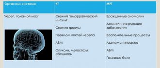
Computed tomography is one of the most informative examination methods. Usually, when it comes to CT scanning, most people first associate it with a brain examination. But in fact, using this technique, you can diagnose almost the entire organism. As a result, the condition of bones, connective and soft tissues, the nervous system, and blood vessels will be visible. Thus, computed tomography is a universal method and is often used in cases where it is urgently necessary to obtain a complete clinical picture of the disease. Therefore, abdominal CT is widely used to examine patients with heart attack, stroke, abscesses, and multiple injuries.
Benefits of CT
- Any internal organ can be examined, including the abdominal cavity;
- the result obtained provides information about the condition of bone, cartilage and soft tissues;
- the final result is presented in 3D format, that is, you can obtain a three-dimensional image of the examined organ;
- it is possible to study the structure of the examined tissues and identify the smallest pathological changes, that is, the disease can be detected at an early stage;
- very reliable diagnostic method - 98%.
Disadvantages of CT
In most cases, during this examination it is necessary to use a contrast agent to obtain a brighter image. And this already imposes a number of restrictions: the patient requires special preparation, and it is necessary to keep in mind the presence of contraindications: severe physical condition (serious illnesses), claustraphobia, severe obesity, allergies to substances containing iodine.
Which is cheaper: MRI or ultrasound?
The cost of abdominal MRI is higher than the cost of an ultrasound . However, comparing diagnostic procedures according to this criterion and choosing a research method in this way is absolutely incorrect. If you independently decide to examine the body for preventive purposes, you can sign up for an ultrasound scan, and then, with the result, go to an appointment with a specialist and consult.
If the doctor told you to do an MRI of the abdominal cavity, then under no circumstances should you replace this method with an X-ray, ultrasound, or. The diagnostic method is selected according to indications. Therefore, the doctor needs to obtain just such a detailed result, which will show the condition of the tissues and make it possible to assess the functioning of the organs. In this case, the price of an abdominal MRI should not be decisive. This is as pointless as being treated with inappropriate drugs simply because they cost less.
X-ray and MRI - what's the difference?
Fluoroscopic methods for studying the abdominal cavity include fluoroscopy of the stomach and small intestine, as well as irrigoscopy - the study of the colon. These techniques allow you to evaluate the pathology of the gastrointestinal tract and are performed using barium. Urography with intravenous contrast with an iodine-containing substance is used to study the kidneys. A regular X-ray of the abdominal cavity is used only to detect stones in the kidneys or bladder; it is not suitable for studying other abdominal organs, since X-rays are absolutely not retained in soft tissues. Thus, X-rays of the abdominal cavity are used only in combination with contrast (iodine-containing or barium), and are not suitable for native examination of other abdominal organs.
Conclusion: MRI is performed only after a preliminary examination of the troubling pathology; in all cases, ultrasound diagnostics or gastroscopy is performed; only in cases where it is impossible to make a detailed diagnosis, more complex methods are used (fluoroscopy, irrigoscopy, colonoscopy), only then CT, MRI or laparoscopy is prescribed.
General conclusions

Both diagnostic methods are highly accurate visualization methods that can be used to obtain photographic images and moving images of internal organs and systems.
MRI and ultrasound are performed without the use of radiation harmful to the human body, and both techniques are non-invasive and painless. As for preparation for the examination, ultrasound often requires dieting, fasting or a full bladder, depending on the organ being examined. MRI does not require special preparation, however, MRI examination has some limitations: the use of pacemakers and other implants, claustrophobia, inability to locate in a motionless state (an almost impossible condition without medicinal support for young children). Diagnosticians usually communicate recommendations before conducting tests.
Advertising