A CT scan uses x-rays to visualize the spine.
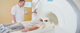
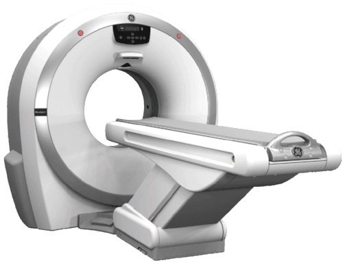
During a CT scan of the spine, the patient will lie on a table that is attached to the CT scanner, which is a large donut-shaped machine. A CT scanner sends X-rays through the body. Each rotation of the scanner takes a second and produces an image of a thin section of the organ or area being examined. One part of the scanning device can tilt to match the curve of the spine. All pictures are saved as a group on the computer. They can also be printed.
In some cases, a contrast agent may be injected into a vein or spinal canal to produce more detailed images of abnormal tissue (tumors, areas of inflammation, or nerve damage).
Indications
A CT scan of the spine is performed for:
- To study the vertebrae.
- Detection of problems with the spine, such as tumors, fractures, deformities, infection, or narrowing of the spinal canal (spinal stenosis).
- Detection of intervertebral disc herniation.
- Detection of compression fractures associated with osteoporosis.
- Identification of congenital problems with the spine.
- Study of problems identified by radiography.
- Monitoring the effectiveness of spinal surgery.
Indications for CT scan of the spine and features of the procedure
To understand when it is necessary to do a CT scan of the spine, let’s consider the main indications for this procedure:
- injuries;
- systematically occurring pain;
- assessment of the condition of the spinal column before or after surgical treatment;
- suspicions of the presence of benign or malignant formations;
- spinal canal stenosis;
- osteoporosis, the need to examine bone tissue;
- osteochondrosis;
- hemorrhage in the spinal cord;
- instability of any parts of the spinal column;
- pinched nerve;
- congenital spinal defects;
- intervertebral hernia;
- abscess.
This is only part of the indications for which the doctor prescribes a CT scan of the spine. They allow us to conclude that diagnostics makes it possible to carefully examine the spinal column, differentiate pathological changes, assess the condition of the vertebrae and the degree of deformation, if any.
How to prepare for the procedure?
Before a CT scan, you should tell your doctor:
- About the presence or likelihood of pregnancy
- Previous history of allergies to any medications, including iodine contrast agents.
- Heart disease, such as heart failure.
- Having diabetes.
- Taking metformin. The patient may need to adjust their medication intake during the day before and after the test.
- Having kidney problems.
- Presence of bronchial asthma.
- Multiple myeloma.
- The patient has had an X-ray with barium contrast agent (eg, barium enema) within the past 4 days. Barium appears on X-rays and makes it difficult to get a clear image.
- The patient is afraid of closed spaces. The patient must lie still inside the CT scanner, so the patient may need sedation to help them relax.
FAQ
How long does the procedure take?
Direct scanning using X-rays takes no more than 10 seconds. Sometimes a repeat image needs to be taken, sometimes doctors need to prepare equipment. Therefore, the entire procedure from the moment you enter the office can take up to half an hour. All this time you need to lie still - so the x-ray technician will definitely tell you how long the study will last.
Why can't CT scans be done for pregnant women?
Computed tomography is not indicated for pregnant women, especially in the first trimester. This is due to the fact that even minimal doses of x-ray radiation can affect the formation of the fetus. Therefore, you must inform your doctor about pregnancy before choosing a diagnostic method. You can feed your child after the examination if no contrast agent was used.
How does CT differ from MRI and ultrasound?
This question interests many patients. In fact, the main difference is that CT uses x-rays to produce images of tissue. In addition, there is a difference in sensitivity. Some tissues are better visible on MRI, others on CT. Ultrasound is the simplest and most accessible method; it is better to check internal organs, but not bone structures.
Answers questions:

Syubaev Roman Borisovich
Doctor - radiologist
18 years of experience
doctor
You can conduct an examination of any part of the spine (by referral or with a preliminary consultation) at the multifunctional CELT clinic. A modern tomograph from a reputable company is the key to a quick and comfortable examination, as well as accurate and timely results. After receiving the results, you can make an appointment with a CELT specialist and develop a treatment regimen. CELT is a place where the latest advances in medicine protect the health of patients.
How is the procedure performed?
A CT scan is usually performed by a radiologist. The images are analyzed by a doctor (radiologist). Other doctors may also view the CT scan.
The patient may have to remove any jewelry. The patient will need to remove all or most of their clothing, depending on which area is being examined. The patient may wear underwear for some scans.
During the test, the patient will lie on a table that is attached to the CT scanner.
The table slides into the round opening of the scanner and the scanner moves around your body. The table will move while the scanner takes pictures. The patient may hear a clicking or buzzing sound as the table and scanner move. It is very important to lie still during the test.
During the procedure, the patient will be alone in the scanning room. But the laboratory assistant will observe the patient through the window. The patient will be able to talk to the laboratory assistant via two-way communication.
The CT scan will take on average 30 minutes. Much of this time is spent preparing for the scan. The actual scan only takes a few seconds.
CT diagnostic services at CELT
The administration of CELT JSC regularly updates the price list posted on the clinic’s website. However, in order to avoid possible misunderstandings, we ask you to clarify the cost of services by phone: +7
| Service name | Price in rubles |
| CT coronary arteries | 16 000 |
| CT scan of the pelvis | 7 500 |
| CT scan of the chest | 7 500 |
All services
Make an appointment through the application or by calling +7 +7 We work every day:
- Monday—Friday: 8.00—20.00
- Saturday: 8.00–18.00
- Sunday is a day off
The nearest metro and MCC stations to the clinic:
- Highway of Enthusiasts or Perovo
- Partisan
- Enthusiast Highway
Driving directions
CT scan with contrast (CT myelogram)
A standard CT scan may be performed before contrast material is administered for a CT myelogram. Contrast is usually injected into the area around the spinal cord. A sample of fluid from the spinal canal (cerebrospinal fluid) may be taken so other tests can be done.
The table may be tilted or the patient may be moved to different positions so that the contrast is moved to different areas of the spine.
The patient must lie very still so that the contrast remains in the right place to produce clear images. Pulse, breathing rate and blood pressure can be checked during the test.
In some cases, contrast may also be injected into a vein in the arm.
A CT scan with contrast material usually takes 15 to 30 minutes. You should drink plenty of fluids for 24 hours after the scan to help flush the dye out of your body.
Decoding the received data
The decoding is carried out by a radiologist or radiation diagnostics doctor. To help the doctor, the patient can provide the results of examinations conducted earlier, opinions of doctors of related specialties, extracts and epicrisis about the treatment carried out or completed. In the photographs, the doctor finds and describes pathological changes in organs and tissues and correlates them with a particular disease. It should be understood that the conclusion of a computed tomography scan of the spine is not a diagnosis. The diagnosis is made by the attending physician, who has the opportunity to question the patient and examine him, as well as various laboratory and instrumental tests.
In terms of time, decoding takes from 30 to 60 minutes . After this, the patient receives pictures and a medical report. Pictures can be transferred digitally, for example on a disk, or they can be pre-printed on paper or film.
How is the procedure tolerated?
- The examination will not cause pain.
- Some people feel nervous inside a CT scanner.
- The contrast may cause the patient to feel warm and red and have a metallic taste in the mouth. Some people feel nauseous or have a headache.
- If a patient is receiving contrast injection into the spinal canal, the patient may feel a burning or pinching sensation when the needle is inserted.
- After a CT scan with contrast, the patient is advised to keep his head up and not bend over or lie flat. This will help prevent headaches and cramps.
Contraindications for examination
Computed tomography of the spine is not performed:
- pregnancy at any stage;
- patient weight exceeding the design capabilities of a particular device (usually up to 120 kg).
The following categories of patients are examined with caution and only if there are serious indications::
- children under 12 years old;
- nursing women;
- patients with renal failure;
- with multiple myeloma.
The use of contrast is prohibited in the following categories of patients:
- pregnant and lactating women;
- patients suffering from diabetes mellitus;
- patients with chronic renal and liver failure;
- persons with intolerance to iodine-based drugs.
Risks
The likelihood of problems occurring with a CT scan is low.
- There is a possibility of an allergic reaction to the contrast agent.
- If the patient is breastfeeding, then in order to prevent the contrast from getting into the baby’s milk, it is recommended to express the milk in advance and not put the baby to the breast for two days after the procedure.
- If the patient has diabetes or is taking metformin, the contrast may cause problems. You should stop taking metformin before the procedure and continue taking it some time after the procedure.
- The patient may experience nausea or vomiting after the test.
- There is a small chance of infection where the needle was inserted or bleeding in the area around the spine.
- An injection into the space around the spinal cord may cause headaches. In rare cases, seizures may occur after contrast material is injected.
- There is a small chance of getting cancer with some types of CT scans.
Spinal osteomyelitis
Osteomyelitis is a relatively rare and difficult to diagnose inflammatory disease of the vertebral bone tissue. It begins with damage to the bone marrow and then spreads to other components of the bone tissue. Leads to destruction of the vertebra with subsequent deformation of the entire spinal column.
Osteomyelitis develops against the background of other diseases. In this regard, the following varieties are distinguished:
- Tuberculous osteomyelitis;
- Luetic spondylitis (complication of syphilis);
- Typhoid osteomyelitis;
- Rheumatic osteomyelitis, etc.
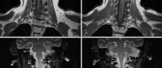
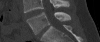
This disease is characterized by local back pain - from such a symptom it is impossible to suspect an inflammatory process and distinguish it from other degenerative-dystrophic diseases of the spine. On tomograms, doctors see a set of symptoms indicating a specific clinical picture of osteomyelitis: foci and abscesses of infection that spreads through the intervertebral disc, erosion (thinning) of the intervertebral disc, destruction of the vertebral bone body with a change in its height.
CT scans also help diagnose other inflammatory diseases of the spine, such as spondylitis and discitis.
Pathological results:
- The spinal bones (vertebrae) are missing, damaged, or misaligned.
- One or more disks may be damaged. One or more hernias are detected.
- The passage of contrast material through the spinal canal is restricted or blocked, indicating a narrowing of the canal (spinal stenosis).
- The vertebrae show signs of arthritis or bone problems caused by osteoporosis.
- Condition that has been present since birth (congenital anomaly)
- An abscess or tumor of the spine has been detected.
Magnetic resonance imaging (MRI)
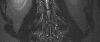
This is a highly informative, absolutely safe for the patient’s health, radiation diagnostic method, which makes it possible to identify and differentiate various pathological processes localized in the bone marrow of the vertebral bodies, the epidural tissue space (located between the hard shell of the spinal cord and the outer wall of the spinal canal and containing connective tissue and venous plexuses) , dural sac (the canal in which the spinal cord and the cerebrospinal fluid protecting it are located), intervertebral discs, paravertebral (paravertebral) soft tissues. In addition, MRI of the spine allows you to assess the condition of the spinal cord and its roots and identify their damage due to injury, tumor, demyelinating or inflammatory diseases. High diagnostic accuracy and reliability are characteristic of the MRI method when examining any part of the spine: cervical, thoracic and lumbosacral.
This type of diagnosis is most preferable for identifying fresh compression fractures of the vertebral bodies, always accompanied by bone marrow edema, the disappearance of which during treatment indicates consolidation (fusion) of the compressed vertebra. MRI reveals metastases in the spine that are localized only in the bone marrow and therefore do not cause destruction (destruction) of bone tissue, which cannot be visualized using CT or radiography.
The great advantage of MRI is its information content: with a relatively small number of contraindications, it provides complete information about the condition of the area under study, and most often, after it is carried out, there is no need for additional examinations. Among other things, MRI of the spine does not require special preparation and has no side effects. During one visit, the patient can undergo MRI of three parts of the spine or any of its parts. In addition, if desired, the patient can undergo an MRI of the coccyx.
What affects CT?
The following may prevent you from getting a CT scan or change the test results:
- Pregnancy. CT scans are not usually performed during pregnancy.
- Barium was used for another test. Barium is detected on CT scan. If a CT scan of the lower back is required, this should be done before any procedures that use barium.
- Metal objects in the body. These objects, such as surgical clamps or metal prosthetic joints, can interfere with a clear view of an area of the body.
- The patient cannot lie still during the examination.
Examination of the lumbosacral region
Computed tomography is also indicated for diagnosing a variety of diseases of the lumbar and sacral region. High detail allows you to notice beginning changes and begin treatment in a timely manner.
Indications for CT scan of the lumbosacral region:
- Infectious diseases.
- Traumatic injury to the spine.
- Pain without an obvious cause.
- Disorders of the pelvic organs.
- Paresis of the lower extremities.
- Rachiocampsis.
The examination will also be required in preparation for complex operations to correct curvature or replace vertebrae.
Conclusions after CT
- Sometimes the results of CT tests may differ from the results of other types of X-ray tests, magnetic resonance imaging (ultrasound), or ultrasound scans because CT scans provide different imaging.
- Children who require a CT scan may need special instructions for the scan. If the child is too young to stand still or is afraid, the doctor may give medicine (a sedative) to help the child relax.
- If your child is scheduled for a CT scan, you should talk to your child's doctor about the need for scanning and the risk of radiation exposure.
- CT scan results are often compared to positron emission tomography (PET) scan results to help detect cancer. Some newer scanners perform both scans at the same time.
- An MRI can provide more information than a CT scan of the spinal cord and spinal cord.
- An MRI of the spine is often performed instead of a CT myelogram.
Thoracic examination
Thoracic CT is a study that is used to identify a variety of diseases localized at the level of the thoracic vertebrae. Allows you to evaluate both the anatomical features of the structure of the spine and identify pathologies of surrounding tissues. The spinal cord continues in the thoracic region, so CT is also used to diagnose diseases associated with it. CT scan shows both bone disorders and pathologies of other tissues - this allows an accurate diagnosis to be made with one examination.
CT scan of the chest is prescribed in the following cases:
- Injuries. As a rule, cracks and fractures in this section are rare. If the spine is nevertheless damaged, the consequences can be very serious. Therefore, timely and accurate diagnosis is extremely important.
- Neoplasms – both benign and malignant, as well as metastases.
- Destructive processes in intervertebral discs.
- Osteoporosis.
- Assessing the consequences of back injuries.
In addition, CT is often prescribed if there are contraindications to MRI. Or if it is impossible to quickly send the patient to a magnetic resonance imaging scanner.