Truths and misconceptions about the dangers of X-rays
There are many opinions regarding x-ray research methods. The main ones are: radiography is extremely harmful, as it is accompanied by radiation; X-ray examinations cannot be performed frequently; X-rays of two or more areas of the body cannot be performed on the same day; X-rays are harmful to pregnant women and should not be used to diagnose pathological changes in them. Let's take a closer look at them.
THE ESSENCE OF X-RAY RADIATION AND THE PATHOGENESIS OF ITS BIOLOGICAL EFFECTS
There are different opinions about the harmfulness of X-ray research methods. X-ray radiation is classified as ionizing radiation. On the electromagnetic wave scale, X-rays fall in the range between gamma rays and ultraviolet. For diagnostics in medicine, “soft” and “medium” X-rays with low energy are usually used. However, such radiation is also capable of ionizing water in the cells of the body, “breaking” molecules into ions, as well as atomic oxygen (H+, OH–, O-2).

The image shows that X-rays are intermediate between ultraviolet and high-energy gamma rays. There is a certain pattern: from right to left, the radiation energy increases, the wavelength decreases and the penetrating ability sharply increases. For example: why does red light not illuminate X-ray film and is used in photography? Because it is located in the row to the left, therefore, it has less energy compared to other light in the visible spectrum.
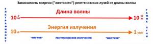
The image schematically shows the relationship between energy and wavelength of X-ray radiation and indicates its position on the electromagnetic wave scale. The more the “wavelength” and “energy” parameters are shifted to the left, the higher the biological impact of these rays.
Atomic oxygen is the most chemically active substance and is responsible for the biological effects of X-ray radiation. It affects almost all tissues of the body, but most pronouncedly on rapidly dividing ones (skin cells, mucous membranes, germ cells). Atomic oxygen “breaks” polymer molecules of nucleic acids (DNA, RNA): as a result of DNA damage, reading information from it either becomes impossible, or reading occurs with errors, which leads to mutations in newly reproduced cells, as a result of which they become unviable (or tumor). In addition to the genetic material, other cell structures (wall components, organelles located in the cytoplasm) are also affected; damage to the wall most often results in the release of the cytoplasm to the outside and death of the cell as a whole.

In the diagram, the letter H indicates the nucleotides that form the basis of two DNA chains that are complementary to each other. The letter P represents X-rays. K5 is oxygen in the water molecule, which does not have free chemical bonds - it is safe. K4 – OH—, oxygen in the hydroxyl ion has one free bond, which it tends to “fill.” K3 – atomic oxygen, the most active. It destroys the chain of the DNA molecule, breaking the bonds between nucleotides. As a result, a defective section of DNA is formed, from which problems may arise when reading information. If the wrong code is copied onto messenger RNA in a cell, there is a high risk of synthesizing defective proteins, which will lead to the development of tumors.
BIOLOGICAL EFFECTS OF X-RAYS
The biological impact (harmfulness) of X-ray radiation depends on the following parameters:
— Wavelength of X-rays (the shorter the wavelength, the more harm to the body);
— Radiation energy (the higher, the more harmful) is a parameter closely related to the wavelength;
— Voltage on the tube and current (kV and mA) - the higher these parameters, the “harder” and more intense X-ray radiation is obtained at the output. " hardness " of the radiation directly depends on the current (kV), the intensity determines the amount of radiation created by the tube and is measured in milliamps.
— Volume of irradiated tissues and organs. Thus, plain radiography of the abdomen will give a larger equivalent dose than radiography of the paranasal sinuses or hand. In addition, to “pierce”, for example, 30 cm of living tissue (stomach), you need a much higher current voltage and intensity, as well as exposure time, than in the case of 2-3 cm (hand);
— Duration of irradiation ( exposure , mA/sec). The higher this parameter, the more harmful the research. For example, with a CT scan of the chest, the duration of the study is 10-15 seconds, while with fluorography it is 0.01 seconds. Accordingly, with fluorography the radiation exposure is hundreds of times lower.
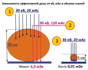
Consider the diagram. It presents several cases. If, as in option 1, the abdominal area (a large array of dense tissue) is irradiated with low-hardness and low-intensity rays, they will not pass through the thickness of the tissue, resulting in one solid white spot on the radiograph. At the same time, such parameters kV and mAs are acceptable for hand radiography (option 3). In order to “pierce” 30 cm of tissue (as in option 2), X-rays of greater rigidity and intensity are required, resulting in a much higher effective dose.
X-ray radiation can cause the following conditions:
— Radiation sickness (occurred earlier at the dawn of X-ray diagnostics, and then during radiation therapy in patients with large tumors);
— Radiation burns as a local manifestation of radiation sickness (usually during radiation therapy);
— Increased risk of neoplasms, including malignant ones;
— Increased risk of deformities in the next generation (in children).
ARE X-RAYS USED IN MEDICINE HARMFUL?
Of course, no one has canceled the harmful effects of X-ray radiation during diagnostic studies. However, it can be minimized if you carry out studies strictly according to indications, exclude “superfluous”, “unnecessary” studies, use “time protection”, i.e. do not perform two or more x-ray studies on the same day, shield adjacent body parts with the area of interest (for example, cover the groin area with a leaded rubber apron when examining the pelvic bones and hip joints).
It is considered undesirable to perform X-ray examinations on pregnant women, especially in the first months of pregnancy. However, in cases where the threat to the mother’s condition prevails, for example, in case of pneumonia or injury, an X-ray can be taken after first protecting the abdomen with special aprons and plates. Of course, the number of pictures for pregnant women should be minimal.
AVERAGE DOSE FOR RADIOGRAPHY OF VARIOUS PARTS OF THE BODY
The most harmful of all x-ray diagnostic studies are fluoroscopic (x-ray of the stomach, lower intestines, chest). The time spent “under the rays” here is long – up to several tens of minutes, and during the study up to 10 x-rays are taken. Urography, both visual and excretory, also carries a large radiation load; a survey X-ray of the abdomen, lumbar spine, pelvis and hip joints, as well as computed tomography, the dose of which can reach 40 mSv! The table below shows approximate radiation exposure figures for radiography or fluoroscopy of various parts of the body. Please note that these values are averaged, obtained on the Unicord-MT device using a standard phantom (corresponding to an “average” adult weighing about 70 kg). On other devices the numbers may differ significantly!
HOW TO REDUCE THE RADIATION DOSE DURING AN X-RAY EXAMINATION?
Proper preparation for the study is the basis for obtaining reliable results. Unfortunately, patients do not always understand that they need to be prepared to visualize pathology. For example, during an X-ray of the lumbar region, gas and intestinal contents, superimposed on the shadow of the spine, can make the image unreadable - it will have to be redone again, and this is an extra radiation dose that could have been avoided.
Preliminary preparation is very important for fluoroscopy of the upper and - especially - lower parts of the gastrointestinal tract. For irrigoscopy, preparation begins three days before the examination: it includes performing cleansing enemas (on the 1st and 2nd days before the examination - once a day, on the day of the examination - an hour before the procedure). In addition, it is necessary to follow a diet that excludes feces and gas-forming foods: fatty meat, beans, legumes, cabbage, milk, etc. Laxatives (for example, fortrans) can also be used for preparation. All this is done with the aim of cleansing the intestines before filling it with a suspension of barium sulfate, so that nothing interferes with seeing possible pathological changes.
During fluoroscopy of the stomach , esophagus, and duodenum, the diet is less strict; it is recommended to have a light dinner on the eve of the examination, and not to eat or drink on the morning of the examination. All this is necessary so that the stomach is empty and nothing interferes with the visualization of changes in it. For excretory and survey urography, it is also necessary to perform a cleansing enema, especially if the patient suffers from constipation and excessive gas formation. In addition, before excretory urography, it is necessary to donate blood for creatinine and urea in order to assess how well the kidneys filter the blood, since this test involves the injection of a contrast agent into a vein, which is then excreted by the kidneys. If kidney function is impaired, the rate of its elimination slows down, which increases the likelihood of toxic effects.
X -rays of the lumbar spine may also in some cases require a cleansing enema to reduce the accumulation of gas and intestinal contents on the spine. During hysterosalpingography , it is first necessary to exclude infectious diseases of the uterus and fallopian tubes (to prevent infection from entering the pelvic cavity). For computed tomography with contrast, the preparation is the same as for excretory urography - examination of creatinine and blood urea, as well as sugar.
All other studies - radiography of the chest, limbs, joints, thoracic and cervical spine, fluorography, mammography, etc. - do not require special preparation. You just need to take with you a referral, an outpatient card, other medical documents, previously taken photographs, a diaper or sheet, replacement shoes or shoe covers.
HOW TO PROTECT FROM THE HARMFUL INFLUENCE OF X-RAYS?
There are several simple rules, the observance of which will help minimize the harm from X-ray radiation during diagnostic studies.
- If possible, try to perform research on more modern devices. Digital X-ray machines can reduce the dose by tens of times compared to film ones.
- Do not perform examinations such as x-rays of the lumbar spine or excretory urography unless absolutely necessary and without a doctor’s prescription. If you have back pain and need to rule out a herniated disc, x-rays are the last thing you should think about. The method of choice is MRI. If urolithiasis is suspected, a kidney ultrasound may be sufficient. However, the final decision rests with your doctor. X-ray of the stomach with contrast can also in some cases be replaced by FGDS.
- Ask the laboratory assistant to cover adjacent areas of the body using protective aprons, vests, and screens. This is a mandatory event during radiography (scopy), and the staff has no right to refuse it.
STANDARD PATIENT QUESTIONS
Let's look at some common questions that patients who are concerned about the dangers of X-rays most often ask.
QUESTION 1. Doctor, I did several studies in a row - for example, an X-ray of the chest, sinuses and X-ray of the stomach. What harm is done to my health? Should I expect any dangerous consequences?
ANSWER. When exposed to ionizing radiation for medical purposes, it makes no sense to talk about direct harmful effects on the body (radiation sickness, burns, leukemia, etc.), since the dose received by the body (the so-called absorbed dose) is much lower than those values that can directly cause what something disease. For example, to get a skin burn, you need to take several thousand x-rays, etc. We can only talk about an increased probability of getting sick in the future (for example, cancer) - this is called the stochastic effect. For example, when undergoing a computed tomography scan, the likelihood of getting cancer in old age increases by a fraction of a percent. In this sense, X-ray examinations are harmful. But a careful examination of the problem shows that similar harm is caused by other types of radiation that we constantly encounter in life. So, we are exposed to radiation when we sunbathe in the sun, fly on an airplane, are high in the mountains, or walk along a pavement made of radioactive (yes!) granite. Finally, there is a normal natural radiation background (it would be better without it). To summarize, the answer is this: don’t worry, there will be no direct harm to health, but it’s still worth thinking about exceeding the dose, and don’t do radiation examinations unless necessary.
QUESTION 2. Doctor, my child had an X-ray of the brain (or chest, or abdomen). Will this affect his health in the future?
THE ANSWER is similar to the answer to the previous question. Don't worry, there will be no health problems. But X-ray examinations should not be done unless necessary, especially for children.
QUESTION 3. What dose is harmless? How many pictures can you take without harming your health?
ANSWER. Any dose of ionizing radiation is harmful; the concept of “harmless research” does not exist. But the harm does not lie in a direct deterioration in health, but in the fact that the likelihood of getting sick in the future slightly increases (read the answer to Question 1 above).
QUESTION 4 . Doctor, they have already taken several pictures of my lungs, and they want to repeat them again. Is it possible to do this? Or is it not worth it?
ANSWER. Any examination is prescribed by a doctor not for beauty, but in order to make an accurate diagnosis. The study needs to be done as many times as necessary for an accurate diagnosis and, therefore, correct treatment - 2 times, 10 times, 125 times. But your doctors must weigh two things: the potential harm from x-rays and the risk of misdiagnosis. If the next x-ray taken does not change anything in the diagnosis and treatment, then it should be abandoned. This is decided each time individually.
QUESTION 5. How often can x-rays be taken? Should you wait some time between tests to reduce harm?
ANSWER. There is no point in increasing the intervals between examinations, since the absorbed dose is still summed up and does not depend on the time of examination. As for frequency, it all depends on the clinical situation: sometimes doctors have to repeat tests dozens of times, sometimes once is enough. X-rays or CT scans should not be prescribed unless necessary; read the answer to the previous question.
QUESTION 6. During the x-ray examination, my genitals were not covered. What does this mean? Is there a risk of infertility/impotence/mutations/receiving sick offspring?
ANSWER. This definitely does not threaten impotence or infertility; it is better to read the answer to question No. 1 here. As for mutations or possible diseases in future offspring, the question is not so simple. The health of future children depends on the state of the genes (chromosomes) contained in your reproductive cells - eggs or sperm. Damage to genetic material when exposed to X-rays is likely only during the maturation of germ cells (when they divide and develop). Therefore, here it is necessary to separate men and women. In women, all eggs mature in utero, so the genetic material that your children will receive is formed even before you are born! Therefore, if you are a woman, then during your life nothing threatens your eggs - the genetic material in them will not change, and you should not think about mutations in children. In men, new sperm appear in the testicles throughout life, and the process of spermatogenesis lasts about a month. Therefore, if you are planning to have children after an x-ray, we advise you to wait at least a month - during this time all the sperm will be renewed, and nothing will threaten the children. Although, even if you don’t do this, the risk of getting mutations in sperm is still minimal.
Vasily Vishnyakov, radiologist
Pavel Popov
Candidate of Medical Sciences, Member of the European Society of Radiologists
What is a visiograph and how does it differ from an x-ray?
This one of the frequently asked questions is akin to the difference between a car and a traffic light... It seems that both concepts have some kind of connection, but it is somehow difficult to compare them. It's the same here. A radiovisiograph is a system that receives x-ray radiation, transforms it into digital form and displays the image on a computer screen. Roentgen (who is Wilhelm Conrad) is a long-dead German physicist who gained worldwide fame for his discovery of short-wavelength rays with enormous penetrating power. The physicist himself called these rays X-rays (in English today they are called exactly that - X-ray), but now we often call them X-rays, and in everyday life simply “X-rays”. The unit of radiation power was also called the x-ray. Now it is clear that a visiograph and an x-ray are completely different things. If we compare the visiograph with anything, it is with x-ray film, which it is universally replacing from all areas of medicine.
Lead Poisoning Symptoms
Lead poisoning can be difficult to detect initially—even healthy people can have high levels of lead in their blood. Signs and symptoms usually do not appear until dangerous amounts have accumulated.
Signs and symptoms of poisoning may include:
- High blood pressure.
- Joint and muscle pain.
- Problems with memory or concentration.
- Headache.
- Abdominal pain.
- Mood disorders.
- Low sperm count and abnormal sperm.
- Miscarriage, stillbirth or premature birth in pregnant women.
Employers have a responsibility to protect workers from exposure to inorganic lead. The employer must also initiate specific compliance actions, including blood lead testing for exposed employees.
Routes of entry of lead into the body
Lead enters the body primarily through inhalation and ingestion. Currently, adults are exposed to lead primarily through inhalation of lead-containing dust and fumes at work or through lead-related hobbies. Lead enters the body through the lungs and can damage many organ systems in the body.
Although inorganic lead is not easily absorbed through the skin, it can enter the body through accidental exposure (eating, drinking, and smoking) through contaminated hands, clothing, and surfaces. Workers may develop various health conditions such as neurological effects, gastrointestinal effects, anemia and kidney disease.
Use of lead in industry
Lead is primarily used in automotive lead-acid batteries, a type of rechargeable electric battery that uses a nearly pure lead alloy.
Lead alloys are commonly found in ammunition, pipes, cable sheaths, construction materials, solders, radiation shielding, collapsible pipes, and fishing weights. Lead is also used in ceramic glazes and as a stabilizer in plastics. Lead was widely used as a corrosion inhibitor and pigment in paints, but the use of lead in residential and public building paints has now declined.
Previously, the organic lead compounds tetramethyl lead and tetraethyl lead were used as anti-knock additives and to increase the octane number of gasoline, but concerns about the environmental impact of lead have led to the phasing out of these compounds. Organic lead compounds are still used in high-octane fuels in the aviation industry.