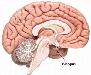
The pituitary gland is a small gland located at the base of the human brain (the fossa of the sella turcica of the sphenoid bone of the skull). Its weight is 0.5-0.6 grams. Despite its small size, the pituitary gland produces 7 hormones that ensure the functioning of the entire endocrine system of the body:
- somatotropin is responsible for growth;
- luteotropic stimulates the formation of milk in the mammary glands;
- adrenocorticotropic - controls the function of the adrenal cortex;
- thyroid-stimulating - affects the growth of the thyroid gland and the production of its hormones;
- gonadotropic - stimulate the production of female sex hormones in the ovaries and the release of a mature egg, and in the male - the secretion of testosterone;
- melanotropin - affects the pigmentation of the epithelium.

Diseases of the pituitary gland manifest themselves as gigantism and dwarfism.
Signs of pathological dysfunction of the pituitary gland may be:
- unexplained, persistent headaches;
- impaired visual acuity or blindness in certain areas of the eye;
- gigantism, dwarfism, enlarged facial features (in the area of the cheekbones, chin, brow ridges);
- dysfunction of erectile function in men, menstrual cycle in women;
- sudden weight gain for no particular reason;
- gynecomastia - enlargement of the mammary glands in men and women (not related to feeding the child);
- deviation of the hormonal levels of the pituitary gland from the norm according to laboratory data.
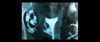
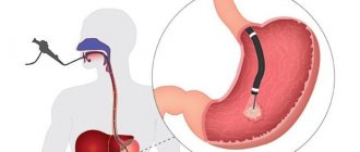
A common cause of gland damage is a benign adenoma tumor. The size of the neoplasm can be significantly smaller than the gland itself, so it can only be seen with magnetic resonance imaging with a contrast agent. The method clearly shows the structure of the pituitary tissue, the presence of a cyst, allows you to recognize the degree of malignancy of the tumor, other diseases of the pituitary gland, and based on the results of the study, draw conclusions that will help provide timely assistance. Correct preparation for MRI of the pituitary gland with contrast is the key to adequate diagnostic results.
MRI of the pituitary gland: the essence of the procedure, preparation and indications for the procedure
MRI of the pituitary gland is a modern and highly accurate diagnostic method, the purpose of which is to identify changes in the structure of the lower medullary appendage due to the development of diseases and pathological processes. This type of diagnosis is considered the most highly informative, allowing one to visualize even minor pathological conditions in which other examination methods will be ineffective.
Indications
There are a lot of hormones produced by the pituitary gland, and with the help of MRI of the pituitary gland, doctors have a chance to understand where the cause of many pathological processes in the body lies. The symptoms of diseases of this organ depend on the production of which hormone is disrupted by the tumor. So:
- Many pathological processes in the brain in the pituitary gland zone change the synthesis of thyroid hormones (T3 and T4).
- Tumors in the sella turcica area can cause overproduction of cortisol, the excess production of which leads to Cushing's disease.
- Growth hormone (somatotropic hormone) is responsible for the growth of the entire body. Overproduction of this substance due to adenoma causes the process of acromegaly.
- A lack of luteinizing hormone and oxytocin, which are responsible for ovulation, causes infertility in women and a lack of testosterone in the stronger sex.
- A pituitary adenoma can affect the production of prolactin, which directly regulates the functioning of the mammary glands.
- Failures in the synthesis of follicle-stimulating hormone lead to a lack of follicle growth in the ovaries in the fair sex and the formation of low-active sperm in men.
- A disruption in the production of antidiuretic hormonal substances leads to problems with urination and can cause the development of diabetes insipidus.
- There is also a hormone of the intermediate lobe of the pituitary gland, the so-called lipotropin and proopimelanocortin, which control the pigmentation of our skin and enhance the metabolism of fats.
What is diagnostics using a tomograph?
MRI uses a powerful magnetic field. Through its effect on the body, radio frequency impulses are formed, the analysis of which is carried out by specialists on a computer. Three-dimensional images can be printed, saved or sent, if necessary, to the subject’s e-mail address.
MRI of the pituitary gland provides an opportunity to fully evaluate existing changes in the functioning of the pituitary gland, as well as examine nearby tissues. Scanning allows you to analyze the state of operation of a separate part of the brain, which ultimately can be used to assess the functioning of many organs and systems.
In the photographs after the diagnosis, you can see in detail the sinuses with the eye orbits. If it becomes necessary to examine other parts of the brain, a full scan is performed to clarify or identify the causes of the pathological condition.
How to properly prepare for an MRI of the pituitary gland
Magnetic resonance imaging is a type of pre-diagnosis for which patients do not need to comply with strict restrictions and extensive preparatory measures. True, there is one exception - if the scanning is planned to be carried out using contrast, in this case some preparation will still have to be completed. So, it is unacceptable to eat on the day of the study, and if the diagnosis is carried out in the evening, you need to wait a 6-hour interval. Otherwise, nausea and vomiting may occur, which will be completely inappropriate.
If an MRI of the pituitary gland is performed without contrast, such restrictions do not apply to the procedure.
Before scanning, all patients must:
- Remove any clothing that has metal inserts. You will have to leave all decorations and gadgets outside the office. Any metal objects can not only affect the operation of the equipment, but also have a negative impact on the quality of the images, which will complicate diagnostic measures.
- The radiologist must be informed about the implants present in the body. Some of them may act as strict contraindications to scanning.
- Women expecting the birth of a baby should not undergo an MRI in the first trimester of pregnancy. Despite the fact that tomography is considered one of the safest examination methods, there is still a potential risk of negative effects on the developing organism.
- As a rule, young children are allowed to be scanned after 5-6 years. MRI is indicated for children only in the most extreme cases and, most often, in a state of medicated sleep.
Preparation and performance of MRI
A little preparation will be required before administering the MRI contrast agent and undergoing the procedure to ensure the results of the examination are as detailed as possible and the patient does not encounter problems
Therefore, it is important not only to familiarize yourself with the features of the study, but also to prepare for it in advance.
Preparation
You should start preparing for an MRI a few days before the procedure.
This is very important to obtain the most accurate results. The first element of preparation is a conversation with the doctor, during which all important information about the patient is clarified, which could become a contraindication for MRI.
Therefore, it is very important to remember whether you have previously had surgery with the installation of implants or other metal elements. This is especially true for dental procedures.
After this, it will be very easy to prepare for the procedure, because... there are no special requirements. You only need to follow these rules:
Prepare psychologically
It is important to set yourself up for the absence of fear, the rapid completion of the procedure, and a positive result. This should be done a few days before the MRI. Stop using makeup
Editorial: Causes, diagnosis and treatment of Parkinson's disease
It is recommended not to use cosmetics before undergoing brain imaging, because this may harm the accuracy of the result. Remove all metal items and electronic devices. It is best not to take anything metal or electronic with you, so as not to accidentally forget about these things. If you have them with you, be sure to remove them before performing the diagnostics.
It is strictly forbidden to drink alcoholic beverages before the tomography. It is also not recommended to smoke, be nervous, or influence your brain or blood vessels in any way.
These rules apply to any type of MRI with contrast.
However, when examining the abdominal cavity, it is important to adhere to a strict diet, avoiding junk food, and also not to eat a day before the procedure, while refusing to drink liquids 5 hours before the MRI
Indications for MRI of the pituitary gland
In most cases, the doctor writes a referral for examination if there is compelling evidence for it. First of all, he conducts a comprehensive examination and issues a referral for preliminary tests.
If the cause of the complaints cannot be identified, the specialist decides on the need for additional examination. The following pathological conditions may be indications for tomography:
- severe periodic pain in the head;
- metabolic disorders in the body;
- existing vision problems;
- menstrual irregularities;
- impotence;
- elevated TSH levels in tests.
Contraindications
Contraindications to tomography are divided into relative and absolute. If a patient has absolute contraindications, then MRI will be prohibited for him, regardless of whether there are compelling reasons for this examination or not. These include:
- metal implants, insulin pumps, pacemakers present in the body.
- the presence in the body of the subject of metal objects received as a result of injury.
Relative or temporary bans on scanning include:
- first trimester of pregnancy. In exceptional cases, diagnostics can still be carried out on expectant mothers if the expected benefit from the procedure for the mother exceeds the potential risk to the fetus;
- the patient is in extremely serious condition;
- epilepsy;
- contrast intolerance;
- disorders of the kidneys;
- fear of closed spaces;
- involuntary movements of the limbs.
If the patient has any doubts or concerns before undergoing a tomography, he can always ask all the questions that interest him to the radiologist.
Referral for MRI and preparation
Most medical centers in St. Petersburg accept patients for tomography of the pituitary gland, both with and without a referral from a doctor. If a person has concerns or doubts about his health, he can independently come to the diagnostic center and voice the problem. Radiologists will help you decide on the area of study, and based on the results of the tomography, advise on further steps. If the patient has any medical history or results of previous examinations, it is better to bring them with you for diagnosis. The tomographic procedure of the head does not require any special preparation from the patient. You can do it any day without having to diet.
MRI of the pituitary gland for young children

This type of diagnosis is prescribed to children, most often after 5 years.
To obtain clear images, the patient should not make any movements during the examination, which is very difficult to achieve from young patients. Children simply cannot be in a calm state inside a confined space due to overwhelming fear. To examine children, doctors use open-type devices. However, even in this case, the baby must remain still throughout the scan. Parents are allowed to be with their child during the examination. If the baby shows excessive anxiety, cries a lot or refuses to go into the office, the doctor may decide on the need to use sedatives or conduct diagnostics in a state of medicated sleep. MRI of the pituitary gland without contrast is most often performed on children.
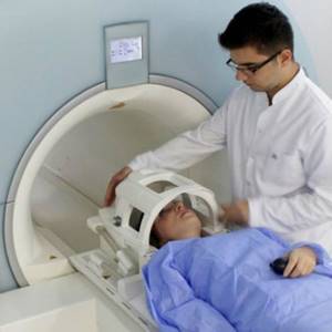
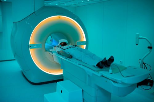
How does the scan work?
MRI is a diagnostic method that is quite simple, and the absence of the need to follow preparatory measures is another big advantage of tomography. The device on which the examination will be carried out is located in a separate room.
Medical staff monitors the examination from the next room, and communication with the subject is carried out using a special speaker. Therefore, you should not worry about the fact that if your health worsens, you will not have the opportunity to inform your doctor about it.
Before being examined, the patient must change into hospital clothes. It is more spacious, comfortable and does not have any metal parts. If the scan is to be performed on a child, he or she enters the office accompanied by an adult. When performing an MRI of the pituitary gland with contrast, the patient is given an intravenous injection of a contrast agent.
Regardless of whether an MRI of the pituitary gland is performed with or without contrast, as soon as the patient is ready to begin the procedure, he will be asked to take a horizontal position on a special extendable table of the tomograph.
The patient's body will be fastened with special straps. There is no need to be afraid of this; this is done to ensure complete immobility and eliminate involuntary movement of the limbs during the procedure.
Actually, to carry out scanning, the table is rolled into the apparatus, which looks like a large tunnel. The tomograph makes specific noises during operation - this is quite normal and does not pose any danger to the patient. The duration of the scan varies from 25 to 55 minutes and depends directly on whether an MRI of the pituitary gland is performed with or without contrast, as well as on the required number of images.
Upon completion of the procedure, the doctor can immediately analyze the resulting images. The evaluation of the obtained images is carried out on a computer monitor using a special program. The doctor evaluates the results obtained and writes a conclusion based on them.
At the time of diagnosis, the patient can await results in the clinic. The results can be sent to him by email. We must not forget that once the results are ready, in order to prescribe treatment, it will be necessary to contact the specialist who issued the referral for examination.
There is no need to observe any restrictions in food or lifestyle after an MRI. After the patient leaves the clinic, he can return to his usual activities without any restrictions.
Decoding
During the examination, the doctor evaluates the following anatomical features:
- The shape of the sella turcica, the edges of its bottom and walls;
- Pituitary gland, dimensions, its contours;
- Gland structure;
- Chiasm and the distance from the upper surface of the pituitary gland to the chiasm and between the mediobasal parts of the temporal;
- Cavernous sinuses;
- Optic chiasm, carotid siphons;
- Adjacent parts of the brain;
- Sinus of the sphenoid bone.
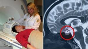
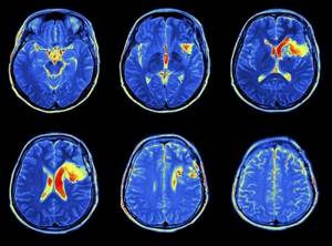
Example of an MRI report
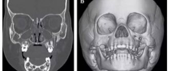
A series of MR tomograms obtained images of the pituitary gland in coronal, axial and sagittal projections.
The pituitary gland in the sella turcica is not enlarged, somewhat asymmetrical in the craniocaudal direction: on the right - 0.8 cm, on the left - 0.7 cm, coronal size - 1.8 cm, sagittal - 1.0 cm. The structure of the pituitary gland is somewhat heterogeneous due to the presence in the central sections of a poorly defined hypointense focus, measuring 0.2x0.2 cm.
The funnel is located in the middle.
The chiasma and suprasellar cistern are without deformations.
After administration of the contrast agent Magnevist 20.0 ml during dynamic observation, the contrast enhancement is expressed unevenly, and there is a lag in the accumulation of paramagnetic by the identified focus.
Conclusion:
— MRI signs of pituitary microadenoma. Recommended: Dynamic MR monitoring in the context of the clinical picture and clinical and laboratory data.
“Second independent opinion” service Medicine is an area where we want to be 100% sure. Therefore, at your request, we will be happy to offer you the service of a second independent opinion from the leading consultant of our clinic, Candidate of Medical Sciences, a doctor of the highest category with 18 years of experience in the field of tomography and radiology Marchenko N.V.
Therapist with 25 years of experience
MRI of the pituitary gland with contrast
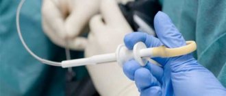
In order to obtain more informative images, a specialist may decide on the need for an MRI with contrast. A contrast agent is administered intravenously to the patient before the procedure begins. Once in the bloodstream, contrast when assessing the resulting images allows the doctor to examine the vascular network in the required area. Almost always, contrast makes it possible to assess the size and location of the pathological process and determine the intensity of blood flow. Scanning using a contrast agent, in most cases, is prescribed to patients before surgery to remove pituitary tumors. The contrast is concentrated in those areas where there is increased blood supply, in particular, in those tissues in which the tumor is located. As a result, it is possible to diagnose even small tumors.
MRI of the pituitary gland without contrast is performed much more often, because pituitary tumors are a rather rare pathological process.
Which is better: MRI of the pituitary gland with or without contrast?
The specialist may prescribe the patient to undergo an MRI of the pituitary gland without contrast, or magnetic resonance imaging using a contrast agent. Most often, paramagnetic substances are used for this purpose, administered intravenously a few minutes before the start of the scan. The dosage of the drug is calculated individually, based on the patient’s body weight. An MRI of the pituitary gland with or without contrast is always decided based on the patient’s medical history. If it is necessary to identify clear boundaries of a neoplasm, its shape, and the condition of nearby tissues, it makes sense to use a contrast agent. In any case, only the doctor decides whether to conduct an MRI of the pituitary gland with or without contrast; the advisability of using contrast is considered on an individual basis.
Why is an MRI of the pituitary gland prescribed?
Among all brain tumors, pituitary adenomas account for about 15-20%, and most often they are detected in patients of working age, mainly from 30 to 50 years. The reasons for the development of this pathology are still reliably unknown.
With questions about the problems of diagnosing and treating pituitary tumors, we turned to the executive director and chief physician of MRT Expert Lipetsk LLC, Oksana Egorovna Volkova.
— Where is the pituitary gland located and what role does it play in the life of the human body?
— The pituitary gland is located in the area of the sella turcica, and the sella turcica itself is at the very base of the skull. The pituitary gland (pituitary gland) is the governing organ of the entire endocrine system of the body. Any dysfunctions and disturbances in its functioning seriously affect the functioning of all organs and systems. The cardiovascular and circulatory systems, muscular system, and musculoskeletal system may be affected. In women, dysfunction of the pituitary gland can lead to menstrual irregularities and infertility.
— Tell us about the types of pituitary tumors and their clinical manifestations.
There are two types of pituitary tumors: hormonally active (prolactinomas, somatotropinomas, corticotropinomas) and non-hormonally active.
Clinical manifestations of pituitary adenoma are varied and depend on the type of tumor, its size and direction of growth in the cranial cavity. The two largest vessels of the brain and many important nerves pass over the tumor itself, including those responsible for facial sensitivity, eye movements, vision and smell. Therefore, the growth of the tumor upward into the cranial cavity leads to compression of the optic nerves. As a result, the person begins to lose vision. With a complaint of decreased vision, the patient comes to an ophthalmologist, who often begins to treat the patient for non-existent ophthalmological problems, for example, optic nerve atrophy or cataracts, which does not lead to an improvement in the patient’s condition. If the pituitary tumor is not removed in time, it can lead to further deterioration of vision, even blindness. Therefore, it is so important to differentiate, using MRI, ophthalmological diseases and pituitary tumors.
Also, a pituitary adenoma can lead to disruption of the outflow of cerebrospinal fluid - occlusive hydrocephalus and death.
As for hormonally active tumors, they grow slowly and in most cases are detected in patients in the early stages of endocrine disorders.
With prolactinomas, women experience symptoms such as menstrual irregularities, while men experience decreased libido and weight gain.
Somatotropinomas, producing growth hormone, cause acromegaly. In children, rapid growth occurs, and in adults, this type of pituitary tumor can cause enlargement of the limbs, nose, or ears.
Corticotropinomas cause Cushing's disease, one of the symptoms of which is weight gain in the upper body and arterial hypertension. Diabetes mellitus and osteoporosis often occur in patients with corticotropinoma.
— Which doctor should you contact if you notice symptoms of pituitary gland disease?
In order to deal with the problem as a whole, patients with pituitary gland pathology are jointly managed by an endocrinologist, a neurologist and a neurosurgeon.
You can find out the cost of an appointment and make an appointment with an endocrinologist here
If alarming symptoms of hormonal imbalance are detected, you should consult an endocrinologist. If your vision begins to deteriorate, go to an ophthalmologist or neurologist.
The doctor will prescribe tests to determine the patient’s hormonal status and refer him for magnetic resonance imaging of the pituitary gland.
You can sign up for an MRI of the pituitary gland in your city here
— Why is it necessary to do an MRI of the pituitary gland? Will an MRI of the brain show pathology of the pituitary gland?
Pathology of the pituitary gland in the practice of doctors “MRI Expert” and “Clinic Expert” occurs quite often. At the same time, patients do not always complain about dysfunction of the pituitary gland. Much more often they come to perform an MRI of the brain. But on MRI images of the brain you can only see a large tumor, a macroadenoma, measuring from 1 cm (at the same time, the normal dimensions of the pituitary gland in women and men on MRI are: vertical - up to 6 mm, in adolescents - up to 1 cm; in elderly patients it may decrease up to 2-3 mm; frontal (lateral) - 1.3 cm and anterior-posterior - on average 1-1.1 cm).
Small microadenomas are not visible on an MRI scan of the brain; the doctor will only be able to diagnose an enlargement of the pituitary gland, which will be reflected in the conclusion, which will say “signs of an increase in the vertical size of the pituitary gland; recommended - consultation with an endocrinologist, clinical and laboratory examination and MRI of the pituitary gland with intravenous dynamic contrast).
If a deformation of the pituitary gland is suspected, it is necessary to conduct a special dynamic study with contrast enhancement in order not to miss even the smallest formations in the pituitary gland. Contrast during an MRI procedure of the pituitary gland is introduced at certain minutes and seconds of the scan. Carrying out MRI diagnostics of the pituitary gland without the use of contrast is of little information.
Why is a contrast agent used in magnetic resonance imaging? The executive director of MRT Expert Lipetsk LLC, Oksana Egorovna Volkova, tells
— How are pituitary tumors treated? Does the diagnosis of pituitary adenoma by default require surgical intervention?
This is wrong. The choice of treatment method for pituitary adenomas is individual and depends on many factors, including neurological symptoms. The indications for removal of an adenoma are quite clearly known. If the patient has visual impairment, then surgery is indicated to save vision. In case of overproduction of aggressive hormones, such as somatotropic and corticotropic hormones, surgical intervention is also indicated in order to reduce the aggression of this hormone on target organs.
For a long time, operations on the pituitary gland remained one of the most difficult in the world of neurosurgery and were associated with a high risk of complications during craniotomy. In this regard, a technique for removing pituitary tumors without opening the skull was developed and implemented. The so-called transsphenoidal method in neurosurgery (removal of pituitary tumors through the nose). The technique is not new. Transsphenoidal surgery of pituitary adenomas has been around for more than a hundred years. During this period, it underwent major changes and became an independent branch of modern neurosurgery. The technique of transsphenoidal access to formations is minimally invasive, which avoids serious complications during surgery. There are practically no contraindications to such an operation. Among the latter are inflammatory processes in the nasopharynx and a large number of somatic pathologies in the patient, which can complicate anesthesia.
Such operations are performed under general anesthesia and last about two hours.
If hormonally inactive tumors or small tumors that do not compress adjacent brain structures are detected, conservative treatment with tablets and dynamic observation are carried out, which consists of MRI of the pituitary gland with contrast, 1-2 times a year.
An MRI examination of the pituitary gland can be performed any day of the week; no special preparation is required. The diagnosis lasts about 30 minutes and does not cause discomfort.
What will the examination show?
MRI of the pituitary gland with or without contrast allows you to visualize any changes in the pituitary gland. Thanks to scanning, it is possible to diagnose any tumors, as well as identify their type and exact location. A radiologist will be able to identify abnormal development of the pituitary gland, its structural changes and changes in size. In addition, MRI provides the opportunity to identify the very cause of the pathological process. It is important to remember that timely diagnosis will be the key to a complete recovery with well-chosen treatment tactics.
Possibility of complications
An absolutely harmless and safe diagnosis by undergoing an MRI of the pituitary gland can still cause unpleasant symptoms in some patients. So, some patients may complain of:
- the appearance of nausea;
- feeling of extreme weakness;
- the appearance of dizziness;
- severe headaches;
- increased heart rate;
- excessive nervous excitability.
As a rule, all such symptoms are caused not because of the tomography, but because of the patient’s suspiciousness and fear of the procedure. MRI of the pituitary gland without contrast should not cause any negative consequences at all, because this is an absolutely harmless procedure, performed even on small children.
But when undergoing an MRI with contrast, it is quite possible that a real complication may arise – an allergic reaction. However, the occurrence of contrast intolerance is extremely rare.
To avoid negative consequences, you need to conduct an allergy test - if its results do not cause concern among specialists, an MRI with contrast can be performed without any fear.
Where to undergo magnetic resonance imaging of the pituitary gland

Every patient who has been scheduled for a scan, regardless of whether an MRI of the pituitary gland will be performed without contrast or with its use, wants to undergo diagnostics quickly, efficiently and without waiting in long lines. It is almost impossible to do this in public clinics. According to the coupon, you will have to wait a very long time for your turn to undergo the examination, and not every government agency has modern equipment that allows you to obtain high-quality images during scanning.
As a result, people are starting to look for a private clinic to undergo examination. The pages of our website present a large number of clinics with detailed information about each of them. All medical institutions provide diagnostics at an affordable cost, which is a key point for patients. In addition, we provide each patient with an additional discount on MRI of the pituitary gland, so contacting us will guarantee a low cost for the examination.
Preparing for diagnosis
A significant advantage of this method of examination is that no significant preparatory measures need to be taken before MRI of the pituitary gland without contrast. All that is required from the patient is to comply with a number of simple requirements immediately before starting the scan.
However, if you plan to perform an MRI with contrast, then it is necessary to adhere to some recommendations. So, the patient must come to the tomography on an empty stomach, or observe a 4-hour fasting break. This is done in order to exclude the appearance of any unpleasant sensations in the intestines during the scan. If MRI of the pituitary gland is performed without contrast, then such restrictions do not need to be observed. Before scanning, all patients, without exception, need to:
- remove items of clothing that have metal parts, and ideally give preference to hospital clothing, which is much more comfortable;
- remove jewelry;
- leave your mobile phone, bank cards, and wallet in the locker room.
Each patient must understand that any metal objects can interfere with the examination, which will negatively affect the quality of the resulting images. Therefore, it is so important to take your preparation seriously and listen to all the advice of experts. Moreover, it does not matter significantly whether an MRI of the pituitary gland will be performed without contrast or with its use.