Diseases that begin with the letter “N”: Narcolepsy, Drug addiction, Corns, Neuralgia, Sciatic nerve neuralgia, Trigeminal neuralgia, Neurasthenia, Neuritis, Neurosis, Miscarriage, Urinary incontinence, Neurodermatitis, Neurosyphilis, Neurocirculatory dystonia, Lactose intolerance, Nephritis, Nephroptosis (wandering kidney, prolapsed kidney).
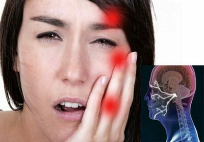
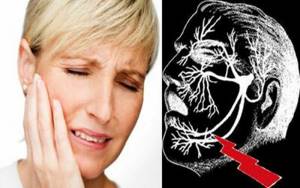
When a nerve in the facial area becomes inflamed, a diagnosis may be made of trigeminal neuralgia. This disease mainly occurs in older people.
Factors provoking the development of the disease
The main reason for the appearance of trigeminal neuralgia is its compression and pinching. Factors provoking this process are:- previous viral diseases (for example, herpes);
- vascular atherosclerosis;
- the presence of a malignant neoplasm in the cerebellopontine region;
- hypertonic disease;
- facial injuries;
- metabolic disorders in the body;
- inflammatory process in the patient’s gums and teeth (for example, periodontitis or pulpitis);
- complication after incorrect dental treatment;
- inflammation of the sinuses.
Signs of development of trigeminal neuralgia
The occurrence of trigeminal neuralgia can be assumed based on the following signs:
- the occurrence of severe aching pain in the face (the eyebrows, lower jaw, cheek and nose hurt especially badly);
- the pain in these areas of the face increases when performing any actions (pronouncing a speech, eating or facial activity);
- in the cold the pain intensifies;
- increased secretion of salivary glands, tears;
- loss of sensitivity or, conversely, the occurrence of heightened sensitivity in the jaw, eyelids and nose;
- uncontrolled movements of facial muscles;
- facial hyperemia.
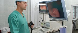
Neurovascular conflict (NVC) is a disease caused by the contact of a vessel with the cranial nerve (CN), causing compression and dysfunction of the latter. Currently, cases of NVC involving all cranial nerves except the olfactory nerve have been described in the literature. NVC is an interdisciplinary medical problem; patients with NVC require careful differential diagnosis and various treatment approaches. The question of neurosurgical intervention in patients with NVC arises when conservative treatment is ineffective or intolerable. The essence of the operation is to separate the compressing vessel from the nerve and install a gasket between the nerve and the vessel, and the operation itself is called “microvascular decompression” (MVD). Before the advent of high-field MRI, the diagnosis of NVC was based on clinical presentation and response to conservative treatment, and surgical treatment was sometimes explorative. The development of MRI and the emergence of MR angiography protocols currently greatly facilitate the diagnosis of NVC. Below we will discuss the epidemiology, clinical picture, diagnosis and treatment of neurovascular conflicts of the posterior cranial fossa. Diseases will be listed in order of frequency of occurrence.
Trigeminal neuralgia (painful tic, Fothergill's disease)
The most common and best-studied neurovascular conflict (NVC) is primary (essential, idiopathic) trigeminal neuralgia (TN), the prevalence of which is 2.5 cases in men and 5 in women per 100,000 population per year. The disease manifests itself in attacks of acute, burning facial pain, lasting from a few seconds to 1-2 minutes, with a frequency ranging from single to hundreds per day. Characteristically, there are trigger zones on the face, touching which can provoke an attack. It can also be triggered by talking, chewing, shaving, or brushing teeth. A painful attack is accompanied by contraction of the facial muscles on the affected side, tearing and salivation. The patient freezes during an attack.
To exclude a space-occupying process, multiple sclerosis, infectious, ischemic lesions, a patient with TN is advised to undergo high-field MRI of the brain with MR angiography. During MR angiography in patients with primary TN, the vessel causing compression of the trigeminal nerve is visualized—most often the superior cerebellar artery. The CISS-3D MRI protocol is highly informative in diagnosing NVC, the image of which is characterized by high contrast between the CN, vessels and cerebrospinal fluid [1, 2]. Combining this protocol with MR angiography allows us to obtain an even clearer picture of the relationship between the vessel and the nerve.
Conservative treatment
The drug of choice for the treatment of TN is carbamazepine at a daily dose of 200-800 mg; treatment is effective in an average of 60% of patients. If carbamazepine is ineffective or intolerable, phenytoin, lamotrigine, clonazepam, and valproic acid preparations are used [3, 4]. Various scales have been developed to assess the effectiveness of treatment. Thus, the BNI (Barrow Neurological Institute scoring system) has five grades of outcome: grade 1 - no pain, no drug therapy required; grade 2 - periodic minor pain, drug therapy is not required; grade 3a - no pain while continuing drug therapy, grade 3b - periodic minor pain, continuation of drug therapy is necessary; grade 4—pain has decreased but persists, drug therapy is ineffective; grade 5 - pain has not changed.
The MMS (Modified Marseille Scale) scale has six grades: grade 1 - no pain, no drug therapy required; grade 2 - no pain while continuing drug therapy; grade 3 - pain intensity decreased by more than 90%; grade 4 - pain intensity decreased by 50-90%; grade 5 - pain intensity decreased by less than 50%; grade 6 - pain intensity increased.
Radiosurgery
In gamma knife treatment, the target is the retrogasser segment of the trigeminal nerve, the radiation dose is 70-90 Gray. B. Young et al. [5] presented the results of treatment of 250 patients with TN 5 years after radiosurgical treatment. In 70% of patients the pain was absent, in 20% it decreased by more than ½, in 5% it was less than ½, and in 5% it became more intense. Among the complications, the authors noted dry sclera (22%), memory impairment (20%), hearing loss (16%), hypogeusia (16%), dysphagia (13%), and weakness of the masticatory muscles (11%).
Percutaneous puncture techniques
This type of intervention involves puncturing the skin under the zygomatic process of the upper jaw, bringing a guide needle to the foramen ovale and entering Meckel’s space. Next, the trigeminal nerve is affected in order to destroy the fibers that conduct pain sensitivity. Currently, radiofrequency trigeminal rhizotomy (RTR), microballoon compression, and glycerol rhizotomy are used. The introduction of alcohol or hot water is not used.
The RTR effect is achieved by heating the electrode to 55–70 °C. The results of treating 1600 patients with the RTR method showed [6] that complete pain regression was observed in 1216 (76%) patients with a single RTR session, and in 24% with its repetition 2-6 times. After 10 years, the analgesic effect remained in 52% of patients with a single RTR and 94% with it performed more than once. The authors note the following complications: decreased corneal reflex (5.7% of patients), development of keratitis (0.6%), dysfunction of the masticatory muscles (4%), painful dysesthesia (1.8%), damage to the abducens nerve (0.13% ), injury to the internal carotid artery with the formation of a carotid-cavernous anastomosis (0.06%).
When a nerve is compressed by a microballoon, it is inflated with a pressure of 1.3-1.6 atm. H. Xiaochuan et al. [7] describe the results of using this technique using intraoperative 3D CT in the treatment of 21 patients. All patients had no pain after surgery and at follow-up at 24 months. In 4 patients after surgery, transient weakness of the masticatory muscles on the intervention side was noted. B. Abdennebi et al. [8] present the results of 200 such operations with compression of the Gasserian node with a microballoon for 6 minutes. When observed for 4 years, there was no pain in 62% of patients, numbness of the face was in 3%, hypoesthesia in 7.5%. Similar studies [9, 10] showed similar results; on average, pain regressed in 70% of patients.
The effect of rhizotomy with glycerol is achieved through chemical action. After treating 2750 patients with this method [11], the recurrence rate of TN was 35%. Recurrence of neuralgia in the 1st year after surgery developed in 0.3% of patients, in 1-5 years - in 21%, 5-10 years - in 7%, 10-15 years - in 4%, 15-23 years - in 3%. During a 3-year follow-up of 1174 patients who underwent rhizotomy with glycerol [12], complete absence of pain was observed in 55%.
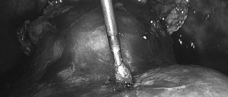
Microvascular decompression (MVD)
MVD of the trigeminal nerve is performed using a retrosigmoid approach. In order to minimize neurological complications, neurophysiological monitoring of auditory and facial CN is performed intraoperatively. Some authors [13] suggest not using spacers between the vessel and the nerve during surgery, but transposing the compressing vessel with further fixation with biological glue or suture. The effectiveness of MVD (no pain and no need to continue conservative treatment) is up to 90% in the 1st year after surgery, decreasing to 70-73.4% after 10-15 years [14, 15]. Complications are cerebellar stroke (1-2% of patients), transient (20%) and persistent (3-8%) hearing impairment, impaired sensitivity on the face (5-30%), transient (5-6%) and persistent (2 %) prosoparesis [16—19]. In the case of forced coagulation of the petrosal vein during MVD, most patients develop vestibuloataxic disorders that regress within a few weeks [20]. Researchers have differing opinions regarding the factors that determine the outcome of surgery. According to a number of authors [14, 21], gender, age, side, duration and frequency of attacks, and the total duration of the disease do not affect the effectiveness of surgical treatment. F. Barker et al. [15] believe that female gender, disease duration of more than 8 years, vein as a source of conflict, as well as the absence of immediate regression of symptoms after surgery are risk factors for relapse of T.N. Intraoperative detection of focal arachnoiditis reduces the effectiveness of surgical treatment. The presence of trigeminal atrophy due to NVC improves the prognosis. R. Sekula et al. [22] performed MIA in 28 patients with TN in whom radiosurgical treatment was ineffective. Over the course of 2 years, 54% of patients had no pain, 7% had moderate pain, and 36% did not decrease; Facial numbness developed in 21%.
Partial rhizotomy (PR)
If the NVC is not detected during surgery in a patient with TN, it is proposed [23] to perform PR of the trigeminal nerve by cutting ¼—1/3 of its sensory fibers. J. Young and R. Wilkins [24] present the results of PR in 83 patients in whom surgery was performed due to the absence (77%) or insignificant (11%) NVC, the impossibility of adequate separation of the vessel from the nerve (12%) [24]. 6 years after surgery, an excellent result was noted in 40 (48%) patients, good in 18 (22%), weak or unsatisfactory in 25 (30%). 1 year after surgery there was no effect in 17% of patients; the number of patients with pain recurrence increased by 2.6% each year.
L. Zhang et al. [25] present the results of MIA in 210 patients for TN, 68 of whom the operation was supplemented with P.R. Pain completely regressed in 90% of patients who underwent only MVD, and significantly regressed in 7%. After 2 years, the number of pain-free patients in this group did not change. Among the patients who underwent MIA and PR, pain completely regressed in 98.5%; after 2 years, the number of patients without pain was 96%.
Facial nerve damage (hemifacial spasm)
NVC with the facial nerve is manifested by hemifacial spasm (HS), the prevalence of which is 9-11 cases per 100,000 in the general population, and is twice as common in women [26, 27]. 0.6-5% of patients have bilateral HS. The disease is manifested by tonic and clonic spasms of the facial muscles on the affected side. In 90% of cases, spasm begins from the orbicularis oculi muscle, in the rest - from the cheek or perioral area. Starting in one area, spasms usually spread to other facial muscles and the platysma. HS can occur in a dream; an attack can be provoked by various facial movements, emotional stress, and fatigue. A characteristic symptom is Babinski-2 (“another Babinski symptom”), which consists of a reflex raising of the eyebrow on the affected side when closing the eyes. The sensitivity of this sign for HS is 86%, specificity is 100% [28]. The clinical picture of HS may include damage to adjacent CNs if they are involved in NVC. An extremely rare form of NVC associated with compression of the intermediate nerve (part of the facial nerve) is manifested by attacks of paroxysmal otalgia, sometimes with pain in ½ of the tongue and in the depth of the jaw [29]. This pain syndrome is observed both in isolation and in combination with HS [30].
Diagnosis of HS requires the exclusion of a number of diseases manifested by facial dyskinesias: blepharospasm, facial tics, ocular myokymia, oromandibular dyskinesia, focal seizures, Tourette's syndrome, synkinesia after Bell's palsy, psychogenic facial spasm. To carry out a differential diagnosis, a patient with HS should be consulted by a neurologist, a specialist in the diagnosis and treatment of extrapyramidal disorders.
A feature of the pathogenesis of HS is the hyperexcitability of neurons in the facial nerve, which is recorded during neurophysiological studies. Thus, when one branch of the facial nerve is stimulated, a pathological muscle response is characteristic in the form of contraction of adjacent facial muscles on the affected side, innervated by other branches. When studying the blink reflex, an increase in the latency and amplitude of the early response component of the R1 reflex is characteristic.
To exclude structural lesions of the brain, a patient with HS is advised to undergo high-field MRI of the brain with MR angiography. N. Galvez-Jiménez et al. [31] described a case of HS caused by a tumor of the parotid salivary gland. MR angiography reveals the vessel causing compression of the facial nerve. In 30-60% of cases, the cause of IVC is the anterior inferior cerebellar artery, in 30-36% - the posterior inferior cerebellar artery, and in 20-30% - the vertebral artery or compression by several vessels [32, 33]. T. Ishimori et al. [34] described a diagnostic technique of virtual endoscopy based on the 3D FASE MR Cisternography protocol. When examining 10 patients with HS, the authors obtained high-quality 3D images. In all cases, the MR image of the IVC according to this protocol coincided with the surgical picture.
Treatment of HS caused by NVC begins with the prescription of antiepileptic drugs (carbamazepine, gabapeptin, etc.), the administration of botulinum toxin, which is most widespread among conservative methods of treating HS. Its effectiveness reaches 90-100% [35-37]. The following local side effects may develop: ptosis (30%), diplopia (8-10%), lagophthalmos (20%), impaired tear secretion (3-7%). The main disadvantage is the need to repeat injections every 3-6 months; with long-term treatment, atrophy of the corresponding muscles and cosmetic defects of the face may develop. The introduction of alcohol and phenol into the branches of the facial nerve is not currently used.
If conservative treatment is ineffective, the next step is stereotactic radiation and MVD. The experience of radiosurgical treatment of HS is currently represented in the literature by isolated observations. Thus, M. Fraioli et al. [38] reported on 3 patients with HS who underwent stereotactic irradiation of the facial nerve at the entrance to the brainstem. The radiation dose was 8 Gray in 1 case and 15 (5 sessions of 3 Gray) in 2 others. The authors note a favorable outcome and absence of complications in all observations.
MVD of the facial nerve during HS is performed using a retrosigmoid approach. In order to minimize neurological complications, neurophysiological monitoring of the auditory and facial nerves is performed intraoperatively. Regression G.S. possible both on the 1st day after surgery and delayed. T. Hatayama et al. [39] analyzed the timing of HS regression after MVD in 4263 patients included in 14 studies. Regression of symptoms within 1 week after surgery was observed in 60-90% of patients, within 1-2 months after surgery - in 7-15%, 3-6 months - in 3-30%, 6-12 months - in 3-24 %. Complications of the operation included prosoparesis in 3.4–4.8% of patients, hearing loss in 7–26% [40]. Saving G.S. after surgery is most often due to insufficient surgical intervention. Thus, in the observation of M. Bigder and A. Kaufmann [41], which included repeated operations in 12 patients with preservation of the GS, all patients had unresolved IVC of the facial nerve.
Damage to the vestibular-cochlear nerve (vestibular paroxysm)
Vascular conflict with the vestibular-cochlear nerve is manifested by attacks of dizziness, which is regarded as vestibular paroxysm (VP) [42, 43]. Among the numerous dizziness syndromes, VP is observed in 4% of cases [44]. Attacks of dizziness during VP can last from several seconds to several minutes, characterized by gait disturbance with deviation in the direction of the lesion (75%), nausea (40%). The intensity of dizziness can change with different positions of the head, intensifying when it is tilted back. 30% of patients with EP have damage to the auditory portion of the nerve, which is manifested by a sensation of ringing in the ears ( eng.
.: typewriter tinnitus; “typewriter ringing”, “staccato ringing”) [45, 46]. If the auditory part of the nerve is damaged, hearing loss may also occur.
A patient with attacks of dizziness should be examined by an otoneurologist, who makes a differential diagnosis between diseases manifested by vertigo (benign positional vertigo, vestibular migraine, vestibular neuronitis, Meniere's disease, vestibulopathy, VP, ototoxic damage, etc.). When recording auditory evoked potentials, an increase in the duration of the response of the auditory nerve and an increase in the threshold of the acoustic response are noted [47]. A hyperventilation test can trigger an attack of dizziness.
To exclude a space-occupying process, multiple sclerosis, infectious, or ischemic lesions, a patient with CAP is advised to undergo high-field MRI of the brain with MR angiography. When performing MR angiography in patients with CAP, a vessel (most often the anterior inferior cerebellar artery - AICA) is visualized causing compression of the vestibulocochlear nerve. An NVC has been described [48], in which a vein in the form of a loop entered the internal auditory canal and caused compression of the vestibulocochlear nerve [48]. Intrameatal compression of the vestibulocochlear nerve of the PICA has also been described [49]. In both cases, MIA was performed, immediately after which the dizziness and ringing in the ears completely regressed.
Treatment of patients with CAP begins with antiepileptic drugs. If conservative treatment is ineffective, MVD is performed from the retrosigmoid approach. Thus, M. Møller et al. [50] when performing MVD for NVC of the vestibulocochlear nerve in 41 patients with positional vertigo, an excellent or good result was noted in 73% of patients. After the operation there was no permanent neurological deficit; 1 patient developed paresis of the superior oblique muscle of the eye, which regressed after 6 months. One patient developed vocal cord paresis after surgery, which also regressed after 3 months. The duration of the period from the onset of tinnitus to surgery affects the outcome of MVD.
L. Yap et al. [51] published a meta-analysis of 22 studies including 545 patients who underwent MVD for NVC of the vestibulocochlear nerve. After the operation, various regressions of symptoms were noted: the disappearance of dizziness - in 75-100% of patients, tinnitus - in 27.8-100%. Hearing impairment was noted in 6.2% of patients.
Damage to the glossopharyngeal nerve (glossopharyngeal neuralgia)
Glossopharyngeal neuralgia (GPN) is characterized by attacks of intense pain in the ear, posterior third of the tongue, and pharynx, which patients describe as sharp, dagger-like, piercing. The prevalence of AGN is 0.4 cases per 100,000 population per year [52]. The disease in 1% of cases is bilateral. Typical provoking factors for a painful attack in AGN are the acts of swallowing, yawning, coughing, and talking. Taking cold liquid can also trigger an attack. A painful attack often occurs at night, its duration ranges from 1 s to several minutes. As a rule, pain begins in the pharynx, tonsil area, and posterior third of the tongue, then spreads along the Eustachian tube to the area of the inner ear and the angle of the lower jaw [53]. A painful attack may be accompanied by copious salivation. With combined damage to the glossopharyngeal and vagus nerves, neuralgia is already vagoglossopharyngeal, and a painful attack can occur with cardiac arrhythmia and/or syncope. Vagoglossopharyngeal neuralgia occurs in 2-3% of patients with AGN [54].
The causes of YAGN can be both intra- (NVH, space-occupying lesion of the cerebellopontine angle, arachnoiditis, MS, etc.) and extracranial (oropharyngeal tumor, stylohyoid syndrome, ossification of the stylohyoid ligament, etc.) [55, 56]. When diagnosing YAGN, consultation with an otolaryngologist is necessary to exclude the cause of neuralgia associated with the ENT organs. After excluding pathology from the ENT organs, as well as intracranial damage to the glossopharyngeal nerve by a tumor, infection, and MS, the most likely cause of neuralgia is NVC. The nature of the pain may indirectly indicate the cause of AGN. With a mass formation or neuroinfection, the pain is less acute and longer lasting than with neuralgia caused by NVC. For instrumental diagnosis of NVC, MRI with MR angiography and 3D image reconstruction is used [57]. The NVC is determined by identifying the contact of the vessel with the trunk of the glossopharyngeal nerve. In 80% of cases, its cause is the posterior inferior cerebellar artery (PICA), in 20% - PICA or PA. Radiation diagnostics of IVC caused by the PICA are characterized by a high origin of the artery, the PICA loop is directed upward, and the passage of the artery in the area of the upper pole of the olive medulla oblongata [58].
Treatment for AGN caused by NVC begins with conservative methods, using antiepileptic drugs (carbamazepine, gabapeptin, pregabalin, etc.), and antidepressants. If conservative treatment is ineffective, gamma knife irradiation, MIA, and rhizotomy are used.
In Gamma Knife treatment, the target is the glossopharyngeal nerve in the region of the jugular foramen. The radiation dose is 60-80 Gray. Currently, data have been published [59] on only 15 patients with AGN who underwent this treatment method. Among these patients, after treatment, complete absence of pain was noted in 11 (73.3%) patients, a decrease in pain by 50-90% in 2 (13.3%), and no changes in 2 (13.3%).
MVD is performed from the suboccipital retrosigmoid approach, extending to the edge of the foramen magnum; access is possible through the area of the condylar fossa. During rhizotomy, the glossopharyngeal nerve is transected. J. Sampson et al. [60] present the results of MVD performed in 47 patients with AGN and NVC. In the immediate postoperative period, there was no pain in 46 (98%) patients. Complications (hoarseness, dysphagia, prosoparesis) were noted in 5 (11%) patients. 29 patients were observed for 12 years, 28 of whom had no pain. P. Ferroli et al. [61] present the results of MVD in 31 patients with AGN. After surgery and over the next 7 years, there was no pain in 28 (90%) patients. The authors indicate that not a single patient had permanent neurological deficit after surgery.
N. Xiong et al. [62] describe a series of observations that included 21 patients with vagoglossopharyngeal neuralgia. The patients underwent MIA and rhizotomy—transection of the glossopharyngeal nerve in the cisternal segment. In all patients the pain completely regressed. The authors note the absence of complications such as dysphonia, dysphagia, or dysarthria. After surgery, all patients had hypoesthesia of the posterior third of the tongue, pharynx, and a decrease in the swallowing reflex, but there was no feeling of discomfort. Y. Ma et al. [63] provide data on 103 patients with AGN who underwent rhizotomy. After 2 years, there was no pain in 94% of patients. Complications were noted in 20 patients: mild dysphagia - in 11, discomfort in the throat - in 8, hoarseness - in 1.
Damage to the accessory nerve (spasmodic torticollis)
The most common manifestation of NVC with an accessory nerve is spastic torticollis (SK), with a prevalence of 1–3 cases per 100,000 in the general population [64]. The disease is manifested by tonic and clonic spasms of the neck muscles on the affected side. As a result of the spasm, a forced turn of the head to the side (torti-), tilt towards the shoulder (latero-), backward (retro-), forward (anterocollis) occurs. Muscle spasms can be painful. A case has been described [65] in which NVC of the accessory nerve caused severe paresis of the trapezius and sternocleidomastoid muscles. Physical activity and emotional stress can provoke an attack of S.K. Differential diagnosis is carried out with extrapyramidal disorders.
Electromyography of the sternocleidomastoid and trapezius muscles in patients with KS in some cases reveals a bilateral nature of disturbances in muscle activity [66]. To exclude a space-occupying process, multiple sclerosis, infectious, ischemic lesions, an MRI of the brain with MR angiography is performed. The most common causes of accessory nerve compression are VA and PICA. C. Alafaci et al. [67] described a case of NVC caused by an abnormally long PNMA loop.
Treatment begins with conservative methods, using dopaminergic drugs and benzodiazepines. If the effectiveness is insufficient, botulinum toxin is administered. Side effects of this treatment method are dysphagia (44% of cases), decreased salivation (in 9%), dysphonia (in 6%), weakness of the neck muscles (in 3%) [68]. The therapeutic effect lasts 10-12 weeks, after which the injection must be repeated [69]. Side effects and the need to repeat the injection are the disadvantages of this technique.
For MVD, suboccipital craniotomy and atlas laminectomy are performed. According to 5-year postoperative follow-up [70, 71], recovery was noted in 80% of patients. The operation is effective in patients with ante-, retro and homolateral torticollis and is ineffective in laterocollis. In some cases, bilateral decompression is recommended. Previously, rhizotomy [72] and partial myotomy [73] were performed in patients with KS. Stereotactic thalamotomy has now been replaced by deep stimulation [74].
Defeat of other CNs
Damage to other CNs as a result of NVC is extremely rare; only isolated observations have been described in the literature. Compression of the hypoglossal nerve is usually caused by PA, which, as a rule, has a developmental anomaly (ectopia, dolichoectasia, kinking). J. Cheong et al. [75] describe a 32-year-old patient with acute difficulties in articulation and swallowing and weakness of the tongue muscles. During examination, the authors revealed only deviation of the tongue to the left with slight atrophy; on MRI, they suspected compression of the hypoglossal nerve roots of the deviated left PA. The patient underwent MVD, and after surgery the neurological deficit regressed.
NVC with abducens nerve is also usually caused by altered VA [76]. H. Kato et al. [77] suggest that this type of conflict may manifest as transient episodes of diplopia due to transient dysfunction of the abducens nerve. Currently, there is only one report [78] of effective MVD in a patient with compression of the abducens nerve by a dilated basilar artery.
Multiple lesions of the CN
In the structure of all NVCs, simultaneous compression of two or more CNs occurs in 2.8% of cases [79]. Both the development of two separate CNs by different vessels, and simultaneous compression of different CNs by one vessel, for example, a tortuous basilar artery, are possible [80]. The most common combination is compression of the trigeminal and facial nerves with the development of TN and HS simultaneously [81]. The tactics of examination and treatment of this pathology are carried out simultaneously, taking into account the characteristics of each individual NVC.
Thus, NVC is a pathology that requires the involvement of specialists of various profiles and, in some cases, complex differential diagnosis. Currently, the most effective type of treatment for drug-resistant forms of such neuralgia remains MVD; if it is ineffective, rhizotomy is indicated.
There is no conflict of interest.
Treatment options
Conservative methods of treating trigeminal neuralgia are:
- administration of an anesthetic drug to reduce pain;
- antiviral and anticonvulsant medications;
- appointment of ultraphonophoresis.
In cases where the patient’s well-being does not improve with conservative treatment, surgical intervention may be required:
- removal of part of the nerve;
- peripheral blockade of the fifth pair of cranial nerves;
- cryosurgical operation.
Why is damage to the trigeminal nerve dangerous?
Untimely or poor-quality treatment of trigeminal neuralgia can lead to the following complications:
- cerebellar hematoma;
- ataxia - disruption of the functionality of the nervous system;
- facial paresis;
- decreased hearing level.
Most often, this diagnosis occurs in older women, as well as in people suffering from cardiovascular diseases.
Read also
Orgasmic or coital headache
Orgasmic pain occurs during sex.
This form of headache occurs more often in males. Symptoms of orgasmic headache: acute (paroxysmal) headache *throbbing… Read more
Temporomandibular joint dysfunction
Symptoms of dysfunction of the temporomandibular joint Among all the lesions of the mandibular joint, one can note myofascial pain click syndrome of the masticatory muscles, which characteristically translates ...
More details
Planning pregnancy in patients with migraine
In recent years, many women suffering from migraine have turned to the prevention and treatment of migraine during pregnancy. Conscious responsibility for one’s health, the desire to reproduce healthy…
More details
Cluster headache
One of the types of primary cephalgia is cluster headache ("cluster" - "bundle"). This type of cephalalgia is characterized by very intense, short attacks concentrated around the eye...
More details
Monoclonal antibodies against migraine
About 11% of people in the world suffer from migraines, and a third of them are forced to stay in bed during attacks. This significantly reduces the quality of life: it interferes with work, communicating with friends and engaging in hobbies.…
More details
Preventive measures
The main recommendations for reducing the risk of trigeminal neuralgia are the following:
- avoid hypothermia of the front part;
- maintain a healthy lifestyle and eat right;
- Seek medical help promptly for viral and infectious diseases of the nasopharynx.
Any disease affecting the nasopharynx can cause a complication such as trigeminal neuralgia. You should be careful about your health.
Reviews
01/15/2018 I am very grateful to all the employees for their attentive attitude towards people and the qualifications of a neurologist.
Povozhilova

05/16/2017 This is not the first time I have contacted a neurologist, I am very pleased with the service.
Thank you very much for your work. I will come to you again. Meshkova
04/21/2020 I visited the orthopedist Alexey Vadimovich Abzianidze.
Doctor is great! An excellent professional and sincere person! T XXX E.V.
Read all reviews