The essence of Doppler ultrasound
Ultrasound Dopplerography, or Doppler ultrasound, of the vessels of the lower extremities is a study based on the physical ability of blood particles to reflect ultrasonic waves. A special ultrasound sensor emits waves that, passing through the blood vessels of the legs, are reflected from the red blood cells. The frequency of the signal changes depending on the change in blood flow speed. It is these changes that are recorded by the ultrasound machine, processed digitally, and the corresponding picture in color is displayed on the screen, which is suitable for analyzing the condition of the blood vessels. As a result of Doppler ultrasound, the doctor can judge the speed of blood flow in the vessels of the lower extremities, the condition of the vessels themselves and the presence of pathologies.
Doppler ultrasound is a non-invasive, painless and harmless procedure. In this regard, it can be prescribed to patients of any age and is carried out with the frequency necessary to make an accurate diagnosis or monitor the condition of the patient’s blood vessels.
Physical basis of ultrasound technology
- The ability of ultrasound to propagate through body tissues.
- Focusing.
- Reflection of an ultrasonic signal, in particular from boundaries between media.

Reflection of the signal on the screen of an ultrasound scanner
The leading position in modern European vein diagnostics is occupied by ultrasound duplex and triplex examination. A good phlebologist today must master the technique of ultrasound examination of veins. This is important not only for high-quality diagnostics; most modern manipulations and innovative surgical interventions are performed under ultrasound navigation.
What does the technique show?
Doppler ultrasound is considered the most informative way to diagnose vascular pathologies of the lower extremities. Using this procedure, various diseases can be identified.
Identify asymptomatic initial vascular lesions
Vascular diseases of the lower extremities in the first stages of their development may not “give” themselves symptoms. Such initial manifestations, for example, of varicose veins or atherosclerosis of the vessels of the legs, such as fatigue, heaviness, soreness, can be attributed by a person to excessive physical exertion at work, stress or other external factors. Patients who are more attentive to themselves with such symptoms go to the doctor, and after undergoing an ultrasound with Doppler, the specialist can tell whether there are varicose transformations of the veins of the lower extremities, whether the tortuosity of the vessels is increased, whether there are atherosclerotic plaques on the walls of the vessels, and so on.

Detect vascular pathologies: atherosclerotic plaques or other pathologies
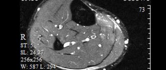
A pathology such as atherosclerosis begins from the moment when cholesterol is deposited on the walls of the arteries of the lower extremities. These deposits narrow the lumen of the artery and begin to interfere with normal blood flow. Over time, if a person does not take care of his health, does not monitor his diet, or leads a sedentary lifestyle, cholesterol build-ups increase and atherosclerotic plaques form. They look like tubercles; in addition to cholesterol, they also contain calcium, which gives them strength.
Doppler ultrasound allows not only to recognize atherosclerotic plaques, but also to determine their size, exact location, quantity, and structure. In addition to plaques, blood flow can also be interfered with by thrombi (blood clots), which are also detected using Doppler ultrasound.
Quantify blood flow (eg, speed of movement)
An important indicator of vascular health in the lower extremities is blood flow velocity. Doppler ultrasound allows you to accurately measure this indicator and track the dynamics of its changes in different parts of the limbs.
Identify segments of arterial narrowing (stenoses) and sizes
Normal blood flow is ensured by the elasticity and smoothness of the inner surface of the vessel - the endothelium. It is also important that the vessel has a normal diameter. Most vascular pathologies are characterized by narrowing of the arteries and most often this occurs in fragments and not throughout the entire vessel. The task of ultrasound examination is to identify segments of stenosis (narrowing) of the arteries, determine their exact location, and understand the possible causes of the pathology.
Advantages of ultrasound examination of veins in our Moscow phlebology center
- Modern expert class equipment.
- High diagnostic information content.
- No contraindications.
- No special preparation is required.
- The procedure is non-invasive and comfortable.
- The study is performed by a phlebologist-expert of the highest level.
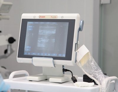
Ultrasound scanner of the latest generation, on which the doctor performs an ultrasound of the veins in Moscow
Ultrasound scanning in the modern phlebological center in Moscow meets the best global and European quality standards.
To whom is it assigned?
Other arteries in the body already have atherosclerosis
If the patient has previously been diagnosed with atherosclerosis, for example, of the abdominal region, renal arteries, upper extremities, experts recommend examining the lower extremities for the same pathology. The fact is that atherosclerosis is a disease that can affect the entire body. It is important to detect it in time and assess the degree of development.
Pain occurs in the calf muscles
Pain in the calf muscles (sometimes turning into cramps) causes serious discomfort to a person. They interfere with normal walking, do not allow you to endure long, intense physical activity, make it difficult to climb stairs, and even at rest constantly remind you of yourself. Often the cause of pain in the calf muscles is a violation of hemodynamics in the vessels of the legs, and pathologies such as atherosclerosis, thrombophlebitis, varicose veins, ultrasound scanning will allow us to find out for sure.
Pain occurs with exertion and walking short distances
When a person begins to feel pain in his legs after just 10-15 minutes of walking, and intense exercise becomes completely beyond his strength (in terms of walking or running), an ultrasound with Doppler will help you figure out what exactly is the cause of the problem. If it is atherosclerosis or venous stagnation (varicose veins), an ultrasound scan will show a disturbance in the speed of blood flow and indicate those segments where patency is impaired. At the same time, pain when walking can be caused by completely different reasons: osteomyelitis, rheumatoid arthritis, osteochondrosis, that is, diseases not related to blood flow. In such cases , ultrasound examination of the vessels of the lower extremities
allows you to exclude those diagnoses that are associated with hemodynamic disorders and prescribe other specific studies (for example, X-rays, MRI, CT, and so on).
Feet turn blue and cold or red and swollen
Swelling of the legs, their redness, blueness or the effect of “cold legs” are symptoms that may indicate the presence of vascular pathologies of the lower extremities. Such pathologies are atherosclerosis, thrombosis, and vascular inflammation.
Vessel nodes are visible on the surface and varicose veins appear
When varicose veins enter the active stage of development, they become visible to the naked eye. Veins appear on the legs (most often under the knee). Seals and nodes may be visible on the veins. Ultrasound examination and Dopplerography of the veins of the lower extremities in such cases can be used both for the primary diagnosis of pathology and for planning surgical intervention.
How is ultrasound scanning of veins performed in Moscow?
In their practice, leading phlebologists in Moscow most often use duplex angioscanning, including:
- B-mode (two-dimensional grey-mirror scanning).
- Color Doppler mapping of blood flow (CDC).
If necessary, the study is supplemented with pulsed spectral Dopplerography. Thus, a triplex scan of the vascular bed is performed. Innovative ultrasound diagnostics in Moscow today is the best technique for studying the venous bed, meeting high European standards. Ultrasound scanning is a good alternative to the invasive methods of magnetic resonance venography and X-ray contrast venography, without being inferior to them in terms of information content.
Indications for research
Ultrasound scanning of the veins of the lower extremities and arteries is a procedure that can be performed for preventive purposes, but most often it is prescribed if there are indications.
Pallor of the legs and cold extremities
Pale and cold legs are a symptom that indicates the presence of a pathology in the blood supply to the skin.
Goosebumps
Often, with vascular disease, people may feel a slight tingling in their legs, it seems that goosebumps are running down their legs. When this symptom is also accompanied by pain in the legs and fatigue, you should not hesitate to visit a doctor.
Legs get tired and tingle quickly
A feeling of fatigue, a buzzing in the legs that does not go away even after a long rest, are alarming symptoms indicating a disruption in the functioning of the blood vessels of the lower extremities.
Bruises appear quickly
When even a small bruise of the legs (or, for example, pressing a finger on the skin) can cause a large bruise, it is worth contacting a specialist and undergoing Doppler ultrasound of the vessels of the lower extremities (deep duplex scanning).
Abrasions do not heal for a long time
If wounds and abrasions on the legs do not heal for a long time, this may indicate that there is a problem with blood flow, the blood does not clot properly, which, accordingly, interferes with rapid regeneration.
Indications for ultrasound of the vessels of the lower extremities
Ultrasound of the arteries is performed:
- for diabetes mellitus;
- pain that occurs when walking;
- high blood pressure;
- elevated cholesterol levels;
- smoking;
- previous myocardial infarction;
- feeling that your feet are freezing even at a comfortable temperature.
Ultrasound of veins is performed:
- if you suspect varicose veins;
- cramps, aches and heaviness in the legs;
- thickening of the skin along the veins;
- visibility of saphenous veins on the legs;
- "spider veins" on the legs;
- swelling of the legs;
- blue skin on the toes;
- trophic ulcers (wounds that arise after the rejection of dead tissue and do not heal for a month or longer).
Preparation and progress of the procedure
Ultrasound scanning does not require special training. The patient is advised not to drink tea, coffee or other stimulating drinks or drink alcohol on the day of the procedure. There is no need to smoke 2 hours before ultrasound examination.
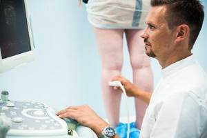
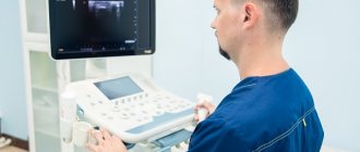
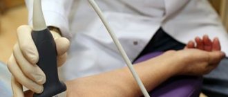
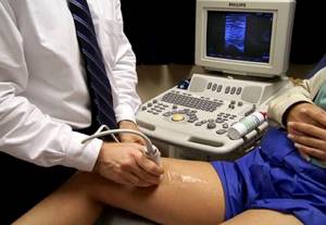
The examination is carried out with the patient standing or lying down. The doctor applies ultrasound gel and moves the ultrasound probe along the legs. Immediately during the procedure, he analyzes and records the received data. How to do an ultrasound scan of the arteries of the lower extremities and veins
, you can watch the video on the Internet.
How is ultrasound performed?
Before undergoing an ultrasound of the blood vessels of the legs, the patient does not require any special preparation; you just need to come to our medical center at the appointed time. A temporary contraindication may be a violation of the integrity of the skin at the examination site. In this case, you need to wait until the wound has completely healed. To undergo an ultrasound, the patient should free the lower limbs from clothing and shoes and lie on the couch with their legs shoulder-width apart. A gel is applied to the skin in the area to be examined to allow ultrasound penetration. In some cases, differential diagnosis may be required (to exclude diseases with similar symptoms).
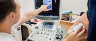
Then the doctor may ask you to take a standing position, hold your breath, etc. After the ultrasound, the data obtained is entered into the medical record within 15 minutes. If the diagnosis requires clarification, you can undergo additional examinations (duplex scanning of veins, phlebography, etc.) in our clinic.
To find out the cost of ultrasound of leg vessels, contact the call center or look at the price list on the website. You can make an appointment online or by phone.
Ultrasound diagnosis of venous pathology at the Moscow Phlebology Center
After a visual examination and palpation, the leading specialist will conduct a full ultrasound examination of the venous vessels of your legs. The doctor will always scan both legs, even if there are unilateral complaints. The phlebologist will use good diagnostic techniques:
- Valsalva maneuver.
- Compression tests.
It is important to know that the diagnosis should be carried out with the patient in orthostasis (standing) in order to obtain the most accurate information. Many specialists in the public sector of medicine in Moscow perform the examination while lying down. This often leads to diagnostic errors.