Table of contents
- What is MRI?
- Applications of MRI
- Benefits of MRI
- Disadvantages of MRI
- Areas of use
Table of contents
- What is MRI?
- Applications of MRI
- Benefits of MRI
- Disadvantages of MRI
- Areas of use
In our age, information technologies and various high-tech techniques have entered our lives so deeply that it is almost impossible to imagine any branch of science where they would not be used. Medicine is no exception, in which such a direction as radiation diagnostics rightfully occupies one of the most important niches in this industry.
Magnetic resonance imaging (MRI) is currently perhaps one of the most informative methods of radiation diagnostics. In this it successfully competes with X-ray computed tomography (XCT), and in some cases even surpasses it in diagnostic specificity, serving as the so-called “gold standard” in identifying a number of pathological changes in various tissues and organs of the human body.
This method, from the moment of its discovery to the present, has gone through many stages of development, each of which was characterized by the transition of this method to a qualitatively new level of diagnostic capabilities.
What is MRI?
First, a little history. In 1946, independently of each other, two American scientists (Felix Bloch and Edward Purcell) described a certain physical effect inherent in the atomic nuclei of certain substances. Subsequently, it was he who became the cornerstone of the entire MRI technique.
It turned out that if the nuclei are placed in a constant magnetic field and then exposed to radio frequency pulses of a certain frequency, this energy will be absorbed by them, as a result of which the entire system will move to a higher energy level. This state is less stable, and therefore, in the future, the absorbed energy will be emitted by the nuclei, and the system will return to its original energy state. This emitted energy carries information about the location of the atom in space, and if we add to this the additional influence of a weaker magnetic field (the so-called gradient), then with the help of a catching device (receiving coil), and subsequent mathematical processing of the received information, it is possible to reconstruct the location of the atoms of any or an object in the form of an image of its cross section on the monitor screen.
In this way, it is possible to obtain layer-by-layer images at different A significant number of such layers, or slices, can be specified, and it is possible to vary their thickness, distance between slices, direction and many other parameters within fairly wide limits, affecting the quality of the resulting tomograms.
In the early 70s of the twentieth century, American scientists R. Damadian and P. Lauterbur independently used the phenomenon of magnetic resonance to obtain an electronic image of the tissues of a living object (including a person) using an MR scanner. It is believed that the first MR scanner was created by R. Damadian and a team of his associates towards the end of the 70s of the twentieth century, at which time he patented his invention. Over time, the technique has become widespread in medicine and is currently successfully used in radiation diagnostics.
Since the advent of the first magnetic resonance scanners for the whole body (early 80s of the last century) to the present day, MRI scanners have come a long way of evolution: software and hardware components have been improved, the image has become better - resolution has improved and the contrast between different tissues has been improved , new techniques were introduced and the boundaries of application of the method were expanded in various areas of medicine, and much, much more. To outline all the stages of MRI development even briefly, one would have to write at least a small book. But since this task is not before us, we will limit ourselves to a short excursion into the main aspects of the application of the method within the framework of medical imaging, where MRI very often becomes the first-line diagnostic method among the impressive arsenal of high-tech tools of modern medicine.
ADVANTAGES OF AN OPEN TYPE MRI DEVICE
As already mentioned, open MRI makes it possible to examine patients of certain groups. These include seriously ill patients, for whom it becomes possible to carry out medical manipulations directly during the MRI procedure. It is also convenient to use an open-type apparatus in the case of examining patients with severe injuries who cannot assume a lying position. Examination of the spine in such a situation becomes possible thanks to open MRI.
Young children who experience stress during any medical procedure can also easily tolerate an open-type MRI because they get the opportunity to be in the room with their parents. It is easier for them to remain calm and still in the presence of loved ones.
Applications of MRI
According to some literature data, the diagnostic accuracy of MRI is 91–99%, and sensitivity can reach 97%.
The main challenges facing medical imaging are generally the following:
- assessment of the spatial location, shape and structure of tissues in organs, the organs themselves, as well as their systems;
- identifying pathological changes of various nature and carrying out their differential diagnosis;
- obtaining diagnostically significant information, which can later be used to plan treatment, including surgical treatment.
MRI as a method of radiological diagnostics has a number of advantages that make Let's look at them in more detail.
Benefits of MRI
- When using MRI, there is no ionizing radiation or radiation exposure to At the same time, there is no need to talk about the possible carcinogenic and mutagenic effects associated, for example, with X-ray radiation, which is also used in computed tomography.
- High image resolution, which is
- The most important parameter when performing various types of tomography is the so-called tissue contrast, that is, diagnostically significant visual differences between tissues with different signal characteristics. This allows you to see differences in the structure of different tissues and organs from each other and unambiguously interpret the pathological changes that appear in them . In MRI, tissue contrast is the highest among currently known types of medical imaging based on radiation effects.
- The MRI method is multi-projection, that is, it makes it possible to conduct research in three projections, as well as
- Radiation diagnostic methods such as radiography and computed tomography often use contrast agents to obtain additional information, which, despite their significance, can have a toxic effect on some organs, as well as cause allergic reactions of varying severity, including such dangerous conditions as edema Quincke and anaphylactic shock. Contrast agents used in MRI do not have cyto‑, hepato‑, or nephrotoxic effects, and also do not cause allergic reactions, which is an advantage compared to contrast agents used in x-ray studies.
- Another advantage of magnetic resonance imaging is the absence of artifacts (interference) from bone structures, which can complicate the interpretation of the image obtained using computed tomography.
Disadvantages of MRI
Like any diagnostic method, MRI also has its disadvantages:
- the need to remain motionless (often for quite a long time) during an MR examination, which is not always realistic for patients with severe pain or in a state of deafness;
- the impossibility of carrying out MR diagnostics in patients with artificial pacemakers in the heart muscle, cochlear implants, implanted spinal cord stimulators, prosthetic joints (especially hips), implanted insulin pumps;
- MRI is also contraindicated for patients with metal fragments in the body, stents, clips on vessels, fixing brackets, plates, knitting needles, bolts made of ferromagnetic materials.
- relative contraindications are claustrophobia, 1st and 3rd trimesters of pregnancy, panic states, functional mental disorders;
- To a certain extent, the relatively high cost of the examination can also be considered a disadvantage. However, recently there has been a tendency to reduce it due to the ever-increasing number of magnets in public and private medical institutions.
Now we will take a closer look at the areas of application of magnetic resonance imaging in clinical practice.
Advantages of MRI diagnostics
The most important advantages of the study:
- When performing an MRI, the patient is not exposed to ionizing radiation. The procedure has no restrictions on the frequency and frequency of implementation. MRI is performed for pregnant women (from the 2nd trimester) and children from one month of age.
- The ability to obtain the thinnest sections allows us to examine in detail the subtentorial space of the brain, the cavity of the spinal canal, detect pathologies of the pituitary gland, neuroma (tumor) of the auditory nerve, etc.
- MRI scanning is the main method for diagnosing foci of demyelination in multiple sclerosis and some congenital defects (Arnold-Chiari anomalies, agenesis of the corpus callosum, etc.).
- Magnetic resonance imaging reveals foci of cerebral ischemia earlier than computer imaging. Abscesses are better visible on photographs of cerebral structures. MRI is informative in establishing the causes of dementia for subsequent determination of treatment tactics.
- Scanning allows you to thoroughly study the structures of the spinal column. Using MRI, you can see prolapsed (bulging) discs, herniation, anomalies in the development of the spinal cord (syringomyelia, etc.) and other pathologies.
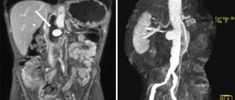
- Magnetic resonance diagnostics makes it possible to assess the condition of blood vessels without the use of special contrast agents. If specific diagnostic options are needed, gadolinium-based agents can be used. Contrast materials are well tolerated by patients and rarely cause side effects.

Preparing your baby for an MRI scan
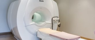
Full MR Scan Process
The MRI procedure does not require special preliminary preparation, so before the examination you can eat and take the necessary medications as usual.
Clothing for diagnostics should not contain buttons, snaps, zippers or other metal parts. The patient can bring suitable clothing with them or use the gown provided at the clinic. Before diagnosis, you must make sure that there are no metal objects and accessories on the body (jewelry, piercings, hairpins, hairpins, buttons, buttons, etc.). Electronic devices, mobile gadgets, credit cards and other personal items should also not be left in the room with the CT scanner.
Best materials of the month
- Coronaviruses: SARS-CoV-2 (COVID-19)
- Antibiotics for the prevention and treatment of COVID-19: how effective are they?
- The most common "office" diseases
- Does vodka kill coronavirus?
- How to stay alive on our roads?
During an MRI of the whole body, the table slides completely into the tunnel, and communication with the x-ray technician is carried out through a mini-headset. The patient is required to remain calm, breathe evenly and not move. The full body scan procedure takes approximately 90 minutes.
To prevent the clicks and taps of the tomograph from causing discomfort during diagnostics, it is recommended to use headphones or earplugs. For moral support of the patient, the presence of a loved one in the room is allowed, with the exception of women in the first trimester of pregnancy.