Abdominal pain is an alarming sign that requires immediate medical attention. After all, it can signal both chronic and acute pathologies, when urgent surgical intervention is indicated. When making a diagnosis, the surgeon will focus on the location of the pain, but the symptomatic picture alone is not enough. Moreover, pain can be radiating, that is, projected to other parts of the abdomen. Then it is necessary to resort to other diagnostic options. One of these hardware methods is abdominal CT. This is a highly informative method that allows you to examine the tissues of organs, vessels and lymph nodes of the peritoneum.
CT helps to obtain a three-dimensional image of the area under study, thanks to the ability to take X-ray images layer by layer, with the thickness of each layer in this case being about several microns. X-rays are emitted in a ring-shaped circuit with sensors, due to which images are obtained in different planes. Entering the computer monitor, they are superimposed on each other, forming a three-dimensional model of the organ being studied.
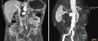
An even more detailed picture can be recreated by a CT scan of the abdominal cavity with contrast. Iodine preparations move along with the bloodstream to the desired area. Thanks to the ability of this drug to increase the contrast of tissues, maximizing differences in the absorption of X-rays, it is possible to most accurately determine the location of pathologies, their structure, shape and diameter.
In what cases is an abdominal CT scan prescribed?
Tomography can show the condition of parenchymal and tubular organs. The reason for its appointment is:
- Acute or prolonged pain in the abdominal area;
- Hyperbilirubinemia (staining of the skin and mucous membranes yellow due to increased bilirubin content in the blood plasma);
- Sudden weight loss not related to diets;
- Nausea, vomiting, flatulence, diarrhea and other disorders of the digestive system;
- Signs of bleeding;
- Upcoming surgery. Scanning is also indicated to evaluate the effectiveness of surgical intervention;
- Problems of the urinary system;
- Suspicion of neoplasms of various nature;
- A strangulated abdominal hernia has occurred;
- Closed injury;
- Interloop abscesses;
- Diseases or injuries of the spleen.
What diseases does an abdominal CT scan detect?
The method allows you to determine the focus of the disease, establish the volume and boundaries of the pathology, check the circulatory and lymphatic system of the area under study, and clarify the homogeneity of the structure. It helps you see:
- Polyps, adenomas, fibromas;
- Abscesses, foci of inflammation;
- Neoplasms of a malignant nature;
- Dropsy;
- Atherosclerosis, lymphadenopathy and other vascular problems;
- Lymphadenitis;
- Tissue necrosis;
- Cirrhosis;
- Cysts;
- Congenital anomalies;
- Hepatosis;
- Gallbladder diseases;
- Pancreatitis;
- Aneurysms;
- The presence of parasites in tissues;
- Chronic and acute hepatitis;
- Appendicitis.
This is not the entire list of diseases that can be detected using computed tomography.
In what cases is an abdominal CT scan with contrast prescribed?
When specialists need to study the smallest structures or identify oncology in the early stages, doctors may recommend performing an abdominal CT scan with contrast. This will allow you to diagnose:
- Degeneration of atypical cells;
- Presence and localization of metastases;
- Vasoconstriction;
- Ureteral obstruction;
- And also conduct a study of the aorta;
- In addition, scanning will help clarify the success of anti-cancer therapy.
How to sign up for a study
Leading expert doctors will perform a CT scan of the abdominal organs according to international protocols, provide a professional opinion and give recommendations on choosing a treating physician. Our specialized specialist will prescribe the optimal treatment for you and develop effective therapeutic programs. To sign up for a study, call us or fill out an application using the form on the website. Before the study, you can sign up for a consultation with a radiologist or the head of the radiology department (regular and extended).
Contraindications to CT scan of the abdomen with and without contrast
Tomography is an X-ray examination method, which means it exposes a person to radiation. In this regard, it has a number of contraindications:
- It is not prescribed to pregnant women during the entire period of pregnancy. This is due to the fact that fetal cells divide at a tremendous speed, forming all body systems, so minor changes at the cellular level caused by X-rays can lead to severe congenital pathologies.
- Since radiation testing methods have a significant impact on children, tomography is not performed on patients until they reach 14 years of age.
- Each tomograph has a maximum permissible load. Depending on the model of the device, it can be 150-200 kilograms. If a person's weight exceeds these values, the scan cannot be performed for technical reasons.
- Since during a tomography it is necessary to follow the diagnostician’s instructions, the patient must be aware of his actions. Therefore, the state of drug and alcohol intoxication, as well as mental illnesses characterized by confusion, are an obstacle to examination.
Abdominal CT with contrast imposes additional limitations:
- The dye is not administered to people with kidney or liver failure. Since the ability to eliminate drugs in people with this pathology is reduced, the use of contrast can lead to intoxication.
- Autoimmune diseases, which include thyroid disorders and diabetes mellitus, especially in severe form, can also become an obstacle to the introduction of radiocontrast.
- It is not advisable to scan people with cardiovascular diseases or plasmacytoma, that is, a malignant brain tumor.
- Patients suffering from allergies of any form must inform their doctor about this. In some cases, iodine preparations can cause allergic reactions. If this happens, the scan will be interrupted and the person will be treated. For this purpose, all medical centers have a stock of antihistamines.
- If a CT scan is to be performed on a nursing mother, then lactation will need to be stopped for several days until the medicine is completely removed from the body. This substance can pass into breast milk and may harm the baby's health. To maintain lactation during this period you will have to pump. In order to avoid introducing artificial formula, you can prepare a supply of breast milk a few days before the scan. It should be stored in the refrigerator in sterile containers.
There are situations when the benefit outweighs the potential harm. In such cases, a council of doctors gathers to prescribe a CT scan of the abdominal cavity. This is especially true for the need to examine the abdominal aorta, which is not done without the introduction of radiocontrast, since without its use the procedure is not sensitive enough.
Indications for use of the method and advantages of the study
Our hospital has a modern multislice computed tomography scanner for scanning the whole body. The device significantly reduces radiation exposure and allows the examination to be carried out as quickly as possible without discomfort. The result of the research is finished images and files recorded on electronic media. Tomography done in our clinic is recognized by all medical institutions in Russia and abroad.
The advantages of multislice tomography performed in our clinic over standard CT:
- a more contrasting picture due to a reduction in the amount of noise using a new generation of iterative reconstruction;
- reduction in radiation exposure by 30-80%;
- possibility of using less contrast agent;
- increasing the area of the study area;
- reducing the thickness of the section, giving a chance to track down even the smallest pathological focus;
- the ability to create 3D models of organs and foci of disease, as well as their multiplanar reconstructions.
Contrast perfusion allows you to cover more parameters, as well as detect bleeding, neoplasms and hemorrhages, so it is recommended for most studies. In 80-87% of cases, the examination is carried out using contrast.
In what cases is a CT scan of the abdominal organs prescribed:
- the presence of pain in the abdomen, bleeding, both due to injury and for unexplained reasons;
- suspicion of tumors;
- the presence of metastases in internal organs, lymph nodes, bones;
- the presence of stones in the kidneys, biliary and urinary tracts;
- upcoming surgery requiring precise examination of the intervention area;
- the need to assess the dynamics of the process and treatment results;
- peritonitis, abscesses;
- liver diseases;
- kidney inflammation;
- the presence of a hernia (area of the anterior abdominal wall, diaphragm);
- developmental anomalies.
First of all, CT scan of the abdominal cavity is aimed at detecting volumetric processes in internal organs, which include polyps, cysts, abscesses, tumors, and stones. CT scan reveals benign and malignant tumors, metastases, stones, cysts, kidneys and liver, echinococosis, atherosclerosis, various changes in the structure of organs (cirrhosis, hepatosis).
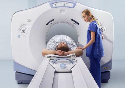
How is an abdominal CT scan performed?
The examination may take from a few minutes to an hour. The patient will have to undress, remove all jewelry, glasses and other wardrobe items containing metal, and lie down on the retractable table of the device. After this, the patient is placed in the tomograph tunnel and the study begins. It is worth noting that the device operates with a characteristic noise that can cause anxiety in the patient. If a person suffers from claustrophobia, panic attacks and anxiety disorder, it is worth discussing this issue with a diagnostician in advance. Perhaps in this case, the use of sedatives will be indicated to prevent an attack.
In a CT scan of the abdomen with contrast, dye injection can be done in different ways depending on the affected area. To clarify the condition of hollow organs and vessels, intravenous administration of the drug is used. If it is necessary to examine the liver, kidneys or pancreas, a person takes a solution of an iodine-containing substance orally in small sips. To detect pathologies of the large intestine, the dye is administered rectally. In particularly difficult cases, the diagnostician may suggest a bolus administration of radiocontrast, that is, it will be introduced into the body by drip using an automatic injector.
After the scan is completed, the diagnostician prepares a transcript of the results. Based on the images obtained, he enters into the research protocol information about the size, shape and relative position of organs, notes congenital anomalies and detected disorders. The conclusion and photographs are transferred to the attending physician or handed over to the patient. Many medical centers are also willing to record the results on a computer disk or flash card, or send them by email. It is worth considering that the final diagnosis is made by a doctor of the appropriate profile based on the images and the research protocol.
Computed tomography of OBP at the Miracle Doctor clinic is:
Safety
The Toshiba Aquilion RXL CT scanner sets the cutting-edge standard for imaging with low radiation dose and full volume coverage of organs. In particular, the equipment allows scanning the heart with a radiation dose of less than 1 mSv.
High-quality photos
The tomograph allows you to obtain 32 “slices” per revolution, which lasts 0.5 s. The detector allows for wide coverage of the anatomical area of study. In addition, the device is equipped with a 3D visualization function in real time. All this allows you to obtain informative images of high definition and resolution.
Wide range of diagnostic capabilities
In our medical center you can do all known types of CT scans. Special packages of MSCT (multispiral computed tomography) programs allow you to conduct virtual endoscopy of hollow organs, studies of the mineral composition of bones, diagnostics of circulatory parameters and much more.
CT with contrast
The high quality of images obtained during computed tomography allows, in most cases, to avoid the use of a contrast agent. Despite this, the method is in demand to clarify the size and boundaries of tumors. If necessary, we perform an abdominal CT scan with contrast.
Suitable for patients weighing up to 300 kg
Toshiba Aquilion RXL equipment allows examination of overweight patients. Devices of previous generations could withstand weights of up to 100–130 kg.
Comfort
The tomograph device provides the ability to communicate with medical staff. A table with a transverse movement function simplifies preparation for the examination. You can make an appointment for a consultation and examination any day of the week.
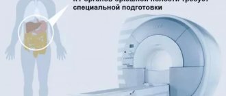
Preparing for an abdominal CT scan
CT scan of the peritoneal organs is one of the few types of examination that requires careful preparation. It will depend on it how well all organs will be visualized in the pictures.
Before the procedure, it is advisable to undergo additional examinations, such as ultrasound of the peritoneum, x-ray or colonoscopy. It is worth consulting with an allergist and anesthesiologist about the possible risks from the introduction of X-ray contrast. If the urinary system is to be examined, laboratory tests must be done for the presence of creatine, urea, AST and ALT enzymes.
Since the main problem that reduces the information content of the examination may be intestinal bloating and fullness, it is necessary to normalize the functioning of the gastrointestinal tract in advance. To do this, three days before the tomography, you need to switch to a gentle diet: exclude not only heavy and fatty foods, but also those foods that cause increased gas formation. These include: cabbage, legumes, beets, celery, fresh and dried fruits, cereals, dairy products, fresh baked goods, confectionery products. It is also undesirable to consume kvass, carbonated water, strong coffee and tea, and alcoholic beverages. It is allowed to eat lean steamed fish, omelettes, light non-rich broths, boiled chicken, and oatmeal with water.
The test is done on an empty stomach, which is why a CT scan of the abdominal cavity is usually scheduled for the first half of the day. The day before, they give a cleansing enema, take a mild laxative, or administer a suppository with glycerin. In addition, 18 hours before you can drink enteric sorbents: activated carbon or smecta.
Two hours before the scan, you should stop drinking any drinks; this may increase intestinal motility.
It is worth noting that the introduction of X-ray contrast without appropriate preparation can provoke vomiting, nausea, and in some cases, intestinal volvulus. If the patient had a CT scan of the abdominal cavity with contrast, then after it it is advisable to drink a liter of clean drinking water. This promotes rapid removal of the dye.
Contraindications
An absolute contraindication to MSCT is pregnancy. In other cases, the doctor considers the conclusion about the admissibility of examination using X-ray irradiation in each situation individually. Relative limitations to computed tomography include:
- the patient’s body weight is more than 200 kg, which is associated with the maximum load on the tomograph table;
- age up to 5 years due to radiation exposure;
- mental illness and other conditions that do not allow the patient to lie still during a CT scan.
Computed tomography of the abdominal cavity with contrast may be complicated by:
- patients with renal failure;
- patients with diabetes mellitus.
How much does an abdominal CT scan cost?
The price range for this type of diagnostics is quite large. It is influenced by a lot of factors: the model of the tomograph, the qualifications of the diagnostician, the location of the clinic. It is worth considering that an abdominal CT scan with contrast will cost 20-30 percent higher. To save money, it is advisable to study the cost of the procedure at various medical centers, as well as find out about the promotions and discounts available there. This is easy to do using our service. Also here you can immediately make an appointment at any diagnostic center at a time convenient for you.
Price
When studying with contrasting, the cost of contrast is added if contrasting is not immediately included in the cost of services (then this is reflected in its name).
You can see prices for services
Preparation for CT scan of the abdominal cavity and retroperitoneum
| Bowel preparation | not required | |
| Diet on the day of the study |
| |
| Drugs | not required | |
| Take with you |
| |
| Keep in mind | the study is almost always performed with intravenous contrast | |
| see also | interview and preparation for intravenous contrast in CT | |
Alternative Methods
A conventional X-ray or magnetic resonance imaging can replace a CT scan. The advantage of an abdominal CT scan over an MRI is the ability to check the structure of hollow organs, including the intestines and stomach. In addition, magnetic resonance imaging is not indicated for patients using pacemakers, insulin pumps and metal implants.
X-ray compared to CT scan of the abdominal cavity has lower informative capabilities. Another examination option is ultrasound. Due to its proven safety and the almost complete absence of contraindications, this method is prescribed quite often in the case of primary diagnosis. And only if it turns out to be uninformative, computed tomography comes to the rescue.
Alternative methods for diagnosing abdominal organs
For patients who, for some reason, are not indicated for abdominal CT, several types of alternative studies are available:
- Survey radiography of the abdominal organs - allows you to detect x-ray positive gallstones and stones in the urinary tract, free gas in the abdominal cavity (with perforation of a hollow organ), signs of intestinal obstruction;
- Ultrasound is a good screening method for most patients, for most of the most common pathologies;
- MRI is a highly sensitive method for diagnosing diseases of the abdominal cavity and retroperitoneal space, which is a full-fledged alternative to CT when performed at a high level. However, a significant limitation of MRI is the inability to detect small stones in the kidneys and gall bladder.
Safety of Abdominal CT
If you follow the diagnostician's recommendations, scanning is a safe method. It does not harm health and has no delayed consequences. It is worth noting that, despite the minimal amount of x-ray exposure during a CT scan, radiation exposure to the body is still present. Therefore, after the tomography, a recovery period is necessary. Experts recommend, if possible, not to resort to this type of diagnosis more often than once every six months. If the examination is required for emergency reasons, it is advisable to replace it with alternative methods, or if this is not possible, do not neglect such protective measures as a lead apron, taking absorbents and maintaining a drinking regime.
In addition, when performing an abdominal CT scan with contrast, the danger is not so much the procedure itself as a possible reaction to the injected drug. Side effects on X-ray contrast may include:
- Joint spasms;
- Slight increase in temperature;
- Headache;
- Damage to the kidneys and urinary canals by toxins;
- Allergic skin reactions, in rare cases - anaphylactic shock.
Advantages of our clinic
- ultra-modern diagnostic equipment - unique Philips digital tomographs, which allow identifying many pathologies, including neoplasms and metastases, with high accuracy without high radiation exposure;
- highly qualified diagnostic doctors - specialists in the field of radiology are trained and trained in leading clinics in Russia, Europe, the USA, Israel on a regular basis;
- automated research quality control system - implemented on the basis of a modern IT platform, the system allows for triple control of results, the research results are sent to a single service center and become available to doctors caring for the patient;
- high-quality equipment of the department - creating a comfortable environment for the patient, the ability to conduct a wide range of studies.
What to take with you to an abdominal CT scan
Medical centers licensed to perform this procedure usually ask you to bring:
- Passport;
- Medical history, results of laboratory tests and previous diagnostics;
- Doctor's referral, if available. Many clinics can provide services without a doctor’s prescription, but to avoid overdiagnosis, it is still better to first consult with a specialist.
Do not forget that timely diagnosis can not only preserve health, but also prolong life. After all, the effectiveness of treatment depends on the accuracy of the diagnosis.