From this article you will learn:
- Differences between MRI of the lumbosacral region and myelography
- Differences between MRI of the lumbosacral region and computed tomography
- Indications for MRI of the lumbosacral region
- Contraindications to MRI of the lumbosacral region
- Preparation for MRI of the lumbosacral region and the procedure
- How MRI of the lumbosacral region will help the doctor
- How much does a lumbosacral MRI cost?
MRI of the lumbosacral region is prescribed to detect various diseases at the earliest stages. Thanks to modern equipment, it is possible to clearly visualize vertebral bodies, intervertebral discs, the spinal canal, blood vessels, ligaments, nerve fibers and detect pathological lesions as small as 0.5 mm.
Like any procedure, MRI has its own characteristics, indications and contraindications. In our article we will dwell on this in more detail, and also tell you how the diagnosis is carried out and how to prepare for it.
Differences between MRI of the lumbosacral region and myelography
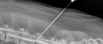
In some cases, it is not enough to do a regular MRI of the lumbosacral region to determine what pathological changes have occurred in the spinal structures. In such situations, myelography is prescribed - screening carried out by an invasive method to examine a certain area of the spine by injecting a contrast agent into the subarachnoid space.
After the injection, MRI is used to monitor how the radiocontrast agent is distributed. This technique has certain advantages compared to conventional MRI:
- the ability to see the contours and nerve roots of the spinal cord, which is not always available with other studies;
- the ability to detect specific neurological disorders, arachnoiditis, injuries to the nerves of the spinal cord;
- the ability to determine the causes of disruption of the normal circulation of cerebrospinal fluid;
- the ability to diagnose spinal canal stenosis;
- the ability to diagnose osteochondrosis and spinal hernia;
- the ability to determine the causes of compression of the spinal canal.
Myelography is performed using the injection of Povidone-Iodine or Lipiodol to obtain a three-dimensional image of the spinal cord. This allows the doctor to see small structures and answer the question of why the patient’s back hurts and what causes health indicators to deviate from the norm. In addition, this technique does not leave traces of radiation in the body. Despite this, such a study has a number of contraindications, the main of which are: arthritis in a severe stage of development, the recovery period after spinal surgery, as well as the anatomical features of its structure.
Recommended articles on the topic:
- How to massage the abdomen for weight loss: different techniques for health and beauty
- Stone massage: description, benefits, methods
- MRI of three parts of the spine: when is it necessary and what are the features of the procedure
The myelography procedure is not performed during pregnancy, high fever, kidney inflammation and diseases of the cardiovascular system in the stage of decompensation, as well as in some other cases. Before the examination, you should not eat for 6 hours. Also, before puncture of the lumbar region and administration of contrast, it is necessary to do an enema. Some patients have a negative reaction to the contrast agent. In this case, you need to drink a large amount of liquid to accelerate the removal of the drug from the body.
Differences between MRI of the lumbosacral region and computed tomography
Computed tomography (CT) is a research method that makes it possible to construct a three-dimensional image of a certain part of the body. This distinguishes a CT scan from a regular x-ray. During the procedure, not one picture is taken, but several from different angles. After computer processing, these images are combined into a single 3D model.
Computed tomography is indicated in the following cases:
- for the study of bones and joints;
- for diagnosing head injuries;
- to identify diseases of the spine, such as hernia, bone fragility due to decreased tissue density, changes in the shape of the spine;
- for the study of blood vessels (aneurysm, atherosclerosis);
- for diagnosing diseases of the chest, abdominal and pelvic organs.
With the help of CT, you can see stones, tumors with accumulation of fluid, and oncological tumors.
What is the difference between MRI and CT:
- MRI is used when it is necessary to examine soft tissues;
- MRI is more revealing when examining joints and blood vessels;
- MRI can detect neoplasms in soft tissues;
- MRI is suitable for examining the brain and cranial structure;
- MRI allows you to study the meninges;
- MRI is prescribed in case of acute cerebrovascular accident;
- this procedure is indicated for persons suffering from neurological diseases;
- MRI allows you to examine the connective tissues that hold bones together and hold organs and muscles in certain positions;
- MRI can be used to diagnose bone and vascular diseases.
However, computed tomography is more effective when examining bone tissue and internal organs. MRI, in turn, is used to diagnose injuries and diseases of muscles, tendons, synovial membranes, fibrous connective tissues, blood vessels and nerves.
It is worth noting: despite the fact that during a CT scan the patient is exposed to x-rays, their negative impact on the body is minimized. The scan takes 60 to 90 minutes, which is not long enough to cause serious harm. As for MRI, it does not have any negative effects and is considered an absolutely safe procedure.
Advantages and disadvantages of the procedure
X-rays for fractures or diseases of the sacrum are the main diagnostic method. This is due to the fact that such a procedure is very informative and accessible. The study allows you to determine the condition of the sacrum. However, x-rays involve irradiation of the body. Of course, radiation exposure standards are not exceeded, but x-rays cannot be taken more than once a year, and this may not be enough to control the disease. In addition, CT and MRI are more informative and will allow you to examine not only bones, but also soft tissues. At the same time, MRI
completely safe.
Indications for MRI of the lumbosacral region
Magnetic resonance imaging of the lumbar region is the most popular examination of all tissues. It allows you to recreate a three-dimensional image and assess the condition of not only bones, as with a regular x-ray, but also ligaments, tendons, muscles, intervertebral discs, radicular nerves, etc.
MRI of the lumbosacral spine is prescribed to identify pathologies and diseases of the spinal column. First, the doctor examines ordinary x-rays, which to a certain extent assess the condition of the intervertebral discs. If there is a suspicion of protrusion, then a decision is made on additional diagnostics, since it is impossible to diagnose “intervertebral hernia” only by X-ray.
MRI is the most effective research method in modern medicine, as it allows us to identify any, even the slightest, degenerative and dystrophic changes in the cartilaginous tissues of the intervertebral discs.

The main advantage of the technique is the modern, latest technology of magnetic radiation. Through it, magnetic waves, colliding with cells, create a layer-by-layer image of tissue. It is completely harmless to health.
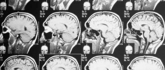
The layer-by-layer image of the tissues is combined into a three-dimensional model, which allows you to see the entire radicular nerve, dura maters of the spinal cord, nerve plexuses, vertebral bodies, arcuate and spinous processes, intervertebral discs, their fibrous rings and pulpous discs. Even minimally sized hernias become visible on MRI.
Note! Only a qualified vertebrologist or nephrologist can assess the condition of the lumbosacral spine using MRI images. Even if a detailed explanation is provided, you will not be able to figure it out on your own and understand what is wrong with your spine and nearby tissues. Therefore, after you have an MRI, be sure to consult a doctor. Only he will be able to make the correct diagnosis and select effective treatment.
MRI of the lumbosacral spine is prescribed to the patient for certain indications. Typically these include:
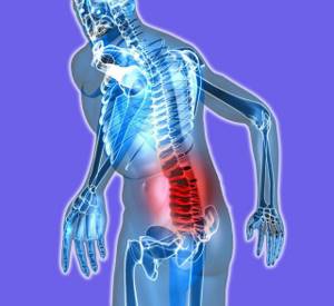
- complaints of back pain;
- severe symptoms of neurological diseases, for example, numbness of the limbs, tingling, pins and needles, inability to perform usual actions, etc.;
- suspicion of osteochondrosis, which is complicated by protrusion, as well as protrusion of the disc;
- examination after physical trauma to exclude compression fractures of the vertebra, splintered injuries of the spinous and arcuate processes, ruptures, tears, displacements;
- the presence of tumors in the spine, including the development of metastases;
- curvature of the spine, incorrect posture, displacement of the pelvic bones;
- deformed osteoarthritis caused by destruction of the iliosacral joints of the bones;
- lack of fixation of vertebral bodies and intervertebral discs, periodic occurrence of vertebral displacement such as retrolisthesis or antelesthesis;
- damage and destruction of the facet joints and intervertebral facet joints;
- disruption of cellular nutrition of the tissues of the spinal cord and dural membranes, which is caused by compression of blood vessels;
- rheumatism, ankylosing spondylitis, systemic lupus erythematosus, scleroderma, polyosteoarthrosis, etc.;
- spinal tuberculosis.
MRI of the lumbosacral spine can also be prescribed for diseases that are characterized by symptoms of osteochondrosis. These include inflammation of the abdominal organs, damage to the hip joints, cauda equina and piriformis syndromes, kidney and urinary system diseases.
Our clinics in St. Petersburg
Structural subdivision of Polikarpov Alley Polikarpov 6k2 Primorsky district
- Pionerskaya
- Specific
- Commandant's
Structural subdivision of Zhukov Marshal Zhukov Ave. 28k2 Kirovsky district
- Avtovo
- Avenue of Veterans
- Leninsky Prospekt
Structural subdivision Devyatkino Okhtinskaya alley 18 Vsevolozhsk district
- Devyatkino
- Civil Prospect
- Academic
For detailed information and to make an appointment, you can call +7 (812) 640-55-25
Make an appointment
We draw your attention to the schedule of technological breaks in the CT and X-ray rooms.
X-ray of the lumbar spine today, when X-ray diagnostic equipment is available in almost every clinic and medical center, is one of the most popular, quick and simple methods of diagnostic research.
If you are looking for a clinic in St. Petersburg with good and safe X-ray equipment, then in the Medicenter network of multidisciplinary clinics you can do an X-ray examination of the lumbar spine. The trauma department of the Medicenter accepts patients seven days a week and on holidays. The department is equipped with high-tech X-ray equipment made in Italy, Clinomat, which allows you to quickly, efficiently and effectively take X-rays of various areas of the body, and study the results obtained in various processing modes. Qualified doctors - surgeons, traumatologists, neurologists, orthopedists and other specialists at the Medical Center will provide you with first aid for injuries, conduct a thorough examination, conduct the necessary examinations and prescribe a course of treatment and recovery.
Contraindications to MRI of the lumbosacral region
MRI is a method of medical examination of the body, which is considered absolutely safe for health and does not entail any negative consequences. However, there are a number of contraindications to the procedure:
- the patient is too overweight;
- claustrophobia;
- inability to remain in a horizontal position for a long time due to anatomical, physiological or other characteristics of the body;
- the presence of metal prostheses and electronic devices in the patient’s body;
- mental illness;
- epilepsy with severe convulsive syndrome;
- pregnancy, especially the first trimester.
The main contraindication to MRI using a contrast agent is the presence of an allergic reaction. It is also not recommended to carry out this procedure for people suffering from acute renal failure.
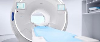
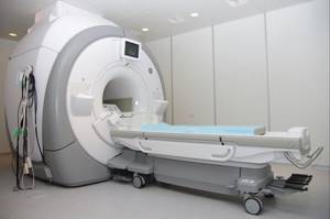
Preparation and performance of MRI of the lumbosacral region
No special preparation is required to perform an MRI of the lumbosacral region. Doctors do not insist on changing the usual daily routine, including not forcing you to give up food, although some recommendations suggest the last meal 3-4 hours before the MRI. Before conducting the test, a woman should make sure that she is not pregnant.
If the patient wishes to undergo the procedure in his own clothing, then it is important that it is loose, does not restrict movement, and does not have metal elements. It is also necessary to remove all jewelry and dentures. If an MRI of the lumbosacral region needs to be done using contrast, then the preparation must be more thorough. It is recommended to do a blood test to rule out allergies.
Patients who suffer from nervous disorders, as well as children, may be offered mild sedatives to calm them down before an MRI.
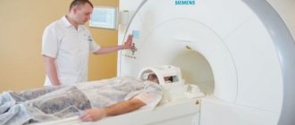
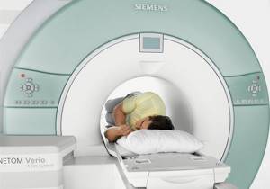
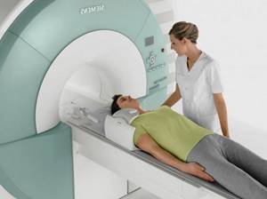
So that the patient does not worry and is not afraid before the procedure, the doctor must explain to him in detail what the essence of MRI is. This is a painless research method that does not involve any unpleasant medical procedures. All you need to do is come to the office, change into hospital clothes, remove excess jewelry and lie down on a special extendable table.
We recommend
Anti-wrinkle facial massage: 10 effective techniques Read more
Next, the patient is fixed with belts in a horizontal position and rollers are placed under those areas of the spine that need to be examined. Before turning on the device, the medical staff goes into the adjacent room. Then the table slides into a special tube and the scanning process begins, during which the tomograph makes loud sounds. You can communicate with staff using a microphone.
The duration of the magnetic resonance imaging procedure ranges from 5 to 30 minutes, depending on the area of study. If contrast is used, the diagnostic time can be increased to 90 minutes. If it is necessary to administer contrast, an MRI without it is first performed. Then a contrast agent is injected into the area under study and it is scanned in three planes.
MRI is a completely safe procedure that has no negative consequences for patients. The only exceptions are those who are registered with a nephrologist or have a severe allergic reaction to a contrast agent.
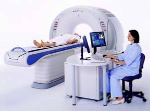
How MRI of the lumbosacral region will help the doctor
Only a radiologist or a functional diagnostics specialist can interpret the MRI results. To do this, images are compared over time to track changes in condition, or compared with an image of a healthy spine to identify possible pathologies.
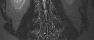
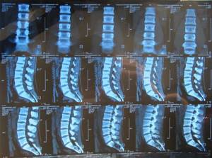
MRI of the lumbosacral spine makes it possible to detect a number of serious diseases and determine the course of treatment. The procedure can be carried out as needed. The images obtained during the diagnosis clearly show the bones, cartilage tissue (highlighted in dark color) and the spinal cord (light color).
This allows the doctor:
- identify defects, injuries and pathologies of the vertebrae;
- detect inflammatory processes or neoplasms;
- find out the size and nature of traumatic injuries;
- examine the condition of blood vessels and nerve tissues in the spine;
- identify osteochondrosis, inflammation of the spinal membranes on an MRI image;
- find out whether the patient has a hernia, which on T-2 weighted images looks like a protrusion with structures, namely the intervertebral disc, muscles, longitudinal ligaments.
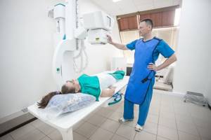
How is an X-ray of the sacroiliac joint done?
It is quite simple to describe how an x-ray of the sacroiliac joint is taken - the patient is placed on his back on the work table of the x-ray machine. Then they point the scanner at the area of interest (pelvic area) and take a picture, this takes 2-3 seconds. The patient must remain still during the scan. There is no pain. To take a lateral view, the patient needs to turn over on his side.
After this, the photograph is developed and a description is made of it.