The outer part of the hearing aid is represented only by the visible auricles, which essentially play the role of locators that capture and amplify sound. The structure of the hearing organs has a complex configuration. The main part of the ear is located deep in the skull, which often makes it difficult to diagnose many pathologies, as they are difficult to identify using classical visual methods.
Often the symptoms of abnormal processes occurring inside the cranial cavity may be mild, but if the root cause of the disease is not determined in time, this is fraught with critical consequences. To help medical specialists, a methodology such as MRI has been developed and has long been used in medical practice. It allows, without instrumental intervention, to visually examine and evaluate both the general condition of the organ and the microscopic pathological processes developing in it.
Tomography, based on the use of such a physical phenomenon as nuclear resonance, is not affected by harmful rays, contrary to popular belief, and does not cause serious consequences or complications for the patient. This method allows you to painlessly carry out diagnostic procedures and obtain highly informative results.
When is an ear MRI prescribed?
Only a professional doctor can refer you for nuclear resonance imaging, since hardware screening is not a cheap procedure and is prescribed only when clearly necessary. Diagnosis of ear anomalies is the specialization of a highly specialized doctor - an otolaryngologist.
Screening is justified both in cases of identifying the cause of diseases, and as a confirmatory or refuting method (when other diagnostic methods have not given an effective result). MRI can be a concomitant method of monitoring during surgery, as well as a method of monitoring in the postoperative period.
Since the ear has channels that go out and are also connected to the upper respiratory tract, the entry of various types of infections into this organ is not a difficult process. The human hearing system is susceptible to multiple pathologies that can be detected through tomography. It is necessary to refer to the technique in the following cases and with the symptoms described below:
- progressive decrease in sound perception;
- constant “interference”, noise;
- causeless dizziness, frequent fainting;
- change in anatomical asymmetry of the face;
- disturbing itching sensations, pain when touched, sharp “lumbago”;
- loss of coordination, balance;
- change in the color of the skin of the ears, the appearance of pigment spots;
- palpable compactions, swelling, narrowing of the external auditory canal;
- chronic pain in the nasopharynx;
- the patient may often feel sick, and the condition does not depend on food intake;
- ineffectiveness of the treatment measures carried out with persistent symptoms;
- the presence of fluid discharge from the ear canal.
If signs of serious inflammatory pathologies or oncological processes are detected, an examination is prescribed using additional enhancement in the form of a contrast agent. This drug acts as a “dye” that is injected directly into the bloodstream, spreads throughout it and accumulates for some time in the soft tissues and vasculature. In the images of the hardware installation, foci of infection, as well as neoplasms, even at the earliest stages of development, become clearly visible.
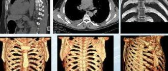
MRI stage of schwannoma
Magnetic resonance imaging of the brain has sufficient resolution to verify the stages of a tumor. Open low-power devices show grade 2-3, so verification is best done with closed (high-field) equipment. To verify tumors, there are several types of head MRI.
Tumor on MRI
The main stages of schwannoma:
- Unilateral hearing loss, mild paresis of the facial nerve, swelling of the affected side, hearing loss - symptoms of stage 1 neuroma (dimensions no more than 2.5 cm);
- Addition of ocular nystagmus, loss of coordination of movements - manifestations of stage 2 schwannoma (the size of a walnut);
- Mental disorders, pronounced nystagmus, optic neuritis (due to compression of the formation) are signs of stage 3 of the formation (more than 5 cm in size).
It is important to determine the nosology before symptoms occur. A lesion of 0.5 cm is characterized by isolated manifestations. Neurologists will not be able to differentiate the pathology, but MRI will allow diagnosis at the beginning of development.
Alternative to MRI examination of the ear
Along with the described diagnostic method, the following diagnostic methods are used to determine abnormal processes occurring in the ear:
- ultrasonography;
- classical radiography;
- vestibulometry;
- otoscopy;
- CT scan.
Ultrasound and standard x-rays can determine the location and extent of obvious disorders, but the techniques are somewhat limited in their technical base. The causes of the pathology, unfortunately, are not available to them. These methods are used at the initial stage of research as a way to detect degenerative processes.
Otoscopy is based on an external visual examination of the ear canal using a special instrument - an otoscope. Visualization of internal structures with such diagnostics is impossible. Vestibulometry is also used to measure the physical data of the human vestibular apparatus, and can only record the presence of a deviation without determining the root cause of the disease.
Both computer scanning and MRI are considered the most informative. Despite the external similarity of the procedures and their results, the methods are based on the use of various physical phenomena. CT is the successor to radiography, as it uses ionizing radiation in its work. Nuclear resonance screening works on the principle of generating a high-frequency magnetic field.
Patients often confuse different types of screening, but methods are even used to study different structures of the same organ. Thus, computer scanning is aimed at a detailed examination of bone fibers, cavity formations and capsules filled with liquid substances. MRI diagnostics sees them worse, but more clearly determines the condition and composition of soft tissues and dense formations than its “rival”.
Computer screening can easily detect disorders such as:
- tumor neoplasms of various nature, cystic capsules;
- foci of infection with the formation of pus;
- hearing loss;
- microcracks, fractures of the bone structures of the skull.
An MRI examination clearly shows:
- dysfunction of the vestibular apparatus;
- inflammatory processes in nerve fibers;
- tumor neoplasms and spread of their processes into adjacent tissues;
- changes in the thickness and volume of the tissue structure, thickening, thinning;
- configuration and diameter of the auditory canals;
- degeneration of the vasculature.
If computer screening clearly sees an anomaly, then nuclear resonance determines the cause of its occurrence even at the very early stage of development. An important advantage of the technique is that it is completely harmless to biological tissues and general human health, since it is not exposed to harmful radiation and is prescribed to people of any age, including children from the first hours of life and pregnant women (from the second trimester of pregnancy).
Causes of acoustic neuroma
The etiology of schwannoma has not been established. Two morphological types of nosology have been identified:
- Unilateral - the result of proliferation of Schwann cells of the vestibular part of the auditory nerve;
- Bilateral – a consequence of the genetic disease “neurofibromatosis type II”.
The anatomical structure of the nerve affects the localization of the formation. MRI often shows schwannomas of the vestibular part of the nerve fiber. The cochlear region is not affected.
Predisposing factors:
- Hereditary determination of neurofibromatosis type 2;
- The influence of chemical compounds (taking pharmaceuticals acting on the vestibular area);
- Mechanical impact.
Neurofibromatosis type 2 is transmitted in an autosomal dominant manner. The presence of the disease in one parent is the cause of pathology in the child. Geneticists set the probability at 50%.
What can be seen with an MRI of the ear?
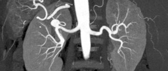
As mentioned above, the MRI hardware installation is based on the principle of generating a magnetic field, the high power of which causes a response in biological tissues at the molecular level. Once within the field's action area, the nuclei of molecules create a certain signature, which is captured and recorded by the machine. Processed in a special computer program, the signals are transmitted to the installation monitor in the form of clear black and white photographic images. High resolution allows you to bring areas with pathologies as close as possible, rotate them at the desired angle, combine them into a multidimensional projection and study the studied area of the ear with the highest quality possible.
Tomography allows you to see such anomalies as:
- areas of infection of nervous tissue;
- oncological and benign formations, migration of metastases;
- aggressive growth of tissue fibers, thickening;
- complications after illnesses;
- changes in the structure and configuration of the ear canals as a result of injury, etc.
The patient gets the opportunity to quickly undergo a full diagnosis, identify the disease at an early stage and prevent the disorder from developing into a more complex anomaly that can spread to adjacent brain tissue. If the cause of the disease is unclear, a comprehensive examination of the head area can be performed. If the picture of the course of the disease is clearly clear, a targeted study of the ear area is carried out, since the study of adjacent areas becomes impractical.
What diseases does tomography detect?
As a result of the study, the doctor receives a series of images that show in detail the structure of all parts of the hearing organ. Each area can be viewed in different projections and in the form of a three-dimensional model. MRI provides detailed information not only about the anatomical structure of the ear, but also about any pathology: inflammation, neoplasms, vascular disorders, deformations, sclerotic processes.
Diseases diagnosed using MRI of the ear:
- sensorineural hearing loss;
- neuritis of the auditory nerve;
- otitis – acute or chronic inflammation of the middle ear as a result of infection or injury;
- barotrauma, which occurs when there is a sharp change in external pressure on the eardrum in combination with insufficiently rapid equalization of pressure in the tympanic cavity (immersion in water or ascent, flying on an airplane);
- labyrinthitis – inflammation of the inner ear due to infectious diseases, transfer of inflammation from the middle ear or meninges, surgical interventions;
- mastoiditis - inflammation of the mastoid process of the temporal bone, most often a complication of otitis media or labyrinthitis;
- benign neoplasms - chemodectomas, hemangiomas, neuromas develop in the middle ear, the mastoid process is affected by osteoma, osteoblastoma, the inner ear is characterized by neuroma of the vestibulocochlear nerve;
- malignant tumors - squamous cell carcinoma, basal cell carcinoma, melanoma of the outer ear, cancer and sarcoma of the middle ear;
- metastases from tumors of another location;
- abscess - a limited accumulation of pus in any part of the ear, occurs as a result of inflammation, can move into the cranial cavity or mastoid process;
- cholesteatoma - a tumor-like formation of the middle ear, consisting of epithelial cells and cholesterol;
- injuries – rupture of the eardrum, traumatic brain injury, blow, burn, bite, foreign body;
- Meniere's disease is an increase in pressure in the inner ear, the causes are not fully understood.
Magnetic resonance imaging makes it possible to clarify the diagnosis, determine the stage of the pathological process, and the localization of the pathology. Detailed information allows you to select the most appropriate treatment strategy and monitor the dynamics of the disease.
MRI diagnostic progress of the ear
Screening is carried out according to a standard protocol and includes the following steps:
- Preparation and instruction. At this stage, the patient undergoes registration at the medical center and a detailed written questionnaire on issues related to the disease. This is necessary for the diagnostician to familiarize himself with the details of the disease and the patient’s medical history. When the area of study and the parameters of the tomography are clear, the medical professional explains to the client in detail what to expect and how the procedure will go.
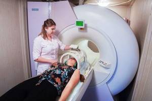
- MRI. The patient is invited into the room with the equipment, helped to position himself on the moving part of the installation, and fixed in a motionless lying position. Next, the conveyor moves into the inside of the device, and scanning begins.
- The machine can slowly change location along the person’s body and make a mechanical hum, do not be afraid of this - this is a standard phenomenon. It is worth remembering that the medical staff fully controls the process; employees are located in the next room and observe the session through a glass partition.
- In the event of an emergency situation, complaints, or severe discomfort, you can always contact the doctor via the public address system and ask to stop the process.
- Waiting for the result. After completing the session, you must wait until professional radiologists examine the images obtained and describe the result in the form of a final report. At this time, the client is in the waiting room or goes about his business, since upon request, you can receive results by email.
Where is the diagnosis carried out?
Scanning of the diseased area can be carried out in any medical organization that has the necessary technical base (high-power MRI equipment) and trained specialists who can correctly interpret the results of the study. There are hundreds of medical institutions in Moscow where you can undergo tomography. To quickly find a diagnostic center, it is recommended to use the unified city service 36go. When you go to the main page of the site, enter the name of the service and select the location.
The list of clinics that appears is differentiated by rating and price, which makes it easier to compare and study information. Read reviews, compare prices and sign up for diagnostics at your chosen center directly from the website. The telephone number for registration is located on each page of the service. When booking an MRI appointment through 36go, portal guests receive an additional 1,000 rubles discount on any type of examination.
When should you not perform an MRI of the ear?
Since the procedure has a medical purpose, it has a list of contraindications, which includes:
- ferromagnetic elements and objects implanted into body tissue (implants, pins, etc.);
- the presence of permanent electronic stimulators;
- during early pregnancy (up to 12 weeks);
- excess body weight from 120-200 kg, which does not allow placement in the inside of the tomograph;
- mental or neurological disorders leading to involuntary motor activity;
- MRI with enhancement (contrast) in the presence of an immune response to medications;
- kidney disease leading to long-term fluid retention (when planning contrast-enhanced tomography);
- asthmatic syndrome.
In some cases, MRI can still be performed, even if there are some contraindications. So, for obesity, you can use open-type installations that have no weight category restrictions. Patients with nervous tics, severe pain or severe fear of closed spaces are sedated or general anesthesia is applied. If you have an allergic reaction to a contrast solution, it is better to use machines with higher power, which can detect pathology without additional staining.
Symptoms of auditory neuritis, neuroma
The course of the disease is individual. The time it takes for schwannoma to grow varies greatly among individuals. The absence of symptoms is typical for lesions up to 1 cm in diameter. For decades, people have not known about the presence of a dangerous tumor. Small nodes require dynamic monitoring using magnetic resonance scanning. It is important to verify a benign tumor early in its development.
Another category - patients with a progressive node measuring about 1.5 cm have local symptoms:
- Loss of sensitivity on one half of the face;
- Pathology of resistance;
- Noise in ears;
- Consecutive decrease in hearing and vision on one side.
Symptoms appear suddenly or gradually get worse.
Neuromas of the second stage cause compression of the brain stem, optic chiasm, and other anatomical structures.
Clinical signs of stage three neuroma:
- Pathology of the vestibular apparatus, hearing disorders – in 60%;
- Unsteadiness, unsteadiness of walking;
- Facial paresis, paralysis;
- Dizziness, vestibular disorders.
The first clinical disorders are diagnosed at different ages. There is no specific period for the formation of a benign neoplasm.
The incidence of neuroma among all intracranial formations is about 14 percent. The tumor is most often localized in the vestibular part of the nerve. The incidence of cochlear lesions is 20%.
Rarely does MRI show malignant schwannomas. Tumors are capable of rapid growth. Tomograms of individual patients showed nodes measuring more than ten centimeters.
Is preparation required for an ear MRI?
Long-term preparatory manipulations of a special nature are not required if the procedure is planned in a standard mode (without contrast). As preparation, it is necessary to prepare documentation that has both a direct and indirect relationship to the disease: a referral from an otolaryngologist, a medical record or an extract from it, pictures of previous MRIs or the results of other examinations, if any.
In addition to documentation, you should take care in advance to undergo tests and tests to identify possible contraindications to the procedure. For example, if a woman is sure that she is not pregnant, it is still worth playing it safe and taking a pregnancy test. Rapid testing to determine the absence of an allergic response to the contrast agent is mandatory.
Before the session, the patient is obliged to warn the radiologist on duty about the presence of metal “parts” in the body, including metal braces in the oral cavity. Women should refrain from applying cosmetics to their faces, as many of them contain metallic inclusions. There should also be no ferromagnetic elements on the body and clothing: chains, earrings, zippers, buckles, etc.
It is advisable to inquire in advance about the specifics of the diagnostic process, since increased noise during the session and the need to remain immobile for a long time can lead to uncontrollable anxiety, which will affect the final results. Even slight movement during the session can negate the quality of the results. If you are not sure of your emotional state, take mild sedatives or ask the laboratory assistant preparing you for the procedure about this.
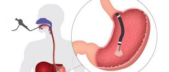
Carrying out the procedure
The patient lies down on a special movable table, which is rolled into the cylinder of the tomograph, where an electromagnetic field is created. The patient must remain motionless during the procedure. If a person experiences discomfort when being in a confined space, taking sedatives that do not affect the diagnosis may be recommended.
The study excludes radiation exposure, so it can be repeated to clarify the diagnosis and monitor the progress of treatment. Based on the results of the examination - a series of layer-by-layer images - an interpretation is performed. To accurately determine the size of the tumor, the doctor may use a contrast agent. The contrast accumulates in the affected area and makes it possible to clarify the location and boundaries of the tumor. A substance based on gadolinium salts is injected into a vein.