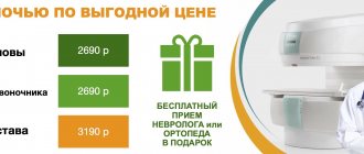
Clinical Hospital City Clinical Hospital No. 71 – Department of Functional Diagnostics – MRI, CT
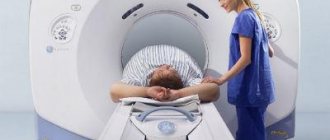
To conduct a complete diagnosis of various human organs, it is not always enough to take tests. Sometimes it is worth conducting a comprehensive examination, including MRI and CT .
MRI and CT are diagnostic methods that provide a complete picture of the condition of the entire body. The question of prescribing a CT or MRI is decided by the doctor in accordance with the diagnostic tasks and depending on the organ or system being examined.
MRI and CT - what are the differences?
What is CT
CT (computed tomography) is one of the modern methods for diagnosing various diseases, in which there is no contact with the surface of the patient’s skin. The CT method is based on the action of X-rays. The device rotates around a person and takes several pictures, which are then processed on a computer and deciphered by a doctor.
Computed tomography (CT) is performed to diagnose the abdominal organs and kidneys, respiratory and skeletal systems of a person. Most often, CT is used to determine the exact location of injuries.
What is MRI
MRI (Magnetic Resonance Imaging) is the diagnosis of internal organs and human tissues using nuclear magnetic resonance. The device makes it possible to obtain a high-quality image of the area of the body being studied, as well as all the changes that occurred in it.
Magnetic resonance imaging (MRI) is performed to identify pathologies in the pelvic organs, diseases of the human circulatory and digestive systems.
MRI is also prescribed for strokes.
Decoding the results

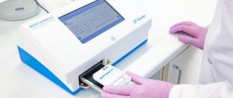
After the scan, the patient must wait 40-60 minutes to receive the examination results. The decoding is carried out by a radiologist and in the conclusion indicates the location and size of organs, features of their structure. When pathological changes are detected, the doctor characterizes them in detail, notes their location and makes a full description. If there is a suspicion of oncology , the specialist will definitely mark the boundaries of the neoplasm , describe the features of the parenchyma and the nature of vascular growth.
You will be given a description, photographs on film, flash media (disk) . Copies must be shown to the attending physician, since the doctor may order an additional consultation with a radiologist or transfer the patient to another institution where a re-analysis of the scan will be required.
Contraindications for MRI and CT
There are several contraindications for CT scanning:
- pregnancy - the effect of x-rays negatively affects the fetus,
- the examination area is in plaster,
- Frequently performing similar procedures,
- lactation period,
- renal failure.
Before undergoing an MRI, you should pay attention to a number of contraindications, in the presence of which diagnosis is impossible.
- These are various electronic devices and metal implants in the studied areas of the body.
- Excess weight. The large size of the patient will not allow him to fit inside the MRI machine.
Indications

Your doctor may order a CT scan if you have the following symptoms:
- abdominal or epigastric pain;
- flatulence;
- increase in abdominal size;
- prolonged absence of stool;
- change in color of stool;
- rapid weight loss ;
- nausea and vomiting;
- constant belching “rotten”;
- change in skin color.
Before scanning, the patient undergoes blood and stool tests, ultrasound or endoscopy. CT is prescribed when the information content of these methods is weak or to clarify the diagnosis.
Which organs and tissues are best visible on MRI and CT?
Computed tomography (CT)
- bones,
- CT is used in the study of patients with head, chest, abdominal injuries, as well as strokes,
- CT is used for pathology of lung tissue.
Magnetic resonance imaging (MRI)
- MRI is used to study soft tissues (cartilage, muscles, ligaments, brain and spinal cord, blood vessels),
- brain,
- Using MRI, the vessels of the brain and neck are examined.
Use of contrast agents
Contrast enhancement expands the range of diagnostic capabilities of CT. The introduction of a contrast agent improves the image quality of the area under study and helps differentiate anatomical structures.
Contrasting is used to study:
- natural cavities, hollow organs (gastrointestinal tract, uterus, bladder, fistulas);
- parenchymal organs;
- brain, spinal cord;
- reproductive organs;
- aorta, coronary arteries, pulmonary arteries, portal, vena cava, iliac veins;
- peripheral vessels, lymph nodes;
- bones, muscles;
- tissue perfusion.
To examine the abdominal cavity, contrast is taken orally on an empty stomach. 30-60 minutes before the procedure, the drug is drunk in small portions, which are divided into 4-5 doses.
Use barium sulfate (barium suspension) or water-soluble agents (“Gastrografin”). Filling the intestinal tube with contrast gives a clear image of the intestinal loops on the tomogram and delimits them from the surrounding tissues.
The condition of the stomach walls can be assessed by filling the organ with water with preliminary intramuscular administration of antispasmodics.
Barium suspension is contraindicated in patients with suspected perforation, when planning operations on the stomach and intestinal loops.
The time for filling the esophagus, stomach, and small intestine with contrast is 20-25 minutes. Contrasting the colon and rectum takes 50-60 minutes.
With intravenous contrast enhancement, the drug accumulates in tissues, which increases density and improves visualization of structures.
A dose of contrast agent is injected manually into the vein of the elbow, or an automatic syringe injector is installed that will dose the substance.
To achieve adequate contrast and prevent unwanted effects from drugs, a strict selection of the dosage of substances is carried out:
| Type of contrast | Dosage | Mode of application |
| Barium sulfate | 250-300 ml per 1 study | The barium sulfate suspension is mixed with water to obtain a total volume of 1 liter. Taken orally. |
| Water-soluble organoiodine compounds: - “Gastrografin” | For the study of the gastrointestinal tract - 10-20 ml, for the pelvic organs - 100-200 ml. | The drug is stirred in 1 liter of water. Taken orally. To contrast the pelvic organs, they are inserted into the rectum. |
| Ionic and non-ionic iodine-containing substances: - "Hexabrix" - "Ultravist" | The general dose for adults is 100-150 ml for intravenous urography, aortography. 80-150 ml of a substance containing 300 mg/ml iodine. | Administer IV as a bolus using an automatic injector. |
Advantages
The MRI (magnetic resonance imaging) diagnostic method has the following positive aspects:
- The obtained results of the MRI study are characterized by high accuracy,
- MRI is the most accurate method for diagnosing diseases of the nervous system,
- MRI accurately determines the presence of hernias in the spine,
- MRI is not dangerous for pregnant women and children,
- There are no restrictions on the number of MRI procedures performed,
- There is no pain during the MRI procedure,
- Information based on the results of an MRI examination is provided in the form of a three-dimensional image,
- Information can be saved to any electronic media or computer,
- It is impossible to make mistakes in the results of an MRI study,
- There is no X-ray exposure.
Carrying out CT (computed tomography) is characterized by the following positive aspects:
- The result of a CT scan is a three-dimensional image,
- Images of bones are obtained with high accuracy,
- The CT scan procedure is absolutely painless,
- The duration of the CT scan is approximately a couple of minutes,
- The information obtained as a result of CT examination is simple and understandable,
- The radiation dose is much less than with an X-ray machine,
- There are no restrictions on undergoing the procedure for people who have metal or electrical devices in their bodies,
- CT scan provides accurate information about the presence of internal bleeding and tumors in the patient,
- The cost of a computed tomography (CT) scan is much less than an MRI.
Alternatives
If it is not possible to perform an abdominal CT scan, the doctor may prescribe:
- Ultrasound is a cheaper examination; scanning produces images with a two-dimensional image. It shows pathologies of hollow organs well, but during analysis you can “overlook” some injuries, perforations, and bleeding.
- MRI uses magnetic waves to produce three-dimensional images. Such a study is informative, is not inferior to CT in price, but is longer and requires mandatory immobility.
- Endoscopy is known among patients as “swallowing the lightbulb.” During diagnosis, a device with a camera is used, which can be used to study the condition of the mucous membrane of the stomach and duodenum. The lower parts of the large intestine are examined with a colonoscope.
chooses MRI as an . Our separate article will tell you about the differences between the two procedures and help you choose what suits you best.
Research limitations
Each diagnostic method has positive and negative aspects.
Disadvantages of magnetic resonance imaging (MRI):
- There is no possibility to conduct a comprehensive study of hollow organs. These include the gallbladder, urinary bladder, and lungs.
- There are restrictions on performing an MRI procedure on patients with metal objects in the body,
- To obtain accurate images, you should remain still and calm during the procedure.
Disadvantages of computed tomography (CT):
- CT scan is dangerous x-ray radiation,
- This procedure provides information only about the structure of organs and tissues, but not about their functioning,
- This type of diagnosis is contraindicated for pregnant and lactating women and children.
Types of computed tomography
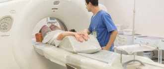
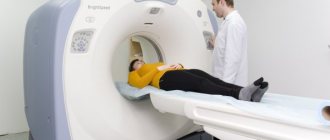
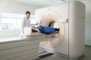
There are several types of CT scans. To understand the differences, you need to understand what the device is on which the diagnostic procedure itself is performed (computed tomograph). It looks like a ring under which a table with the patient lying on it slides. Each type of CT scan works differently.
In spiral tomography, the tube rotates continuously and the table moves at the same time. This allows you to increase the speed of organ scanning and reduce the radiation dose.
Multislice tomography is similar to spiral tomography, but the radiation comes from two sources. This achieves an increase in the resolution of the resulting image, while reducing the procedure time. This type of CT is used to obtain images of small vessels and a detailed scan of a small area of tissue.
Multilayer tomography is distinguished by the fact that the speed of wave emission and rotation of devices is increased, and the sensors are arranged in several rows. This type of study provides a representation of the work of an organ in dynamics.
CT with contrast involves the introduction of a contrast agent, which enhances the contrast of the image of organs against the background of adjacent tissues. Thanks to this technique, the images in the photographs are clearer and easier to decipher for making an accurate diagnosis.
Difference between MRI and CT
Computed tomography (CT) is the best way to visualize bone structures. Therefore, CT is good to use to identify bone injuries and various injuries.
MRI clearly shows soft tissues: muscles, blood vessels, cartilage, spinal cord and brain. Therefore, MRI is better used to identify tumors and pathologies in neurology, neurosurgery, and endocrinology.
Under the influence of X-ray radiation, a computer tomograph (CT) produces a series of images of the object under study.
A magnetic resonance imaging (MRI) scanner operates based on the magnetic field in which the patient is placed.
MRI helps to obtain more accurate results in the following cases:
- the body’s response to the injected contrast agent was detected during computed tomography,
- you should check the condition of the brain, the condition of soft tissues,
- musculoskeletal diseases in children,
- it is necessary to check the condition of the pituitary gland, the condition of nerve cells in the brain,
- for damage to cartilage, joints,
- for suspected cancer.
CT is effective for:
- any mechanical damage, head injuries,
- damage to bones, their deformation under various influences,
- examination of blood vessels, heart,
- suspicion of the development of purulent diseases - sinusitis, otitis media,
- pathologies in the abdominal cavity,
- respiratory problems,
- suspicions of cancer, changes in the chest and its organs.
MRI is a harmless diagnostic method and does not irradiate the body, as with computed tomography. It is a good replacement if the body is intolerant to the contrast agent that is administered for CT scanning.
CT has a more intense effect on the body.
It is impossible to compare these two procedures, since they are completely different. Their main differences are contraindications, indications, and method of exposure. Therefore, the doctor himself makes a decision in favor of choosing one or another research method.
Carrying out
Despite the fact that diagnostics require complex and expensive equipment, the procedure is quite simple to perform and does not require any effort from the patient. The list of stages describing how a computed tomography is done can include 6 points:
- Analysis of indications for diagnosis and development of research tactics.
- Preparing and placing the patient on the table.
- Adjustment of radiation power.
- Performing a scan.
- Recording the received information on removable media or photo paper.
- Drawing up a protocol describing the results of the examination.
On the eve or on the day of the examination, the patient’s passport data, medical history and indications for the procedure are recorded in the clinic database. The results of computed tomography are also entered here.
Important! Carrying out a CT scan does not require special preparation from the patient, with the exception of the need to conduct an examination of the gastrointestinal tract. In this case, you should come to the procedure on an empty stomach, limiting the consumption of foods that stimulate gas formation in the intestines the day before.
It is quite difficult to cover all areas of development and diagnostic capabilities of CT, which are still expanding. New programs are appearing that make it possible to obtain a three-dimensional image of the organ of interest, “cleared” of extraneous structures that are not related to the object under study. Developments of “low-dose” equipment that provide results of similar quality will be able to compete with the no less informative MRI method.
Rules for preparing for CT and MRI procedures
To perform the procedure, a referral from a doctor is required. Because the doctor must know your diagnosis in order to perform the correct procedure. When undergoing repeated examinations, it is recommended to take past results with you.
It is recommended that you come in comfortable, non-restrictive clothing so that you feel comfortable during the procedure.
On the eve of these procedures, it is recommended to drink a sufficient amount of liquid. Since when examined using a contrast agent, its removal from the body will occur much faster.
You can get detailed information about MRI or CT scanning by phone or write to us by e-mail