Radiography is a quick method for studying anatomical structures, identifying joint damage and fractures in traumatology. The resulting image is displayed on a special film or digital media.
Situations arise when X-rays need to be taken on a part of the body that is damaged and fixed with plaster. Then the question arises: is it possible to take an x-ray through a cast?
Alternative to traditional plaster
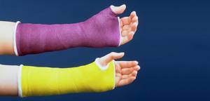
However, the fracture site must first be fixed and immobilized, that is, immobilized. In emergency rooms and hospitals, they offer classic plaster in the old fashioned way, in the absence of other alternatives. In medicine, doctors use new generation materials - polymer immobilizing bandages, also called “plastic casts”.
Plastic plaster is a bandage impregnated with a special polymer composition.
Plastic plaster is used not only in case of a broken arm or leg. It is used to fix feet, elbows, knees, a dislocated shoulder or ankle, and is also used for sprained muscles or ligaments.
However, in order to apply or remove polymer plaster, the doctor must have special knowledge, tools and materials. Medical is one of the few in the city where doctors can apply modern orthopedic bandages made of plastic.
Equipment for the radiography room at clinic No. 1 ViTerra Belyaevo
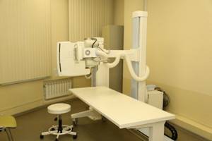
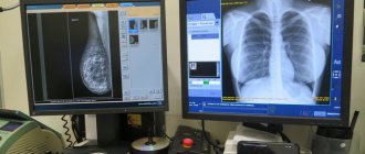
Clinic No. 1 ViTerra Belyaevo has the most modern digital X-ray equipment from leading companies in its arsenal: an X-ray tube from the company VARIAN medical system (USA) model - DIAMOND , equipment from the companies Sirona (Germany) , X-RAY (Galaxy) . This, combined with the highly qualified radiologists, allows us to diagnose a wide range of diseases of the musculoskeletal system, respiratory system, gastrointestinal tract, ENT organs, and genitourinary system.
- Our X-ray allows us to obtain high-quality images of any organ or area with minimal stress.
- X-ray examinations carried out at clinic No. 1 Viterra Belyaevo are processed by the SITEC FEEL 2.0 fine VIEW (KOREA) . The images are recorded on digital media and can be viewed by any specialist in a medical institution.
Advantages of artificial plaster
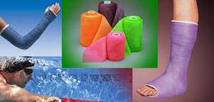
Unlike classic plaster, plastic weighs several times less and does not interfere with movement. At the same time, the material is very durable, which guarantees proper healing of broken bones.
Modern plastic plaster is not afraid of water, you can safely swim with it both in the bath and in the sea, and if it gets dirty, you can simply wipe it with a damp cloth.
The porous structure of the material allows oxygen to pass freely, so the patient does not experience itching, irritation or other skin reactions. In addition, polymer fixatives are non-toxic and hypoallergenic.
Due to the elastic material, the plaster fits tightly to the site of injury, and at the same time allows you to simulate any shape for complex fractures.
During an x-ray, the plastic cast does not need to be removed; it does not block x-rays. The doctor can see all the details in the image, which means there will be no difficulties in assessing bone fusion.
The plastic plaster looks neat even after wearing it for a long time, and the patient feels it like a regular bandage and does not feel discomfort as when wearing a regular plaster.
This type of retainer is removed using a special saw and, due to its smooth texture, does not injure the skin and hair. This compares favorably with classic plaster, which, when worn for a long time, almost fuses with the skin.
X-ray with contrast
In radiology practice, in addition to standard examination methods, studies are also carried out using radiopaque agents.
, such as:
excretory (intravenous) urography
and
hysterosalpingography
.
Urography (intravenous excretory urography)
- This is an x-ray of the urinary system. A radiopaque contrast agent is first administered intravenously.
Hysterosalpingography (HSG)
is a method of X-ray diagnostics of the condition of the uterus and fallopian tubes, based on the introduction of a contrast agent into them.
In our clinic you can sign up and have both of these studies done with a detailed transcript of the results from the specialist doctors.
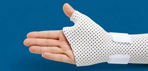
What types of artificial plaster are there?
There are several types of polymer gypsum. The most famous are scotchcast, softcast and turbocast.
Scotchcast is a polymer plaster, strong and rigid, due to which it reliably immobilizes the fracture. The lightest of all artificial bandages. A special lining stocking is worn under it so as not to injure the skin. Scotchcast comes in different colors, which will appeal to children and lovers of bright colors.
Softcast allows you to create bandages of varying stiffness. After hardening, the material remains semi-rigid, which allows you to maintain muscle mobility in the damaged area. At the same time, the material is flexible, but not stretchable, which helps maintain its original shape. It is used not only for fractures, but also for sprained limbs.
Turbocast is the most famous polymer gypsum made of thermoplastic. It makes it possible to give the bandage any shape, which makes it indispensable for the most complex fractures, including in children and adolescents. The special design allows the patient to independently remove and put on the bandage (with the doctor’s permission).
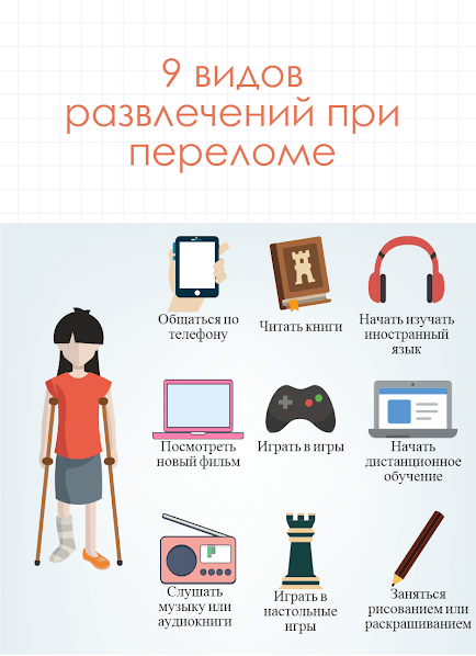
One more thing about rehabilitation. What to do after removing the cast? Recovery process.
This article contains important information about how the recovery process proceeds after plaster removal. Information for you was provided by Nadezhda Epishina, a physical therapist at the Foundation and head of the Mobile Rehabilitation Service project. So, the injured limb is finally “free”, and the main task now is to restore muscle function in order to gradually return sensitivity and mobility to the body. But more about restoration later. First, it’s worth talking about why we need striated muscles at all and what functions they actually perform.
- The main function of muscles is motor (dynamic and static). With their help, we move in space and maintain our body in an upright position.
- In addition, muscles perform a thermoregulatory function; they help the body maintain the desired temperature. In “fragile” people, this function is often impaired, and muscles also need to be developed in order to restore normal thermoregulation.
- It is also the muscles that give us the ability to breathe.
- Another important function is receptor function. Muscles help us feel our body. When a limb is left without movement for a long time, deep sensitivity decreases, and it is imperative to help it recover.
- Finally, skeletal muscle helps the heart function. They are even called the second heart, because when their mobility is impaired, blood microcirculation is also impaired.
When a person remains motionless for a long time (for example, in a cast after a fracture or surgery), the muscles weaken and the normal course of natural processes in the body is disrupted. That is why restoring muscle mobility is the main goal of rehabilitation. It is worth remembering that you need to start small, gradually increasing the load. Typically, the first exercises at this stage include passive movements - when the person himself does not move the limb, but other people or a special apparatus do it for him. To develop joint mobility, it is good to use a portable exercise bike. And yet, the specifics of osteogenesis imperfecta require that a “fragile” child or adult perform as many movements as possible independently. In the first stages, you can support and help - make sure that the limb is bent at the correct angle, and the speed of the exercise. At all stages of recovery, you need to exercise slowly, listening to the signals that the body gives. Children (especially very young ones) often want to show how hard they are trying to their parents or specialist who works with them, and perform exercises too quickly or jerkily. It is very important to ensure that the child does not “overdo it” and harm his body.
Consult your doctor
Each type of polymer plaster is indicated for certain injuries and fractures. Only your attending physician can determine which one is right for you.
A plastic bandage does not cancel visits to the doctor and does not speed up the healing of broken bones, but allows you to make treatment as comfortable as possible.
In medical matters, traumatologist-orthopedist Natalya Vladimirovna Sokolenko (link to doctor) will help you determine the type of polymer plaster that is right for you.
You can find out more detailed information or make an appointment by phone. +7 (4712) 46-03-03.
What are emergency rooms for?
Traumatology is one of the oldest areas in medicine. After all, even in ancient times, people who were injured during hunting, war or careless movement turned to healers and chiropractors for help.
During Soviet times, free government emergency medical care centers appeared. Such emergency rooms operated on the basis of clinics or hospital emergency departments. In trauma centers, traumatologists examined the injured person, provided him with first aid and determined the direction of further therapy. Unfortunately, in those days there was no alternative, and in order to receive the necessary treatment, one had to wait in long lines at the traumatologist’s office.
Now there are emergency rooms on a paid basis, where you will be helped with injuries, poisoning and other emergency conditions (for example, dog bites, ticks, burns, frostbite, etc.).
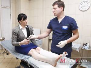
1 emergency room. Plaster application
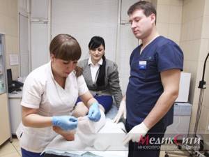
2 Emergency room. Plaster application
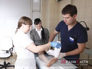
3 Emergency room. Plaster application
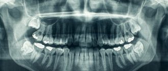
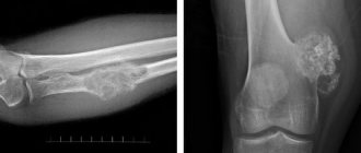
In what cases is a hand x-ray prescribed?
It is worth consulting a doctor if you experience swelling, deformation and hyperemia (redness) of the hands . These symptoms can signal various pathologies: lymph stagnation, allergies, heart disease, kidney disease, liver disease, etc. With such pathologies, the structure of bone tissue can be disrupted. To exclude pathology of bone tissue, it is necessary to take an x-ray of the hands and wrists.
The most common types of hand deformities are the so-called Heberden's nodes (formed on the joints of the outer phalanges of the fingers) and Bouchard's nodes (formed on the middle phalanges of the fingers).
Therefore, the procedure can be prescribed not only by a traumatologist, but also by a rheumatologist.
X-ray of the hands allows you to diagnose:
- arthritis
- arthrosis;
- tendon inflammation;
- osteophytes (these are pathological growths that form along the edges of the joints).
X-rays of the hands are also prescribed if osteomyelitis is suspected. With this pathology, bacteria enter the bone tissue, causing it to rot and collapse. The cause of the development of such an infectious disease can be injuries, as well as diseases such as purulent tonsillitis, pneumonia, measles, and furunculosis.
In our multidisciplinary clinic, traumatologists, radiologists, ENT doctors and other specialists are ready to see you. As a team, treatment is quick and convenient; you can take all the tests and get the necessary advice from us.
You should also consult a doctor if you have a tumor on your hand. One of the most common is hygroma.
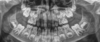
When children experience a delay or, on the contrary, an advance in growth and development, an x-ray is taken to determine bone age. The study is prescribed by endocrinologists.
Despite the fact that the dose of X-ray radiation in modern X-ray machines is minimal, there are a number of restrictions under which X-rays should be avoided unless absolutely necessary.
Contraindications to x-rays of the hands:
- pregnancy;
- age up to 15 years (although x-rays can determine bone age, this study should only be performed as prescribed by a doctor).
A pregnant patient can undergo ultrasound and MRI instead of radiography.
What to choose: radiography or MRI?
As we have already said, x-rays allow you to see the bone structure, detect pathologies, injuries and displacements.
MRI allows you to evaluate the condition of bone tissue, muscles and ligaments. The study is prescribed if pathologies such as cancer, abnormalities of the hand, etc. are suspected.
Depending on the medical history and examination results, the doctor will refer you to the necessary specialist.
How is the research conducted?
No special preparation required. The main thing is to remove all metal jewelry before the examination, because... they can affect the information content of the images. The radiologist will tell you about this.
X-ray of the hands is a painless, non-invasive examination that is performed in a few minutes or even seconds. Those. the radiation dose is minimal. The patient first bends his arm at the elbow and then places it on the tabletop with the back side down. The radiologist takes 2 pictures.
We conduct research using a modern digital X-ray machine ITALTAY CLINODIGIT COMPACT. It is produced in Italy jointly with the USA.
The development and description of x-ray images, as a rule, takes no more than 15-20 minutes. The images are immediately handed over to the patient.