Endoscopy is one of the most informative methods of instrumental diagnostics, which allows you to study the structure and structure of internal organs, detect bleeding, neoplasms, and foci of inflammation.
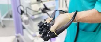
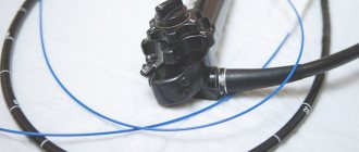
Endoscopy is performed using an endoscope - a long flexible plastic or metal probe with an LED and a lens or camera at the end. The endoscope is inserted into the patient’s body through the natural cavities of the body: for example, to examine the esophagus and stomach through the mouth, and during bronchoscopy through the respiratory tract. The device can be equipped with tools for additional surgical procedures: taking a tissue sample (biopsy), removing polyps.
What is endoscopy? What types of examinations are included here?
Endoscopy allows the doctor to visually assess the condition of various organs and tissues: bronchi, gastrointestinal tract, bladder, joints, and so on.
Depending on the type of cavity being examined, the procedure is divided into:
- bronchoscopy. The only method for studying the respiratory tract that allows you to examine the relief of the bronchial mucosa, assess the condition of the vessels, and, if necessary, perform a biopsy. An examination is prescribed if the patient has a cough of unknown etiology, blood in the sputum, constant relapses of pneumonia and bronchitis;
- esophagogastroduodenoscopy. One of the most informative instrumental studies of the upper gastrointestinal tract, in which the endoscopist alternately examines the esophagus, stomach and duodenum. During probing of the stomach, the doctor assesses the current state of the mucous membrane, clarifies the presence of a tumor or inflammatory pathology, and if a focus of the disease is detected, clarifies its location. Usually a stomach check procedure if the patient complains of difficulty swallowing, frequent heartburn, a regular feeling of heaviness and pressure in the stomach after eating;
- colonoscopy. An examination of the colon that allows you to visualize the presence of various lesions: areas of erosion, inflammation of the mucous membrane or damage to its integrity. Colonoscopy also shows the presence of congenital anomalies in the structure of the colon and is prescribed to patients experiencing digestive problems: frequent diarrhea and constipation, pain in the lower abdomen, a feeling of pressure in the anus.
These are the main types of endoscopy used in pulmonology and gastroenterology. But the possibilities of the method are much wider. For example, it can be used to diagnose diseases of the uterus (hysteroscopy), assess the condition of the outer ear (otoscopy), and detect pathologies of the bladder (cystoscopy). Depending on the type of examination prescribed, the indications for the procedure and preparation for it will differ.
Modern endoscopy – not scary, not painful, informative
At the Regional Consultative and Diagnostic Center (Rostov-on-Don, Pushkinskaya St., 127)
In the endoscopy department, patients are offered diagnostic and therapeutic procedures using high-tech expert-class video information systems.
At the request of the patient, most studies are performed under intravenous anesthesia.
Endoscopy is a method of examining certain internal organs using an endoscope, which is inserted into the cavities through the patient’s natural pathways. Modern endoscopy allows diagnosticians to obtain images of the smallest details, identify and classify structural changes in the mucous membrane of internal organs for the purpose of early detection of precancerous pathology.
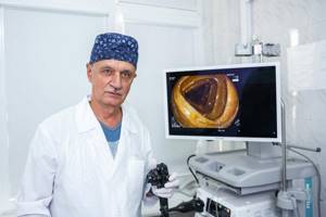
“The equipment of our Center allows us to significantly expand diagnostic capabilities,” says Leonid Petrovich Kudryavtsev, head of the endoscopic department of the OKDC, Honored Doctor of the Russian Federation.
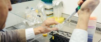
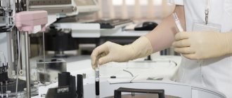
– We pay special attention to the morphological diagnosis of the detected pathology. In almost all studies, a mucosal biopsy is performed for cytological and histological analyses. In addition, our systems are equipped with a photo archiving function. The results are stored in the Center’s databases for decades, making it possible to trace the dynamics of disease development.”
Endoscopic tissue biopsy is usually performed from any suspicious area. This is a standard that allows you to detect early gastrointestinal cancer and successfully treat it. The study is necessary to make sure that there are no signs indicating a malignant process. If cell degeneration is detected at the very beginning, then early endoscopic removal without surgery is possible.
The latest electrosurgical complexes and highly qualified endoscopists allow excision of pathological areas during the examination.
What endoscopic examinations are performed at the OKDC? — gastroscopy (VGDS) — assessment of the condition of the mucous membrane of the esophagus, stomach and duodenum; — bronchoscopy (VTBS) — examination of the trachea and bronchial tree; — colonoscopy (VCS) — assessment of the condition of the colon mucosa; — endosonography – endoscopic and ultrasound examination performed simultaneously with one device. Allows doctors, under visual control, to bring the ultrasound sensor as close as possible to the object of study and get a clear picture of the pathology of the gastrointestinal tract; — enteroscopy is a method of examining the small intestine, in which an endoscopist performs an examination, biopsy and therapeutic manipulations.
Endoscopic examinations at the request of the patient
carried out using sedation. Before the procedure, you must consult with an endoscopist and follow his recommendations on preparing for the study, and if you decide to use intravenous anesthesia, a consultation with an anesthesiologist is required.
For anesthesia at the Regional Consultative and Diagnostic Center
The latest anesthetics, modern high-tech anesthesia and respiratory equipment and monitoring tools from leading manufacturers of medical equipment on the world market are used.
Regional consultative and diagnostic. Rostov-on-Don, st. Pushkinskaya, 127. 8 (863) 227-0000. Website rokdc.ru
share information
0
Social buttons for Joomla
What diseases can be found using endoscopy?
One of the key benefits of endoscopy is its ability to detect cancer of the respiratory and digestive system in its early stages. Visualization of organs allows the doctor to find foci of pathology in real time and take a sample for further histological analysis. Other pathologies that may be detected during the examination depend on the type of procedure.
For example, gastroscopy shows:
- diverticulitis;
- varicose veins of the esophagus;
- stomach ulcer;
- esophagitis;
- esophageal stenosis.
Endoscopy allows not only to detect a disorder, but also to carry out targeted treatment: apply medications directly to the affected area.
Colonoscopy allows you to diagnose disorders such as:
- ulcers;
- polyps;
- colitis;
- intestinal bleeding and so on.
Bronchoscopy shows the condition of the respiratory system. With its help, you can detect inflammatory diseases (for example, pneumonia), confirm cystic fibrosis, bronchial asthma, tuberculosis. In general, the procedure allows you to assess the tone of the bronchial tree, the diameter of the lumens, and the presence of structural abnormalities.
Advantages and disadvantages
Endoscopy under anesthesia has the following advantages:
- there are no painful or unpleasant sensations;
- During the study, extensive surgical procedures can be performed;
- the doctor can fully concentrate on his actions and not be distracted by the patient’s anxiety;
- the study can be carried out longer.
Disadvantages include an increase in the cost of the examination due to the involvement of additional specialists, the need for follow-up, and more complex preparation.
The existing disadvantages of this procedure are insignificant compared to the significance of the method in diagnostic practice. In many developed countries, endoscopy of the large intestine is a mandatory method for medical examinations after 40 years, as it plays an important role in the prevention of cancer. Detection of tumors in the early stages and precancerous processes, such as polyps, allows timely treatment to begin, which significantly increases the life expectancy of such patients.
The Medicenter network of clinics performs a wide range of endoscopic examinations using general anesthesia. Experienced anesthesiologists and modern medications will help you get rid of discomfort and fear before the examination.
I'm afraid of "swallowing the tube." What to do?
In gastroenterology, to make a diagnosis, the doctor may order the patient to have an esophagogastroduodenoscopy (also called an EGD or gastroscopy). This procedure has received the common name “probe swallowing”, since during the examination the patient will need to swallow the probe so that it passes through the esophagus into the stomach. The study has given rise to many fears and myths: many people who have never undergone FGDS are afraid of this procedure and refuse to perform it. How reasonable are these fears and concerns, and when is FGDS necessary?
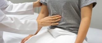
Endoscopic examination of the upper gastrointestinal tract is prescribed for patients experiencing the following symptoms:
- regular pain in the abdominal area;
- heartburn and belching after meals;
- feeling of heaviness in the stomach;
- nausea and vomiting;
- weight loss for no apparent reason;
- sudden change in appetite.
During gastroscopy, pathologies such as reflux disease, gastritis, gastric or duodenal ulcers can be detected. Also, checking the stomach allows you to find foci of inflammation, detect polyps, ulcers, erosions, and internal bleeding. Today, this is one of the most informative instrumental diagnostic procedures for visual assessment of the condition of the upper gastrointestinal tract. Therefore, if a gastroenterologist has referred you for an endoscopy, you must undergo the procedure.
Gastroscopy is performed as follows: an assistant sprays a local anesthetic onto the root of the tongue. This is necessary to relieve the gag reflex and freely insert the endoscope. Then the patient is asked to lie on the couch on his left side and hold the mouthpiece between his teeth - it will prevent him from reflexively closing his mouth.
The probe is then slowly inserted. At this time, the person needs to breathe relaxed and deeply, and follow all the recommendations of the endoscopist. The doctor will ask you to make several swallowing movements to pass the endoscope. At this time, the patient will feel a sensation of pressure in the throat and esophagus.
When the tube reaches the stomach, air is supplied to straighten its folds and carefully examine the inner surface of the organs. If necessary, a biopsy and pH tests can be performed. The probe is then removed. The entire procedure takes 10-15 minutes and is absolutely painless. The only unpleasant sensations that a person may encounter are pressure in the throat when the endoscope moves and distension in the stomach when air is supplied.
If you are still afraid to undergo an EGD, follow these recommendations:
- sign up for a gastroscopy at a medical center with good reviews - an experienced endoscopist will conduct the examination quickly and delicately;
- During the procedure, try to relax, breathe through your nose slowly and as deeply as possible;
- follow all the specialist’s recommendations - swallow only at his command.
Is endoscopy painful?
Most fears of patients undergoing endoscopy for the first time are associated with the myth that the procedure is painful. The progress of the study depends on what type of endoscopy the doctor prescribed, but all of them are performed without pain.
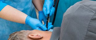
For example, during bronchoscopy, an anesthetic spray is used to suppress the gag reflex. Then a thin probe is inserted into the respiratory system, which does not interfere with natural breathing. There are no nerve endings in the bronchi that are responsible for the perception of pain, so the movement of the tube does not cause pain. The only thing the patient can feel is a slight pressure in the throat from the device. Also, on the eve of the procedure, the specialist may recommend that the patient take a sedative. This will help relieve anxiety and relax during the manipulations themselves.
Patients are no less concerned about the upcoming colonoscopy. This examination allows you to examine the large intestine, while a probe is inserted through the anus. Discomfort may occur when air is supplied: it is necessary to straighten the folds of the intestine and provide the doctor with a full view of the internal cavities. But at the same time, the sensations are not strong, comparable to spasms that occur during the urge to defecate.
During the procedure, thin and flexible endoscopes are used, in which there are no protruding sharp or cutting parts, so the movement of the tube cannot injure the mucous membrane - there is no irritation after the end of the examination.
In most cases, endoscopy uses local anesthesia, but in some cases it is possible to perform the examination under general anesthesia. It is used if the patient cannot remain still due to neurological disorders or experiences panic fear that prevents the examination.
Are there any contraindications for endoscopy?
Endoscopic examination is an informative and safe diagnostic method used in gastroenterology, pulmonology and other areas of medicine. Although most patients tolerate the procedure well, some patients are contraindicated for endoscopy.
Absolute contraindications include:
- general serious condition of the patient;
- previous myocardial infarction;
- disruption of blood supply to the brain;
- congenital or acquired anomalies of the esophagus that interfere with the insertion of the probe.
People suffering from severe cardiopulmonary disorders should not undergo endoscopy. There are no other absolute contraindications. But doctors also identify relative contraindications, in which the study is carried out only if the potential benefit outweighs the possible risk.
Among the relative contraindications:
- blood diseases;
- mental disorders;
- aneurysms;
- respiratory infection in the acute stage;
- chronic heart disease.
To identify contraindications, the doctor examines the patient, collects anamnesis, and prescribes additional tests before prescribing endoscopy. If the specialist decides that the patient should not undergo endoscopic examination, he will select alternative diagnostic methods.
Features of anesthesia during endoscopy
As a rule, endoscopic examinations are short-lived and are therefore performed under light intravenous anesthesia. In exceptional cases, when there is a risk of gastrointestinal contents entering the respiratory system, endotracheal anesthesia may be used. In this case, a special tube is installed in the trachea through which the drug is supplied.
The choice of the type of anesthesia and the specific drug is the task of the anesthesiologist. The specialist directly administers anesthesia and monitors the patient’s condition during and after the examination. Accordingly, the endoscopy room must be equipped with a workstation with all the necessary equipment.
Endoscopy under anesthesia requires more careful preparation. In addition to general recommendations (not to eat before the procedure), the patient must be consulted by an anesthesiologist, who may prescribe a certain examination (blood tests, ECG, coagulogram, etc.).
After the procedure, the patient continues to remain under the supervision of the clinic staff, since the effect of the anesthetic drugs continues to persist for some time. For this reason, it is not recommended to drive a car on the day of the examination.
What can replace endoscopy?
Endoscopic examinations allow a specialist to visually assess the degree of damage to the mucous membrane, examine the condition of blood vessels, and detect foci of inflammation and erosion. In addition, additional manipulations are possible during endoscopy: taking a biopsy sample, conducting a pH test, additional contrast staining to detect cancer at an early stage. Therefore, there is no complete alternative to endoscopy.
If a gastroenterologist or supervising physician has referred you for an endoscopy, it must be performed. But in some cases, the patient is contraindicated to undergo this examination due to medical contraindications. In this case, the specialist selects other diagnostic procedures and may prescribe:
- radiography. This procedure allows you to diagnose diseases such as hernia, ulcers, stenosis, and structural abnormalities. However, the doctor cannot see the current state of the mucous membrane. Therefore, in most cases, radiography is carried out as an auxiliary study and, if it detects any abnormalities (for example, the contrast agent has not filled a certain area), gastroscopy will be needed;
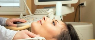
- Ultrasound. Ultrasound examination of the gastrointestinal tract is not informative enough, since it only shows the contours of filled organs and denser structures (for example, bones). The photo turns out to be insufficiently informative. However, if endoscopy is not possible, an ultrasound may be prescribed to assess the general condition of the stomach and intestines. The main advantage of the method is maximum safety and complete painlessness;
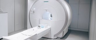
- CT and MRI. Tomography is another modern diagnostic technique that allows you to obtain a three-dimensional image of internal organs. CT and MRI differ from each other in the type of radiation: in the case of computed tomography (CT), the image is obtained due to exposure to x-rays. In magnetic resonance therapy (MRI), the tomogram is the result of exposure to an electromagnetic field. The images give an accurate idea of the presence and location of foreign bodies, neoplasms, and foci of infection. These techniques make it possible to replace not only gastroscopy, but also bronchoscopy and other types of endoscopic examinations.
The doctor may also prescribe a capsule gastroscopy. In this case, the patient will be required to swallow a small capsule containing an LED and a camera. After a day, the capsule will leave the body along with the feces, and a specialist will be able to view the image captured by it: for example, to detect an ulcer.
How to prepare for a stomach and intestinal check?
Preparation for endoscopy depends on which organ the endoscopist will study. However, it should be remembered that all studies are carried out on an empty stomach - on the day of the procedure you must refuse to eat. Also, many doctors recommend taking a sedative the day before - this will help you relax and relieve stress.
How to prepare for endoscopy? High-quality preparation facilitates the conduct of surveys and increases its information content and accuracy. The patient should warn the endoscopist about medications and allergies. No special diet is required, but the last meal should be no less than 6 hours before the procedure. After endoscopy, you can eat after 2 hours, if the procedure was accompanied by taking a biopsy sample; for the first day, it is recommended to refrain from eating fatty, salty and spicy foods, and alcohol.
Colonoscopy requires more careful preparation. 5 days before the study, the patient should exclude from the diet foods that cause increased gas formation: for example, coarse fibrous foods, bran bread, legumes. It is recommended to consume pureed soups and dishes made from boiled or stewed vegetables 1-2 days in advance. It is better to avoid flour, spicy and smoked foods.
Before a colonoscopy, it is necessary to cleanse the intestines: this can be done with laxatives (drugs and dosage are prescribed by the attending physician), or with an enema. The specialist who issued the referral for the examination will tell you in detail about the rules of preparation for endoscopy.
0
0
0
Article rating:
4 out of 5 based on 2 ratings
Author: Digtyar Gennady Ivanovich
Endoscopist. Highest category. Work experience over 25 years.
2. Taking cleansing and auxiliary drugs (defoamers) according to the doctor’s recommendations
Read the instructions for using the cleansing (laxative) drug (the instructions are inside the medicine package). It is strictly forbidden to reduce the volume of the drug liquid in preparation for the study.
Any cleansing (laxative) drug acts individually: on average, after 1-2 hours from the start of administration, the first stool appears, intense loose stools persist for 2 hours, and by the time preparation is completed, the stool becomes a clear or slightly colored liquid.
It is necessary to stop taking the drug 3-4 hours before the scheduled time of the study. For example: if the study is scheduled for 14.00, then you should stop taking the drug between 10.00 and 11.00.
It is not recommended to carry out colon cleansing procedures with enemas or synthetic oils in addition to or instead of medications.
For chronic constipation, 3-5 days before preparing for the study, you should start taking laxatives of non-herbal origin, preferably on the recommendation of a gastroenterologist.
If you are constantly taking any medications, then take them as usual, but not earlier than 1 hour after finishing taking the cleansing (laxative) drug.
If you are constantly taking any medications
5 days before the test, avoid taking iron-containing drugs, adsorbents, and bismuth-containing drugs.
If you have diabetes, sign up for a test in the morning and take your medications with you; check your blood glucose levels on the morning of the test.
In all other cases, take medications 4 hours before the test with a small amount of water (up to 50 ml).
If you have bronchial asthma, take with you an inhaler that you use regularly.
Tell your doctor if you are taking blood thinning medications.
The study is carried out with the obligatory availability of the following research results:
— HIV, syphilis, hepatitis B, C (period: no more than 30 days)
And also if there are results of additional studies, if they were prescribed by a doctor:
— general blood test (term: no more than 14 calendar days);
— biochemical blood test (term: no more than 14 calendar days);
— coagulogram (term: no more than 14 calendar days);
— electrocardiogram (term: no more than 30 calendar days).
Recommendations after the study
If the study is supposed to be carried out under intravenous anesthesia, then on this day it is not recommended to drive a car, operate any mechanisms, or make important decisions. It is advisable to be met after the examination and escorted home.