A medical term such as “abdominal x-ray” is used to designate the most well-known and widespread diagnostic method for gastrointestinal pathologies. It allows you to examine the organs of the gastrointestinal tract, kidneys and digestive system and is a priority when a patient is admitted to the hospital with complaints of stool disorders and abdominal pain.
The process involves an X-ray machine that generates a beam of rays and passes them through the part of the body that needs to be examined. When the rays pass through the anatomical structures of the human body, corresponding images are obtained. The technique is absolutely painless and allows you to see the first results quite quickly, which is its significant advantages. You can take an x-ray of the abdominal cavity at the diagnostic center of the multidisciplinary clinic CELT. We have several mobile and stationary X-ray machines made in Japan and Germany, which allow us to carry out the procedure in accordance with international standards.
X-ray of the abdominal cavity - 1,000 rubles.
X-ray of the abdominal cavity (survey) - 2,400 rubles.
15 - 20 minutes
(duration of procedure)
Types of radiography of the abdominal organs
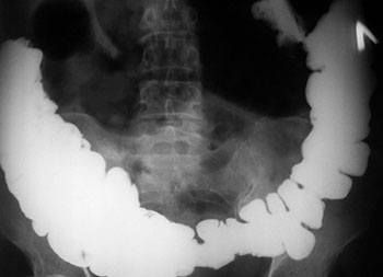
There are two main types of this procedure: overview (standard) and contrast. The first allows you to assess the condition of the abdominal organs and see foreign objects or pathological changes. X-ray diagnostics are prescribed along with a number of other studies in order to clarify the diagnosis. The procedure takes only a few minutes, is inexpensive and is absolutely safe. Abdominal X-rays with contrast require the use of a special barium sulfate solution, which the patient must drink before the procedure. Sometimes it is given through a tube. The use of barium is necessary in order to obtain more accurate information.
The fact is that barium does not dissolve in the physiological fluids of the human body, but at the same time it absorbs X-rays well. By illuminating the solution that moves through the digestive organs, it is possible to obtain information about the relief of the mucous membrane, examine the functioning of the gastrointestinal tract and identify neoplasms, damage or obstruction.
Indications for abdominal radiography
| Disease groups | Pathologies and symptoms of their manifestation on x-ray |
| Stomach pathologies | A contrast x-ray is performed, before which the patient drinks a drug based on barium sulfate. X-ray can reveal:
|
| Pathologies of the urinary system | The technique makes it possible to identify foreign bodies, including stones in case of cervical stones. The use of contrast makes it possible to determine inflammatory processes, pathologies of renal vessels, as well as various abnormalities of organ development. |
| Intestinal pathologies | On a regular x-ray, manifestations of obstruction are clearly visible. The procedure with contrast makes it possible to see inflammation and neoplasms on the intestinal walls. |
| Abdominal injuries | X-ray of the abdominal organs determines the presence of foreign bodies, gas or liquid. It allows you to determine the consequences of penetrating wounds. |
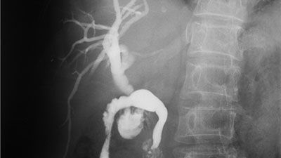
| Pathologies of the liver and gallbladder | X-rays are always performed with contrast and can detect cholelithiasis, hepatitis, cirrhosis, and cholecystitis. |
| To main |
(Radiation diagnostic news 1999 1: 4-6)
X-ray signs of pathology of the digestive and abdominal organs on plain radiographs of the chest organs.
Filippovich N. S.1, Koretko M. V.2
1Institute for Advanced Medical Studies, 2Minsk Diagnostic Center.
Plain chest x-ray is the most commonly performed x-ray procedure. It accounts for at least 40% of all procedures performed by the radiology service. A correctly taken picture includes in the field of view some area of the abdomen below the diaphragm. The intrathoracic location of the esophagus and the natural contrast of the digestive tract with air creates the opportunity to obtain additional information about the gastrointestinal tract. Changes in plain radiographs may be due to pathology of the esophagus, diaphragm, abdominal organs and retroperitoneal space.
Diseases of the esophagus.
Changes in the contours and thickness of some mediastinal structures may reveal processes affecting the esophagus, for example, secondary dilatation not associated with a stricture, or increasing weakness of the esophageal wall. At certain stages in the development of achalasia, the dilated esophagus moves to the right, pushing the trachea forward. If the image is taken with the patient in an upright position, retained food debris may give a picture of a horizontal fluid level. Another symptom of achalasia that can be visualized is the absence of a gas bubble in the stomach ( Figure 1
).
In half of cases of systemic sclerosis, the esophagus is involved and radiographs show a dilated, air-filled esophagus without peristalsis and without a horizontal fluid level because there is no obstruction. In these diseases, in 25-50% of cases, lung abnormalities are also detected, most often represented by interstitial fibrosis.
Esophageal diverticulum can be localized in the cervical (Zenker's diverticulum), middle or distal esophagus near the esophagogastric junction (epiphrenic). When large, they can simulate a tumor and displace mediastinal structures.
Leiomyoma is the most favorable of the esophageal tumors. If the tumor is large, it may be visible as an additional shadow in the posterior mediastinum.
When diagnosing carcinomas of the esophagus, the following signs are useful: - widening of the mediastinum - pushing the trachea forward and/or to the side (upper and middle thirds) - thickening of the posterior wall of the trachea more than 4.5 mm (upper and middle thirds) - abnormal gas bubble of the stomach (esophagogastric connection) - prestenotic dilatation of the esophagus with or without a horizontal fluid level.
Thickening of the posterior tracheal wall may be caused by periesophageal lymphatic infiltration, tumor invasion, or the tumor mass itself and is considered an early sign. In almost half of the cases it is determined up to six months before the final diagnosis is made.
Duplications of the esophagus occur in 20% of all cases of duplications in the gastrointestinal tract. About 23% of duplications are located in the cervical, 17% in the middle and 60% in the distal third. Duplications in the distal third of the esophagus are often asymptomatic and are detected as incidental findings during X-ray examination. They appear as shadows of soft tissue density in the posterior mediastinum. When communicating with the esophagus, a horizontal level of fluid may be detected in them.
With portal hypertension, changes are detected on direct plain radiographs of the chest cavity in approximately 8% of cases. These include shadows in the posterior mediastinum, uneven contour of the descending aorta, and decreased radiodensity in soft tissue areas adjacent to the descending aorta.
After a radical or partial esophagectomy, the reconstructive tube may include displaced portions of the stomach, large intestine, or small intestine. Typically, colonic (substernal, subcutaneous or middle mediastinum) and gastric interposition (middle mediastinum or substernal) operations are performed.
Esophageal perforations: iatrogenic injuries (dilatation or endoscopy), spontaneous ruptures (Boerhaave syndrome), tumor and trauma. Evidence from plain radiographs can be suggestive of perforation in at least 90% of cases. These include pneumomediastinum, subcutaneous emphysema, and mediastinitis. ( Fig. 2
) Additional signs may include pleural effusion, pneumothorax and hydropneumothorax. With perforation of the middle third of the esophagus, the pleural effusion is usually right-sided, while with perforation in the lower third it is left-sided.
Diseases and damage to the diaphragm.
Hiatal hernia (HH) is a relatively common finding on x-ray examination. Among all diaphragmatic hernias, this pathology accounts for 90%. On a direct plain radiograph, one can see an additional shadow in the cardiodiaphragmatic angles, with a clear outer contour, one or two gas bubbles in the form of an hourglass against the background of the heart. On the lateral radiograph there is an additional shadow in the posterior mediastinum with clearing in the center or a horizontal level of fluid, which requires differential diagnosis with pulmonary pathology ( Fig. 3
).
Morgagni-Larrei hernias occur as a result of unsuccessful fusion of the fibrous elements of the sternal and vertebral parts of the diaphragm. They usually involve the omentum and colon, but the small intestine, stomach, and liver may also extend into the hernial sac. Appear in adults, most often right-sided. Radiologically, they appear as an intense shadow in the right cardiophrenic angle; gas may also be present in it.
Bochdalek's hernia is a congenital postlateral defect in the diaphragm, occurring in one in 2,200 live births. It is a consequence of non-closure of the pleuroperitoneal canal by the membrane of the same name. The stomach, spleen, large and small intestine may be involved. In 90% of cases, the hernia is left-sided. There may be pulmonary hypoplasia, which will be a more serious and earlier manifestation of a hernia during fetal development. In adults, it is usually asymptomatic and appears as a shadow in the posterior mediastinum.
Traumatic diaphragmatic hernias are left-sided in 90% of cases. Plain radiographs of the chest cavity are informative in 95% of cases. Changes in them can be regarded as suggestive and diagnostic. Suspicion of a hernia can be caused by a high standing dome of the diaphragm, pleural effusion, disc-shaped atelectasis over an unclear contour of the diaphragm, and contralateral displacement of the mediastinum. Suggestive findings include loss of normal contour of the diaphragm and gas bubble, horizontal fluid levels, and other unusual shadows above the diaphragm. Pathognomonic symptoms include the presence of the stomach or intestines above the diaphragm.
Other pathology.
Relaxation of the diaphragm is a developmental anomaly manifested by a high dome (complete) or an additional rounded shadow in the cardiophrenic sinus, usually on the right (partial). At the border between relaxed and unchanged areas of the diaphragm, you can see a symptom of the crossing of two arches.
Tumors and cysts of the diaphragm appear as an additional shadow against the background of the lungs and are not separated from the diaphragm during polypositional examination ( Fig. 4
).
| Rice. 1. Achalasia. The dilated esophagus expands the mediastinum to the right. | |
| Rice. 2. Pneumomediastinum as a consequence of perforation of the esophagus. A strip of gas in the mediastinum on the right (arrows). | |
| Rice. 3. Hiatal hernia. (a) Direct chest radiograph shows an additional shadow at the right cardiophrenic angle. | |
| Rice. 3. Hiatal hernia. (b) Right lateral chest x-ray shows an additional shadow in the posterior mediastinum. | |
| Rice. 4. Tumors and cysts of the diaphragm (arrow) appear as an additional shadow against the background of the lungs and are not separated from the diaphragm during polypositional examination. (a) Direct chest radiograph. |
| Rice. 4. Same patient. (b) Left lateral radiograph. |
Hepatomegaly and focal liver lesions are detected if they are large enough to manifest visible signs (raising the dome of the diaphragm, displacement of the gas bubble of the stomach). Calcifications and abscesses can produce focal shadows. Gas in the portal vein and bile ducts appears as expanded linear clearings in the hepatic parenchyma. Calcified gallstones and a “porcelain” gallbladder may be visible.
In acute pancreatitis, changes on a chest x-ray are detected in up to 49%. The most common are discoid atelectasis and pleural effusion on the left. Other signs may include diaphragmatic infiltration, pulmonary infarction, pulmonary edema, left dome enlargement, and adult distress syndrome.
The stomach and colon may be visible on chest films due to the gas they contain. As a result, it is possible to detect thickening or thinning of their walls caused by inflammation or a neoplastic process. The distance between the vault of the stomach and the diaphragm increases.
In patients with an acute abdomen with suspected visceral perforation, plain chest radiography is the first imaging modality. Pneumoperitoneum is visible as a curvilinear strip of clearing under the dome of the diaphragm ( Fig. 5
).
Upper abdominal abscesses may also be found. As a complication of surgical interventions, they are often located in the right subphrenic space. Pleural effusion and a raised dome of the diaphragm are the most reliable signs, present in 85% of cases. A horizontal liquid level is observed in 70% of cases ( Fig. 6
).
Thus, plain chest radiography may be useful in identifying certain pathologies of the gastrointestinal tract, abdomen, or secondary complications. Interpretation of data obtained by this method should be critical in cases of initial detection of pathology or in the absence of suspicion.
| Rice. 5. Pneumoperitoneum - a strip of clearing under the right dome of the diaphragm - a consequence of perforation of the duodenal ulcer. | |
| Rice. 6. Left lateral chest radiograph. There is a liquid-gas level (abscess) under the right dome of the diaphragm. 1 - right dome of the diaphragm, 2 - left dome. | |
Contraindications for abdominal radiography
| Groups of contraindications | Reasons and specific cases |
| Contraindications to the standard procedure |
|
| Contraindications to the procedure with contrast |
|
Preparing for the study
Preparation involves following a diet with the exclusion of foods that cause flatulence. 12 hours before the event, you should stop eating and cleanse the intestines with an enema or laxatives.
Immediately before the procedure with contrast, the colon is cleansed and a barium suspension is administered through a tube, orally or retrogradely. A standard study is performed in a standing position, with a contrast agent - standing or lying down, in several projections.
How is an abdominal x-ray performed?
A survey x-ray allows you to image all the organs of a certain anatomical area. It is carried out, first of all, in order to exclude most diagnoses and determine tactics for further diagnosis, and sometimes treatment.
The procedure is performed in an X-ray room. The patient is asked to undress to the waist and remove all metal objects, as they may interfere and distort the results. After the patient takes a standing position in front of the X-ray screen, the emitter is installed at the same level with his abdominal cavity.
The recording lasts for several seconds, during which the patient needs to hold his breath. After this, the procedure is considered completed. If it is difficult for the patient to stand, he is placed on the X-ray table and the procedure is performed in this position. When using a contrast agent, the procedure takes much longer and is more complex. It requires taking several pictures, which are taken after a certain time as the barium moves through the gastrointestinal tract. So, after one hour the solution ends up in the stomach and small intestine, after three it completely enters the small intestine, after six, nine and twelve it moves through different parts of the intestine, after a day it enters the rectum.
Preparation and technology for conducting the survey
During routine plain radiography with the administration of barium suspension (orally or retrogradely), the patient is advised to follow a slag-free diet for several days. The night before, a fat-soluble contrast agent is taken, and a cleansing enema is performed immediately before the examination.
The patient is positioned in a specially designated area, the radiologist adjusts the position of the device depending on the patient’s height, after which he is asked not to move for several minutes.
Advantages of abdominal x-ray in CELT
The multidisciplinary clinic CELT has been providing services in the capital's paid medicine market for more than 25 years. We have a powerful diagnostic base, which includes:
- Mobile X-ray machines made in Germany “Ziehm Imaging” with the ability to create three-dimensional images;
- Mobile device made in Japan “Shiamdzu”;
- Universal digital device "SoniavisionVersa" made in Japan "Shiamdzu".
The procedure is carried out by experienced specialists with over 15 years of experience. You can make an appointment with us for a consultation online or by phone: +7.