This information will help you prepare for an image-guided biopsy of the lung, pleura, mediastinum, or adrenal gland and help you know what to expect after the procedure.
Your healthcare provider may recommend a biopsy (small samples of organ tissue) from the lung, pleura, mediastinum, or adrenal glands (see Figure 1). These organs are located in the chest. The lungs are surrounded by 2 layers of tissue called pleura, which protect the lungs. The space between these two layers is called the pleural cavity.
- The pleura is 2 layers of tissue that cover and protect the lungs.
- The mediastinum is the area in the middle of the chest, between the lungs.
- The adrenal glands are hormone-producing glands located above the top of the kidneys and hidden under the ribs.
Figure 1. Organs that can be biopsied
to come back to the beginning
About the procedure
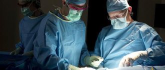
During a biopsy, an interventional radiologist inserts a needle through the skin to remove a tissue sample. An interventional radiologist is a doctor who specializes in performing image-guided procedures. Computed tomography (CT), magnetic resonance imaging (MRI), ultrasound, or fluoroscopy (real-time x-ray) may be used to determine the biopsy area. The sample is sent to the laboratory for testing.
Leakage of air from the lungs
This type of biopsy can cause air to leak from their lungs and accumulate in the pleura. The increased amount of air in the pleura prevents the lungs from expanding fully. About 30 out of 100 patients who have this type of biopsy experience a small leak of air from the lungs into the chest cavity. Most people do not experience any symptoms and feel better without any treatment. We will monitor your condition after the procedure.
Hemoptysis
Hemoptysis (coughing up blood) occurs as a consequence of bleeding from the biopsy site. It may make it more difficult for your healthcare provider to perform the procedure. It usually lasts no longer than a few minutes. In this case, you may be asked to lie on your side.
to come back to the beginning
Indications for transthoracic biopsy
Transthoracic biopsy is the definitive method for diagnosing many lung diseases. First of all, we are talking about malignant neoplasms, when morphological confirmation is necessary to confirm the diagnosis and choose further treatment tactics.
In addition, a biopsy is necessary for interstitial and disseminated processes, when the diagnosis cannot be made by other research methods.
Before the procedure
Ask questions about your medications
You may need to stop taking certain medications before the procedure. Talk to your healthcare provider about which medications you can stop taking. Below are some common examples.
Blood thinners
If you are taking blood thinners (which affect blood clotting), ask your healthcare provider what is best for you. Doctor contact information is listed at the end of this material. Whether your doctor recommends stopping this medication depends on the type of procedure and the reason you are taking the anticoagulant.
Do not stop taking your blood thinner medications without talking to your healthcare provider.
| Examples of blood thinning medications: | |||
| apixaban (Eliquis®); | dalteparin (Fragmin®); | meloxicam (Mobic®); | ticagrelor (Brilinta®); |
| aspirin; | dipyridamole (Persantine®); | nonsteroidal anti-inflammatory drugs (NSAIDs), such as ibuprofen (Advil®, Motrin®) or naproxen (Aleve®); | tinzaparin (Innohep®); |
| celecoxib (Celebrex®); | edoxaban (Savaysa®); | pentoxifylline (Trental®); | warfarin (Jantoven®, Coumadin®); |
| cilostazol (Pletal®); | enoxaparin (Lovenox®); | prasugrel (Effient®); | |
| clopidogrel (Plavix®); | Fondaparinux (Arixtra®); | rivaroxaban (Xarelto®); | |
| dabigatran (Pradaxa®); | heparin (subcutaneous administration); | sulfasalazine (Azulfidine®, Sulfazine®). | |
Read the resource Common Medicines Containing Aspirin and Other Nonsteroidal Anti-Inflammatory Drugs (NSAIDs) or Vitamin E. It contains important information about the medications you should not take before your procedure and what medications you can substitute for them.
Medicines for diabetes
If you take insulin or other medications for diabetes, ask your healthcare provider who prescribed the medication what to do the morning of your procedure. You may need to change your dose before the procedure. Your healthcare provider will monitor your blood sugar levels during the procedure.
Diuretics (diuretics)
If you are taking any diuretics (which cause you to urinate frequently), ask your healthcare provider what to do next. You may need to stop taking them on the day of your procedure. Diuretic medications are sometimes called diuretics. Examples of such medications include furosemide (Lasix®) and hydrochlorothiazide.
Removing devices from the skin
If you wear any of the following devices on your skin, the manufacturer recommends removing them before undergoing a scan or procedure:
- Continuous Glucose Meter (CGM)
- Insulin pump
Contact your healthcare provider to schedule an appointment closer to your scheduled device replacement date. Make sure you bring a spare device with you that you can wear after your scan or procedure.
If you are unsure how to monitor your glucose levels when the device is turned off, talk to your diabetes care provider before your visit.
Arrange for someone to take you home
You must have a responsible companion to take you home after the procedure. A responsible companion is someone who can help you get home safely and report problems to your healthcare provider if necessary. Agree on this in advance, before the day of the procedure.
If you are unable to find a responsible escort to take you home, call one of the agencies below. You will be provided with an escort who will take you home. There is usually a fee for these services and you will need to provide transportation. You can take a taxi or rent a car, but you need to have a responsible escort with you.
| Agencies in New York | Agencies in New Jersey |
| Partners in Care: 888-735-8913 | Caring People: 877-227-4649 |
| Caring People: 877-227-4649 |
Ask about air travel
After a biopsy, there is a risk of air leaking from the lungs and accumulating in nearby areas. If this happens, you will not be able to travel by air until the air leak stops and your healthcare provider confirms it is safe to fly. If you plan to travel by air 2 weeks after your biopsy, talk to the healthcare provider who will perform the procedure.
Arrange for someone to drive you home and stay with you overnight.
You must have a responsible chaperone who will take you home and stay with you overnight. Agree on this in advance, before the day of the procedure. If you have no one to contact with this request, please tell your interventional radiology nurse.
Let us know if you are sick
If you are sick before your procedure (for example, have a fever, sore throat, cold or flu), call your Interventional Radiology provider. Doctor's working hours: Monday to Friday from 09:00 to 17:00. If you call after 5:00 p.m., or on weekends and holidays, dial 212-639-2000 and ask for the Interventional Radiology specialist on call.
Record your appointment time
Interventional Radiology will call you two business days before your procedure, which is Monday through Friday. If your procedure is scheduled for Monday, you will be called the previous Thursday. If you are not contacted by 12:00 noon on the business day prior to your procedure, please call 646-677-7001.
A staff member will tell you when you should come to the hospital for your procedure. You will also be reminded how to get to the department.
Write down the date, time and location of the procedure in this column.
If for any reason you need to cancel your procedure, please notify the healthcare provider who scheduled it.
to come back to the beginning
What will happen during a bronchoscopy?
The procedure takes place in an endoscopy room, which meets all sterility standards accepted for operating rooms. It can be performed on an outpatient basis.
1. The insertion of a bronchoscope is often accompanied by a gag reflex and pain when passing through the pharynx and nasal passage. If the patient does not have an allergic reaction to anesthetics, the endoscopist uses it to irrigate these surfaces a few minutes before the procedure.
2. Research is carried out in a sitting position: the patient sits on a chair and tilts his head slightly back. Your hands can be placed on your knees or between your legs.
3. The endoscopist gradually begins to insert the device through the lower nasal passage into the nasopharynx. When the device reaches the vocal cords, the doctor sprays them and the epiglottis with an anesthetic. If the patient is prone to nosebleeds or narrowing of the nasal passages, the bronchoscope is passed through the oral cavity.
4. The airways are devoid of pain receptors - bronchoscopy will not cause pain.
5. Using a bronchoscope, the doctor carefully examines the condition of the mucous membrane of the bronchial tree. If the diameter of the bronchoscope and the patient’s condition allow, then, in addition to the large bronchi, it is also possible to examine the branches.
6. If it is necessary to obtain material (bacteria, respiratory tract cells) for subsequent laboratory research, the doctor performs bronchial lavage. To do this, 20-100 ml of sterile liquid is injected into the lower respiratory tract, after which it is removed. Lavage does not interfere with the patient’s spontaneous breathing.
7. If it is necessary to obtain tissue samples for histological analysis, the doctor performs a biopsy. The tissue is removed using miniature forceps through the instrumental channel of the bronchoscope. The procedure does not cause pain.
Day of the procedure
Instructions for drinking before your procedure
You can drink no more than 12 ounces (350 ml) of water between midnight and 2 hours before you arrive at the hospital. Don't drink anything else. Do not drink any liquids two hours before you are scheduled to arrive at the hospital. This also applies to water.
What you need to remember
- Take only the medications your healthcare provider tells you to take the morning of your procedure. Wash them down with a few small sips of water.
- Do not apply cream or Vaseline® to your skin. You can use deodorants or light lotions to moisturize your skin. Don't wear makeup on your eyes.
- Remove all jewelry, including body piercings.
- Leave all valuables (such as credit cards and jewelry) at home.
- If you wear contact lenses, wear glasses instead if possible.
What to take with you
- A list of all the medications you take at home, including patches and ointments.
- Case for glasses or container for contact lenses.
- A health care proxy form, if you have completed one.
What to expect
Upon arrival at MSK, doctors, nurses, and other medical staff will ask you several times to say and spell your name and date of birth. This is necessary for your safety. People with the same or similar names can undergo the procedure on the same day.
After you change into a hospital gown, a nurse will meet with you. You will have an intravenous (IV) catheter inserted into one of the veins, usually in your arm or hand. You will first be given IV fluid, and then a medication will be given through the same IV to make you drowsy and more relaxed during the procedure.
You will talk to your healthcare provider before the procedure. They will explain the procedure to you and answer your questions.
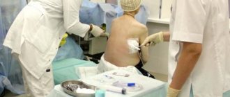
During the procedure
When it is time for your procedure, you will be taken to the treatment room and assisted on the examination table. You will be connected to equipment to monitor your heartbeat, breathing and blood pressure. In addition, you will receive oxygen through a thin tube that is located under your nose.
The healthcare provider will clean the skin around the biopsy site and cover it with a cloth. A local anesthetic (medicine that numbs the area of the body) will be injected into the area that the healthcare professional will work on.
Your interventional radiologist will insert the biopsy needle through the skin and check its position using MRI, CT, ultrasound, or fluoroscopy. Once the needle is in the right place, the radiologist will perform a biopsy. The sample will be checked to ensure that the amount of tissue collected is sufficient. If it is sufficient, the interventional radiologist will remove the needle. If there is not enough tissue, another sample will be taken.
After the procedure is completed, the healthcare professional will clean the site and apply a bandage.
to come back to the beginning
Why is bronchoscopy performed?
The study is carried out for both therapeutic and diagnostic purposes.
Diagnostic bronchoscopy helps to clarify the diagnosis if the patient is suspected of having tumors of the respiratory tract (lung cancer), or it is necessary to decide on a treatment plan for an already confirmed process, or to understand why the patient has a narrowing of the respiratory tract. During this procedure, the doctor takes tissue samples (performs a biopsy) for subsequent histological examination.
The use of bronchoscopy and bronchial lavage makes it possible to extract the substrate (contents) from the alveoli and small bronchi.
A bronchoscope helps to find and remove foreign objects from the airways, wash out mucus plugs; it is used if it is necessary to install a stent - a structure that strengthens the airways and prevents them from collapsing.
Therapeutic bronchoscopy is necessary for patients on mechanical ventilation (artificial ventilation), it helps maintain the breathing tube in the correct position.
After the procedure
In the hospital
After the procedure, you will be taken to the recovery room. You will have at least 2 chest x-rays to check for air around your lungs. The first one will be done right away. The second photo will be taken approximately 2 hours later.
While you are in the recovery room, tell your nurse if you have:
- shortness of breath or trouble breathing;
- pain or discomfort;
- any symptoms that bother you.
You may be given oxygen through your nose during this period. You will not be able to eat anything immediately after the procedure.
After 2 hours, you will have another chest x-ray. If the results are normal, you can go home. You must travel home accompanied by a responsible companion who will stay with you overnight.
If air collects around the lungs, your healthcare provider will decide whether additional X-rays need to be taken to monitor the lungs. You may have a small chest tube inserted to allow your lungs to expand again. A chest tube allows air to drain around the lung until the leak stops. You may be hospitalized while your lungs expand again. Such consequences of undergoing a biopsy occur in approximately 8 out of 100 patients.
At home
- You must have a responsible escort who will stay with you until the morning. This is necessary for your safety.
- After you are discharged from the hospital, you can return to your normal diet.
- You may shower or bathe the day after your biopsy. After showering, remove the bandage.
- The day after the procedure, you can return to your normal lifestyle.
- If you do not have an air leak in your lungs and plan to travel by plane, you are allowed to fly 2 days after your biopsy. If an air leak is visible on a chest X-ray, you will not be able to travel until your healthcare provider confirms that it is safe to fly.
- To find out the results of your biopsy, call your doctor who ordered it a few business days after your procedure.
to come back to the beginning
Contraindications to transthoracic biopsy
Contraindications to puncture biopsy:
- Clinical cases when the result of the study will not affect the treatment of the disease or its prognosis, for example, in case of metastatic lung disease.
- The patient's inability to maintain a static position for the time required for the examination. Such situations arise in cases of mental disorders, severe pain, etc.
- Uncontrollable cough.
- Severe respiratory failure.
- Some diseases of the cardiovascular system - uncontrolled arrhythmia, unstable angina, severe heart failure.
Contraindications to open biopsy methods are:
- Severe cardiovascular diseases.
- Decompensated respiratory failure.
- The presence of “honeycomb lung” signs on X-ray images, which indicate the final stage of diffuse lung pathology.
What is a puncture biopsy for nodular formations in the lungs?
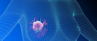
Nodes in the pulmonary parenchyma are round formations or foci of pathologically altered lung tissue.
Most often, nodules are detected on a chest x-ray and are usually not accompanied by pain or any other symptoms. As a rule, nodular formations of any localization are detected during instrumental examination. However, imaging methods do not always allow us to talk about the nature (benign or malignant) of the detected changes.
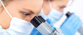
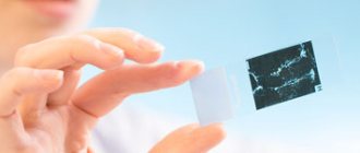
A needle biopsy, or aspiration, involves removing a small sample of cells from a suspicious area using a hollow needle and then examining the resulting tissue under a microscope to make a diagnosis.
A puncture biopsy of nodular formations of lung tissue is often performed under visual control, for example, using MRI or fluoroscopy, which allows the doctor to bring the instruments precisely to the site of pathological growth.
Up
When is a puncture biopsy performed for pulmonary nodules?
Despite the fact that more than half of single (solitary) nodes in the tissues of the chest organs ultimately turn out to be benign, they are initially treated as malignant formations until the contrary is proven. This is exactly where a puncture biopsy helps.
After detecting a nodular lesion, an instrumental examination is carried out, which in most cases allows us to talk about the nature of the formation: benign or malignant. In cases where this is not possible, a puncture biopsy is necessary.
If the doctor prescribes a biopsy, this indicates that other diagnostic techniques, such as bronchoscopy, do not allow examining the nodular formation.
Up
When are chest interventions used?
Pleural puncture
used for the following purposes:
- Reducing pressure on the lungs
- Treating symptoms such as shortness of breath or chest pain
- Determining the cause of the accumulation of excess fluid in the pleural cavity
Pleurodesis
performed after thoracentesis to prevent further fluid accumulation.
Pleural biopsy
is prescribed in cases where pleural puncture does not allow determining the cause of fluid accumulation in the pleural cavity. In this case, a sample of pleural tissue is removed and examined under a microscope for the presence of the following conditions:
- Tuberculosis
- Malignant neoplasm
- Viral, fungal or parasitic disease
Up
Benefits and risks of chest interventions
Advantages:
- In general, thoracentesis and other chest interventions are a safe procedure.
- No surgical incisions are required.
Risks:
- Any procedure that involves violating the integrity of the skin carries a risk of developing infection. However, in this case, the chance of developing an infection that requires antibiotic treatment is less than 1 in 1000 cases.
- The following complications may develop: Pneumothorax or partial collapse of the lung caused by injury to the lung tissue and air entering the pleural cavity.
- Pulmonary edema, which can occur when too much fluid is removed.
- Infection and bleeding.
- Serious difficulty breathing.
Up
What does the diagnostic equipment look like?
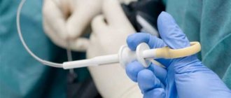
The biopsy needle is usually several centimeters long, and the syringe barrel is the diameter of a wide paper clip. The needle is hollow inside, allowing the tissue sample to be grasped and removed.
Several types of needles are used when performing a biopsy:
- Thin needle attached to a syringe: Thinner in diameter than needles used to draw blood from a vein.
- Automatic Cutting Needle with Spring Mechanism: Consists of an inner “thick” needle that is inserted into a locking cell covered with a cutting sheath and attached to a spring mechanism.
- Vacuum aspiration biopsy device: allows you to obtain a fairly large tissue sample.
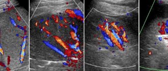
A puncture biopsy is often performed under the guidance of CT, fluoroscopy, ultrasound or MRI.
CT scan
A CT scanner is usually a massive, rectangular machine with a hole, or short tunnel, in the middle. During the procedure, the patient is placed on a narrow table that slides inside the tunnel.
The X-ray tube and electronic X-ray detectors are located opposite each other inside a ring-shaped structure called a gantry. In a separate office there is a computer workstation where the resulting image is processed. There is also a doctor or technologist here who monitors the operation of the tomograph and the progress of the examination.
Fluoroscopy
Fluoroscopy typically uses an X-ray tube, a patient table, and a monitor located in the radiologist's office. To monitor the process and control the doctor’s actions, a fluoroscope is used, which converts X-ray radiation into a video image. To improve image quality, a special amplifier is used, suspended above the patient's table.
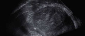
Ultrasonography
An ultrasound scanner consists of a console that includes a computer and electronic equipment, as well as a video display and an ultrasound transducer used for scanning. The sensor is a small, portable device that resembles a microphone and is attached to the scanner using an electrical cord. The ultrasound sensor sends high-frequency sound signals inaudible to the human ear and picks up echoes reflected from the internal structures of the body. The principle of operation of the device is similar to sonars that are used on submarines.
In this case, an image instantly appears on a monitor resembling a television or computer screen. Its appearance depends on the amplitude (strength), frequency and time it takes for the sound signal to return from the patient's body to the transducer, as well as the type and condition of the tissue through which the sound travels.
Up
Proper preparation
Before performing endoscopy of the bronchi and lungs, the patient must remember the basic rules of preparation for the procedure:
- The examination is carried out on an empty stomach. Otherwise, food or a drink will accidentally enter the respiratory tract due to an accidental cough or vomiting.
- If a course of medication is prescribed, it is recommended to talk with your doctor; you may need a break for one day.
Before performing a pulmonary endoscopy, it is necessary to plan so that the last meal takes place no later than 21 hours before the start of the procedure.
What procedures are meant by interventions on the chest organs?
Chest procedures include minimally invasive procedures that are used to diagnose the causes and treat pleural effusion, which is a collection of fluid in the pleural space.
The pleural cavity is a slit-like space inside the chest that surrounds the lungs. Pleural effusion occurs in a variety of conditions, including infections, inflammation, heart failure, and malignancies. Excessive fluid accumulation around the lungs makes breathing difficult.
Interventions on the chest organs include:
- Thoracentesis
(thoracentesis, pleural puncture): removal of fluid from the pleural cavity using a needle attached to a syringe, followed by examination of the resulting sample under a microscope. - Pleurodesis
: the introduction of drugs into the pleural cavity in order to reduce its volume and the amount of accumulated fluid. - Pleural puncture biopsy
: involves removing a sample of pleural tissue, a thin layer of tissue that covers the outside of the lungs and lines the inside of the pleural cavity. In this case, a hollow needle is used, and the resulting sample is examined under a microscope.
Up
Our prices
| Specialist consultation | |
| Repeated appointment with a surgeon | 2700 |
| Primary appointment with a surgeon | 3200 |
| Appointment with surgeon K.M.N. repeated | 4700 |
| Appointment with surgeon K.M.N. primary | 5300 |
| Repeated appointment with the oncologist | 3200 |
| Initial appointment with an oncologist | 3700 |
| Appointment with oncologist K.M.N. repeated | 4200 |
| Appointment with oncologist K.M.N. primary | 5300 |