CT scan of the jaw is an x-ray method for studying the condition of the bone and soft tissue structures of the lower part of the facial bones. CT scan of the upper jaw or CT scan of the lower jaw can detect diseases of the joints, ligaments, maxillary sinuses, bone tissue and teeth. However, this examination should not be confused with a dental CT scan, which is a targeted method of scanning the dentition of the jaw area.
CT scan of the upper and lower jaw has a number of advantages in comparison with x-rays of the jaw area and MRI of the jaw joint:
- minimal time spent on CT scanning, in contrast to the longer MRI of the temporomandibular joint;
- higher image quality and detail compared to radiography;
- absence of pain and discomfort;
- issuing results on the same day.
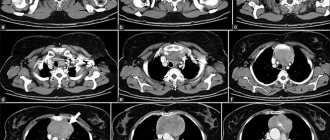
High quality of reproduced images is ensured by layer-by-layer scanning of the selected area in sections up to 1 mm thick.
The main disadvantage of computed tomography of the jaw is the presence of radiation exposure to the body. On average, it ranges from 1-3 mSv per scanning session.
Free diagnostic consultation
If in doubt, sign up for a free consultation. Or consult by phone
+7
All about diagnostics
Prices for computed tomography of the jaw
I can immediately say that the cost is considerable, in our LeaderStom clinic the price for a CT scan of one segment (with recording on disk) is 2000 rubles, and for a CT scan of both jaws also with recording on disk it will be 3000 rubles.
Research methodology
- Supine position with head fixed;
- The direction of the scanning plane is parallel to the occlusal cavity;
- Closing the jaws in a state of habitual occlusion;
- Cut thickness 1 mm
- Scanning of the TMJ is carried out in the position of habitual occlusion and with the mouth as open as possible.
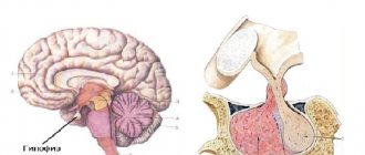
Computed tomography gives an idea of the location of the mandibular canal, incisive canal, and the relationship of pathological changes with surrounding anatomical structures. The method allows you to assess the prevalence and type of bone pockets and identify diseases of the temporomandibular joints.
Contraindications
A CT scan of the jaw can be performed on an adult patient if there is a referral from a doctor or on personal initiative. For children, due to the presence of radiation exposure to the body, computed tomography is performed strictly on the direction of a doctor and exclusively for strict medical reasons.
When scheduling a CT scan, patients should consider the following contraindications:
- the patient is pregnant at any stage;
- The age of the subject is up to 3 years.
CT scans of the maxillofacial area are used in various fields of dentistry, for example:
- in surgery for the diagnosis of post-traumatic and congenital deformities of the facial skull;
- periodontitis and periodontal bone changes;
- cyst;
- neoplasms;
- inflammatory processes of the jaw;
- diseases of the maxillary sinus;
- during implantation and orthodontic treatment;
- for analysis of endodontic treatment.
Preparation
Before performing a CT scan of the jaw, no special preparation is required, with the exception of studies with contrast. Before conducting a contrast study, it is recommended to refrain from eating 6-8 hours before the procedure.
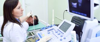
During the procedure, the patient lies on the scanner table. The head can be fixed to achieve immobility during the examination. The duration of the procedure is on average 20 minutes. If contrast is used, which is administered intravenously before tomography, the duration of the study can increase to 30 minutes.
Indications for CT scanning:
- clarification of radiology data;
- analysis of the condition of the maxillary sinuses;
- identification of fractures of the facial skull bones;
- prosthetics of dental defects based on the use of implants;
- assessment of congenital and acquired defects.
CT creates a unique opportunity to study the condition of the mucous membrane of the paranasal sinuses and the ethmoidal labyrinth, since radiography and layer-by-layer studies suggest the presence of additional shadows simulating the pathology of the sinuses, including the presence of fluid levels.
Contraindications to CT scan of the jaw
There are a number of contraindications for which the procedure is not recommended.
These include:
- weight more than 150 kg or girth more than 150 cm (the device is designed for a load of up to 150 kg);
- pregnancy (the risk of developing defects in the fetus increases);
- lactation period (during the procedure, rays can directly affect the composition of a nursing woman’s milk);
- severe mental illness (during the procedure the person must be at rest);
- the presence of metal objects in the area of interest, as they can reduce the information content of the results;
- children under 5 years old;
Difference between CT and MRI?
MRI and CT differ in the technique of execution. The difference with CT is the nature of the waves: with MRI they are electromagnetic. Under their influence, different areas of tissue give a different “response”, which is recorded by the receiving device of the device. And then, just like with CT, the signals are processed and converted into an image. But the main difference is that computed tomography is usually used to look at hard tissues (teeth and bones), and magnetic resonance imaging is used to look at soft tissues ( tongue, mucous membrane, muscles, salivary glands).
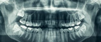
What is a CT scan of the jaw and what does it show?
A CT scanner consists of a ring through which a table with the patient lying on it passes while a series of sequential images are taken.
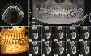
Computed tomography shows all the necessary structures:
- body and all four processes of the maxilla;
- body and branches of the lower jaw;
- adjacent bone and cavity structures - maxillary sinuses, nasal bones.
Variant of the norm on a CT scan of the jaw
Soft tissues are capable of transmitting X-rays, and they appear in gray shades on the image. Dense tissues, on the contrary, absorb the rays and appear white on the tomogram.
The intensity of darkening of individual areas is the main diagnostic indicator, by analyzing which the doctor will be able to determine the presence or absence of pathologies in your body.
Indications
CT scanning of the maxilla and mandible is indicated for various conditions where visual information is needed to confirm the diagnosis. CT is indicated in the following cases:
- Implantation planning
- Diagnosis of pathological changes in the jaws
- Abnormal arrangement of teeth requiring extraction
- 3D reconstruction and measurement of deformities and developmental anomalies in the lower and upper jaw
- Planning of surgical intervention for jaw tumors (lower and upper)
- Injuries of the maxillofacial area
- Sinus infections (sinusitis)
- Tumors
- Facial nerve damage
- Pathology of the mandibular joint
- Pathology of the salivary glands
CT scan for dental implantation
Having a computer model of the jaw in hand, the implantologist will be able to select the correct shape and size of the dental implant. The success of the operation depends on this, as well as the degree of healing and service life of your new tooth.
In addition, the jaw bone tissue is assessed: height, volume, density. For example, when the height is less than 1 cm, implantation is simply impossible. The image will also show if bone resorption has occurred. Then a sinus lift is prescribed, that is, augmentation with bone material.
What does a CT scan reveal?
In therapeutic dentistry, CT scanning of the teeth and jaw makes it possible to determine the structure of a particular tooth, the number and shape of root canals, and the location of the nerves in them. The doctor will be able to see any changes and pathologies:
- cracks in the roots
- cysts,
- tumors
- granulomas, etc.
In surgery, computed tomography gives a clear picture of the shape of the roots of a complex tooth, such as a wisdom tooth, and determines the further course of action during extraction.
For implantologists , CT scanning is essential to plan implant surgery and accurately position the artificial root in the bone. This way, the doctor can fully see the width and height of the bone tissue, its thickness and density, the proximity of the maxillary sinuses, and the location of the mandibular nerve to eliminate any risks during surgery and subsequent serious complications.
In orthodontics, CT scans provide a high-quality three-dimensional image of the bite and the position of all teeth to accurately predict their movement and alignment. Also, when treating children and adolescents, you can see unerupted teeth, the direction of their growth, etc.
In addition, with the help of CT, doctors effectively identify:
- periodontal diseases, periodontal diseases,
- complications after injuries to the jaw or teeth,
- structural anomalies of the temporomandibular joints.
Get a CT scan of your teeth in St. Petersburg: price of the service
The cost of computed tomography of teeth in dentistry in St. Petersburg may vary, because the price of the service will depend on:
- On the type of equipment used in the study;
- The price of the service will also be affected by the size of the jaw area that needs to be examined;
- The need for printing the resulting images will increase the price of CT scanning.
It is worth considering the fact that the price of a CT clinic may include various services, but usually it includes the examination itself, recording of finished images on a digital medium and a specialist’s report, which is handed to the patient in writing.
When is a dental CT scan indicated?
It is often quite difficult to visually determine damage resulting from trauma. Computed tomography of the teeth must be carried out after any injuries, because it is necessary to identify in time whether there is a fracture or displacement of the jaw, as well as damage to the roots of the tooth, which can lead to its loss. The list below gives a complete picture of the indications for CT scanning:
- Any mechanical damage, the consequence of which may be a fracture or displacement of the jaw. Invisible during external examination, these injuries are revealed by tomography. Injuries can also damage the roots of the tooth, which subsequently leads to its loss. Tomography will allow you to detect damage and prescribe timely treatment
- Deviations in the development of teeth, anomalies of the dentition. The orthodontist prescribes a tomography before starting treatment for the patient. Determine the ability to withstand the load of the bracket system. CT scan of teeth allows you to accurately determine the time and direction of growth of wisdom teeth
- If it is necessary to create a bracket structure, it is important to correctly model the future systems. A special role is played by spatial modeling in the manufacture of internal systems and aligners. Individual production of such products is provided, and it is tomography that gives a complete picture of the structural features of the teeth and jaw of each patient. This contributes to the speedy achievement of positive results from orthodontic treatment.
- If there is a suspicion of complications after endodontic treatment. The doctor cannot visually check the quality of the therapy performed. Only a dental CT procedure makes it possible to monitor and timely detect relapses or improper treatment
- If tumors are suspected or already present, the use of computed tomography will help to detect a tumor in time or clarify the condition of an existing one. The jaw bones and soft tissues of the oral cavity are susceptible to the appearance of neoplasms after injuries and other damage. To avoid the development of a tumor and the negative consequences of this, it is recommended to remove the tumors
- A planned surgical operation must be carefully prepared, and computed tomography is of great importance in this regard. If implantation or particularly complex tooth extraction is necessary, a CT scan will help the doctor identify factors that complicate the operation and determine how long the operation will take, given its complexity
- Monitoring and checking the effectiveness of treatment in many cases is impossible without CT scanning. Just as it is sometimes difficult to diagnose a disease without tomography, after a course of treatment it is not always possible to correctly evaluate the treatment. It will help determine whether all damaged tooth tissues have been removed or whether partial roots remain after a complex tooth extraction
- If sinus lifting or bone grafting is planned, then a high-quality 3D image is necessary. Bone tissue can also be seen on an x-ray, but the condition of soft tissues, mucous membranes, canals and blood vessels can only be seen on a three-dimensional high-quality image
How is a CT scan of the lower and upper jaw performed?
Before a CT scan, the patient must remove any metal objects, including removable hearing aids and dentures. In addition, he must provide the diagnostician with a referral from the attending physician. He will be asked to take a lying position on the tomograph table, which will move into the scanning frame, after which the scanning procedure will begin.
During this procedure, the device will emit clicks and characteristic crackling sounds, which are normal and should not cause discomfort to the patient. He must remain motionless for 15 to 20 minutes while the shooting is taking place.
Medical personnel are located in an adjacent room during scanning. The patient has the opportunity to communicate with him through a public address system and can contact him if he experiences severe discomfort.
During a procedure using a contrast agent, the patient may experience a moderate burning sensation in the injection area, mild dizziness, hot flashes, or nausea. These signs are normal and disappear upon completion of the procedure. If they are too pronounced, the patient has the opportunity to inform the diagnostician about this.
How often can you do it
Cone beam tomographs have been developed for dentistry and maxillofacial surgery. They have a clearly directed narrow beam of rays, which allows you to speed up the procedure and reduce the radiation dose to 40-70 microsieverts.
According to sanitary rules, a person can receive a total dosage of 1000 μSv in a year without health consequences. Therefore, tomography is allowed more than 10 times a year.
3DCT is performed for pregnant and nursing mothers according to strict indications. The decision on how often a dental CT scan can be done during pregnancy and breastfeeding is made by a consultation of a gynecologist, pediatrician, and dental specialists.
Why is the CT method better than others in diagnosing these diseases?
Benefits of CT
- Dentists or maxillofacial specialists can obtain complete information about the shape of the jaw, its size, assess the density of bone tissue, the location of the nerve canals, and the condition of the paranasal sinuses.
- Unlike 2D X-rays, which overlay images of tissue, 3D jaw CT provides distortion-free images and precise measurements down to fractions of a millimeter of jaw and tooth structures.
- An accurate three-dimensional image and analysis of the area where the implant is planned to be installed allows you to select the most optimal positioning of the implant, especially if there are areas with rare bone tissue
Contraindications and restrictions
The patient’s weight – heavy weight may not allow the examination to be carried out since CT scanners have weight restrictions.
CT scanning is not advisable for persons under 14 years of age, since at this age the body is actively growing and radiation exposure can be risky.
The presence of kidney disease, decompensated heart disease, and pregnancy are contraindications for CT scanning.
The presence of an allergy to iodine-containing products often does not allow CT scanning with contrast, since contrast agents have iodine atoms in their structure, and this sharply increases the risk of dangerous allergic conditions such as anaphylactic shock.
Advantages of CT of the upper and lower jaw in CELT
Our clinic is multidisciplinary and is deservedly considered one of the leaders in the domestic market of paid medical services. We have been working for more than twenty years and during this time we have restored health to hundreds of thousands of patients. We employ leading domestic specialists, doctors of science, who are active in scientific work and create effective treatment methods.
The specialists of our diagnostic radiology department have modern equipment that allows us to take computed tomography to a new quality level. In this they are helped by a thorough knowledge of the technology and decades of experience. They know how to identify injuries to the alveolar process, identify neoplasms of any etiology and inflammatory processes even in the early stages of their development. By providing complete information to the attending physician, they allow him to accurately diagnose and develop appropriate treatment tactics.
Computed tomography is performed using modern foreign-made equipment. The process uses modern contrast agents that minimize the negative impact on the body. Before the procedure, a diagnosis must be carried out to exclude the presence of contraindications in the patient.
You can find out the price of a CT scan of the jaw by reading the price list presented in this section of our website. Despite the fact that we regularly update it, in order to avoid misunderstandings, to clarify the data, please contact our operators by phone: +7 (495) 788-33-88.
Benefits of CT
Computed tomography has a huge advantage over classical x-ray examination. After all, the doctor receives a three-dimensional three-dimensional image, and not a planar one, as with an x-ray. An X-ray image is static, whereas with a CT scan, the 3D model can be rotated at any angle to see the smallest details. There are no distortions or errors on the computed tomogram. For the patient, an additional benefit is a much lower radiation dose, which makes tomography possible even for children.
The Gendex GXDP-700 3D tomograph used in the dental department of our clinic allows you to create accurate 3D models of the jaws, without the slightest inaccuracies or errors.
Advantages of a dental tomograph:
- minimal radiation exposure (6-10 times less than with conventional spiral tomography). The effective dose of an average shot is comparable to a three-hour plane flight.
- fast scanning time - up to one minute.
- scanning is performed in a standing position. No special preparation for the study is required.
- high quality of the resulting image, which provides a lot of diagnostic capabilities.