The most common symptoms of damage to the vascular system of the brain and neck are headaches, unsteadiness of gait or weakness in the arm or leg. If symptoms are present, the patient should be examined using MRI of the brain vessels. The purpose of diagnosis is to exclude congenital pathology, namely:
- arterial aneurysm;
- arteriovenous malformation (AVM).
Moreover, symptoms can go undetected for too long, up to 45 years.
The essence of the technique
The study is based on hydrogen atoms, which are affected by a magnetic field, forcing them to be an “exchanger” of energy. This is due to the fact that hydrogen atoms are part of any human organ, including liquids.
Hydrogen atoms rotate around a magnetic axis. When a person is placed in a tomograph, energy is absorbed by hydrogen atoms, and upon completion of magnetic resonance imaging, energy is released. The degree of release by different tissues is somewhat different, since their chemical composition and density play a role. The computer captures this emitted energy, amplifies and re-digitizes it in various degrees of gradation in brightness. Next, the information is displayed on the monitor and recorded on all supported media.
The study is harmless to the patient's health. This is the fastest diagnostic method that painlessly displays all structural and morphological changes in the vascular system of the brain, head and neck.
MRI of vessels with contrast agent
To increase the information content of the study of the circulatory system, intravenous injections of a “staining” drug are used. For magnetic resonance scanning, a solution of gadolinium salts is used. The paramagnetic allows you to visualize the vascular network with the smallest branches, shows the narrowing/expansion of the lumen, violation of the integrity of the walls, and intravascular objects.
Contrast is administered intravenously. More often, a catheter connected to an automatic injector is used. Bolus administration ensures a constant rate of drug entry into the blood.
Gadolinium chelates (soluble salts) are characterized by low toxicity and hypoallergenicity. The substance leaves the body naturally, through the kidneys and liver.
Contraindications to the use of contrast enhancement during MRI are:
- pregnancy;
- severe pathologies of the kidneys and liver;
- individual intolerance to the drug.
For children under 12 years of age, bolus boosting is available in a hospital setting, which is due to the peculiarities of intravenous injections for children.
Diagnostics MRI of cerebral vessels
MRI of head and neck vessels is an informative neuroimaging method. Give a chance:
- assess the condition of the cerebral hemispheres;
- identify the presence of space-occupying formations;
- determine the presence and development of focal pathological changes;
- assess the condition of the cerebellum and brain stem.
During MRI of the vessels of the head and neck, the entire structure is analyzed with the exception of the skull bones. At the stage of such a procedure, the following occurs:
- assessment of the structure of the cerebral vascular system;
- the presence of an aneurysm in the substance is excluded or confirmed;
- confirmation or refutation of the pathological state of the structure of the vascular system.
Modern MRI techniques
Depending on the preliminary diagnosis, as well as on the set of symptoms, the doctor prescribes the necessary magnetic resonance imaging method. For example, after a stroke, a functional MRI is performed to assess the extent of the lesion. The doctor has the opportunity to observe a detailed picture of the work of brain areas, each of which is responsible for a specific function in the human body. During the study, the patient must carefully listen to the doctor’s commands, for example, speak, sing, at this time the device will read the brain’s response to stimulation.
MRI with contrast, which is administered by intravenous infusion, significantly increases the efficiency of the study. This is especially true when examining for the presence of oncology and differentiating its nature, as well as for determining congenital or acquired anomalies, determining the zone of inflammation, and so on. It should also be said that the use of a contrast agent significantly increases the effectiveness of the examination if the doctor needs to evaluate the functioning of the vascular network of the brain.
The doctor chooses a survey tomography if it is necessary to assess the overall picture of the interaction of the nervous system organs or to track the entire route of movement of the brain fluid.
Possible diagnosable diseases, pathologies
The diagnostic method will help confirm or refute:
- development of deviations due to previous traumatic brain injuries;
- any possible brain infections;
- tumor formations;
- stretching of the walls of the artery with subsequent deformation (most often, immediately after the bed);
- non-standard connection, as well as the location of the capillaries of the vascular system;
- autoimmune diseases;
- carotid artery syndrome;
- artery abnormalities;
- blockage of deep veins.
Such diagnostics can be carried out without the introduction of a contrast agent.
MRI of the vessels of the head and neck is relevant in cases where the patient has been experiencing for a long time:
- headache;
- dizziness;
- attacks of loss of consciousness;
- epilepsy attacks;
- other convulsive conditions.
The patient needs to contact a neurologist with this problem. The latter, using generally accepted medical techniques, will be able to determine how important MRI of the vessels and arteries of the neck is for the patient.
MRI of vessels and arteries of the neck and MRI of brain vessels: differences
When performing MRI of cerebral vessels, there is no direct possibility of assessing vascular structures. If a patient has a large aneurysm, most likely this will be visible during a routine native examination of the head.
As for the need to visualize vascular structures, for example, the demanded analysis of blood flow through the vascular system, excluding possible narrowings or other defective variants of the structure of the vascular bed, it is necessary to conduct an examination of the head. Also, if the patient has any ischemic conditions or strokes, with the help of additional brain diagnostics, it is possible to determine at the level of which of the arteries the “catastrophe” occurred.
MRI of neck vessels: who will this diagnosis help?
If a person is about to undergo an MRI of the neck vessels for the first time, he may have various questions about this procedure. Why and how is it done? Is any special preparation needed? What is better - MRI or ultrasound of neck vessels?
Vladimir Vladimirovich Tulinov, radiologist, executive director of MRI Expert Lipetsk, answers these and other questions.
— Vladimir Vladimirovich, how often do patients come to your center to undergo an MRI of the neck vessels?
- Yes, relatively often. As a rule, this study comes as an addition to the examination of the soft tissues of the neck.
— Is MRI of the vessels of the neck an independent study or is it included in some other one?
- Yes, this is an independent examination.
— Why is an MRI of the neck vessels done?
— To exclude any organic changes and congenital pathology, including aneurysms and arteriovenous malformations, as well as areas of stenosis.
— What complaints and symptoms in a person can be a reason to refer him for an MRI of the vessels of the neck?
— The doctor may prescribe this study if the patient has headaches, noise in the head, dizziness, episodes of loss of consciousness, memory impairment, and some other manifestations.
— What does MRI of the neck vessels show?
“With the help of magnetic resonance imaging, the doctor cannot see all the vessels - only the arteries, the veins are not visible. Moreover, only the main arteries are visible: the vertebral arteries (segments V1, V2 and V3), the external and internal extracranial (extracranial) sections of the carotid arteries on the right and left. The doctor sees these vessels in a 3D projection and can examine them in detail, from different angles. The doctor can, in particular, evaluate the course of the vessels, their pathological tortuosity, and the connection between the vessels.
— What does the procedure look like and how long does an MRI of the neck vessels take?
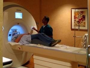
“The patient lies down on the tomograph table face up, a special coil is placed around his neck, the table is brought inside the machine, and the examination is carried out within 20 minutes. The patient's task is to lie down without moving. Since this particular type of study is characterized by a large number of slices, on the basis of which a 3D image model is then built, it is very important to make swallowing movements as little as possible. Otherwise, the picture may become blurred and turn out to be of poor quality. If a person still wants to swallow saliva, it is advisable to do this carefully, with a low amplitude.
— What is an MRI of neck vessels with contrast, and in what cases is this study prescribed?
— In principle, MRI of neck vessels, unlike, say, computed tomography (CT), is a non-contrast method. This is one of its advantages. That is, we can build a 3D image based on the physical properties of flowing blood. But in some cases, contrast is required - in particular, if thrombosis is suspected. In their normal state, the vessels allow the contrast agent to pass through, and the place of blockage by thrombotic masses - the thrombus itself - is, as it were, enveloped by the contrast. Without contrast, we will not see this picture. This is important for subsequent treatment: the surgeon will decide whether to remove this clot, or use bypass surgery to bypass the clot.
The contrast agent is made from gadolinium salts. It is administered intravenously, does not carry any radiation exposure and is completely eliminated from the body within 4 hours naturally, along with urine.
For more information about why contrast is needed during MRI, read our article
— Do I need to somehow prepare for an MRI of the neck vessels?
— No, no special preparation is required from the person. But there is one nuance here. It happens that the patient cannot withstand the entire 20 minutes of the study, lying still, and asks to pause (in order to give a signal, he has a special pear in his hand). Then the examination is considered failed, and the entire procedure will have to start again.
— A common method for assessing neck vessels is ultrasound. Why then does the doctor refer for an MRI? Is this method better than ultrasound?
— These are two completely different methods, they complement each other. That is, it is impossible to say which is better. Ideally, in order to fully understand some pathology, you need to do both. If, using MRI of the vessels of the neck, their anatomical state is assessed, congenital or acquired organic changes are detected, then through ultrasound we evaluate functional changes - the speed and volume of blood flow, the thickness of the vessel wall, the presence of pathological turbulent flows (vortices). In short, when a doctor wants to get as much information as possible, he, as a rule, gives the patient a referral for these two types of research at once.
You can read more about ultrasound of neck vessels here
— Does it happen that, based on the results of the study, you see that the patient may need other diagnostics - in particular, computed tomography of the neck vessels? Or can MRI answer all the questions?
— CT, like ultrasound, is a complementary diagnostic method. With the help of computed tomography, for example, atherosclerotic plaques are better visualized. But let me remind you this. MRI of neck vessels is done mainly without contrast enhancement, and if contrast is used, it is a substance based on gadolinium salts. During a CT examination, there is radiation exposure and an iodine-containing contrast agent is administered. It may be noted that iodine-containing drugs very often cause allergic reactions, including anaphylactic shock and, as a consequence, death.
The choice of one or another research method depends on the specific situation and remains with the doctor.
You can sign up for an MRI of the neck arteries here. ATTENTION: the service is not available in all cities
Interviewed by Igor Chichinov
The editors recommend:
MRI of the neck: who needs it and what will it show? Why does the cervical spine hurt? We are convening a medical consultation. How will MRI of cerebral vessels help a patient?
For reference
Tulinov Vladimir Vladimirovich
In 2010 he graduated from Ryazan State Medical University named after. Academician I.P. Pavlova with a degree in General Medicine.
In 2010-2011 – internship in the specialty “Radiology”.
Since 2011, radiologist at the federal network “MRI Expert”.
Since 2015, director and chief physician of MRI Expert Yelets. Since April 2021 - Executive Director of MRT Expert Lipetsk and MRT Expert Lipetsk II.
In 2021, he received a diploma from Moscow State University of Education and Science of the Moscow Government with the qualification “Master of Business Administration (MBA).”
Decoding of received images
MRI of the head and neck vessels is a complex diagnostic method and requires special skills that only a radiologist and a neurologist can have. A few hours after MRI of the cerebral vessels, you can get a detailed answer from a specialist about the current situation. Also receive recommendations regarding treatment or further hospitalization of the patient.
The examination is carried out using modern equipment, so in addition to the conclusion, as well as photographs, the patient receives a CD with a recording of the examination. It is the use of the latter medium that makes it possible to find abnormal channels of the vascular system of small sizes.
What does it show
The price of MRI of the brain is relatively low, given that it is one of the most accurate and effective diagnostic methods. Scanning provides information about the following conditions:
- presence of brain injuries;
- infectious diseases;
- presence of tumors, brain metastases;
- some diseases of the central nervous system;
- emergency conditions;
- neurodegenerative conditions.
The cost of a brain MRI with contrast is higher, but this procedure is more often recommended because it produces more accurate results.
Preparing for an MRI of the brain
MRI does not require special preparation. The patient needs to change into hospital clothes, leave jewelry, keys, mobile phone, other gadgets and metal objects.
The operating principle of brain MRI without contrast is based on a magnetic field created by radio waves. Side effects have not been proven, so this diagnostic method is also used for pregnant women and children.
Examination with contrast
An MRI examination of the vessels and arteries of the neck using contrast is carried out exclusively according to the doctor’s indications. As a rule, injection of a dye into a vein is not required. At the time of examination, attention is first paid to the native part of the procedure. If at this stage any pathology or mass formation is detected, the radiologist may decide to carry out such a manipulation.
Despite such a simple procedure, there are a number of diseases that require the introduction of a contrast agent for better diagnosis. These include multiple sclerosis.
How often should the examination be carried out?
MRI of the vessels and arteries of the neck is a completely safe diagnostic method and does not leave any traces in the body. It can be carried out countless times, namely as many times as the patient’s condition requires until a complete diagnosis is completed. For some, one study without follow-up is enough. A patient who needs a follow-up examination, for example, after removal of large tumors, undergoes MRI of the vessels and arteries of the neck after 6-12 months.
Contraindications
MRI of the head and neck vessels has virtually no contraindications. The only exception is the presence of metals of a certain group in the body. These include products such as pacemakers or other electronics in a metal case. A complete exception may be the presence of titanium structures after osteosynthesis, fractures or joint surgeries. Each situation is considered individually. As a result, each patient can learn about the presence of contraindications.
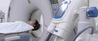
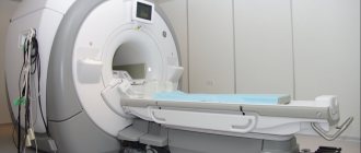
How is magnetic resonance imaging of blood vessels performed?
For MR angiography, open-type devices and tunnel devices are used. The latter are more powerful and provide high-resolution photographs. The magnetic field strength of tunnel devices is 1.5-3 Tesla.
Open tomographs are used to examine patients with a body weight of more than 130 kg and an abdominal circumference of 150 cm. A scanning system in the form of a frame allows the procedure to be carried out for patients suffering from claustrophobia.
Stages of MR angiography:
- The patient is positioned on a mobile table. The conveyor is equipped with fastenings and bolsters for fixing the body and limbs. Random movements during scanning lead to defects in the images.
- The doctor and x-ray technician move into the next room. Visual control of the procedure is carried out through a transparent partition; communication with the patient is maintained using an intercom.
- The table is rolled into the tomograph tube and a series of native photos are taken.
- A contrast agent is administered through an intravenous catheter.
- Scanning is continued after 10-15 minutes. During this time, the gadolinium solution fills the vascular bed in the area of interest.
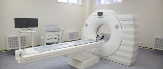
Examination of the vessels of the head on a closed tomograph
As a result of MR angiography, photographs of axial, sagittal and frontal sections of the scanned area are obtained. Three-dimensional reconstruction of the vascular network in a specific area is possible.
How long a vascular MRI will take depends on the area of study. The average session duration is 45 minutes.
What is more informative: MRI of head and neck vessels or computed tomography
CT angiography with the introduction of a contrast agent still remains a more informative procedure. This level of detail also has one significant drawback - the absorption of high load rays by the body. The latter can aggravate the development of cancer and completely excludes dynamic monitoring of certain pathological conditions.
The iodine-containing contrast agent administered during CT angiography is quite allergenic. Therefore, not everyone can conduct computer research. The examination itself is variable. At the time of assessing the clinical picture, the neurologist already understands which study will be more relevant for the patient: MRI of the vessels and arteries of the neck or CT angiography.
WHAT ARE ANGIOGRAPHY AND VENOGRAPHY?
MR angiography is essentially the same as MRI: angiography is a method of scanning blood vessels using an MRI scanner. This method reveals both functional and anatomical features of the structure of blood vessels.
Venography is considered a type of angiography and examines only the venous blood supply. The patient's attending physician usually tells you what type of examination is needed.
The main thing is not to be afraid of complex terms, because in essence there is nothing scary or unusual in these procedures!
Choosing a clinic, specialist and price of service
The mrt-mozga service is a full-fledged resource with a database of diagnostic clinics in Moscow (about 300 centers). The resource’s simple and intuitive interface will suggest the best offer. The patient can sort clinics according to various parameters: geolocation of the clinic, price, type of tomograph used, availability of contrast agent, operating hours, promotional offers. The information on the site is constantly updated and expanded.
You can make an appointment by calling 8 (495) 032-07-59 from 09:00 to 00:00. By taking advantage of the site's offer, you can not only make an appointment for free, but also get the best price offer.