Method for obtaining an x-ray image (options)
The invention relates to medicine, in particular to radiology, and can be used to diagnose diseases of internal organs. The method of obtaining an X-ray image consists of generating an X-ray radiation flux, passing it through a filter that passes predominantly the high-energy part of the radiation spectrum of the X-ray tube, installed in front of the volume under study, sending it to sensors - X-ray quantum recorders, reading information from them and obtaining an image using a computer. Additionally, the X-ray radiation flux is passed through a filter that transmits predominantly the low-energy part of the X-ray tube radiation spectrum, installed in front of the volume under study. In the second embodiment of the method, a scanning X-ray radiation flux is formed, the high-energy part of the radiation spectrum is directed to a line of sensors - X-ray quantum recorders, and additionally the X-ray radiation flux is passed through a filter that passes predominantly the low-energy part of the radiation spectrum of the X-ray tube, installed in front of the volume under study, and sent to an additional a line of sensors - X-ray quantum recorders, installed parallel to the existing line. The method makes it possible to improve the quality of diagnostics through the use of high- and low-energy X-ray radiation to obtain an image based on the values of attenuation of radiation by calcium and other substances. 1 c.p. f-ly.
The invention relates to medicine, in particular to radiology, and can be used to diagnose diseases of internal organs.
There is a known method for obtaining an X-ray image of a volume under study, in which an X-ray radiation flux is formed, it is passed through a filter that passes predominantly the high-energy part of the radiation spectrum of the X-ray tube, which is installed in front of the volume under study, and the radiation flux passed through the volume under study is directed to sensor-recorders of X-ray quanta. After reading information from the sensors using a computer, an X-ray image of the volume under study is obtained. In this case, the dimensions of the total surface of the sensors correspond to the dimensions of the investigated part of the volume. The disadvantage of this method is that the resulting image is summative, representing a set of projectionally superimposed images of the internal structures of the volume under study. This complicates image analysis and forces the use of tomography, a more expensive method with increased radiation exposure to the patient. In addition, the cost of full-scale panels with sufficient spatial resolution is high due to the complexity of their production technology. (N.N. Blinov. Equipment for radiation diagnostics in 2001. “Radiology - practice” No. 3. 2001). Simpler full-scale digital recording systems (for example, CCD matrices) produce lower-quality images.
There is a known method for obtaining an X-ray image of the volume under study, in which a scanning X-ray radiation flux is formed, it is passed through a filter that passes predominantly the high-energy part of the radiation spectrum of the X-ray tube, which is installed in front of the volume under study, and the radiation flow passing through the volume under study is directed to a line of X-ray sensor-recorders quanta Next, an image is obtained using a computer. The advantages of this method are that it requires fewer sensors and there is no scattered radiation that negatively affects image quality. But at the same time, the image of the studied volume is also summative, the examination time becomes longer, which contributes to distortion of the shape of highly mobile structures of the studied volume, for example, the heart.
The proposed methods for obtaining X-ray images, in contrast to the above prototype methods, make it possible to exclude the projection overlap of structures that differ in their calcium content.
The technique of digital radiography uses the method of so-called energy subtraction, which uses the difference in the attenuation properties of X-ray radiation by the structures of the human body and the iodine contained in X-ray contrast preparations. (X-ray technology, edited by V.V. Klyuev, M.: Mechanical Engineering, book 2 1992. p. 176.) This method is applicable only to improve the image quality of artificially contrasted structures of the studied volume. To carry it out, two images of the volume under study with the contrast agent contained in it are formed. When forming images, X-rays of different energies are used; for one, slightly lower, and for the other, slightly higher than the “K-jump” region - a violation of the uniformity of the dependence of the radiation attenuation coefficient by iodine on the radiation energy. Next, both initial images are processed using a computer, obtaining an image of iodine with an almost eliminated image of the volume under study.
Another research method is more widely used, which also uses x-rays of different energies - digital absorptiometry. (“Bulletin of Radiology and Radiology”, No. 2, 1993, p. 4.) The method, which is not related to either contrast research methods or methods of obtaining X-ray images, allows you to determine what part of the radiation is retained by calcium, fatty and soft tissues contained in the studied volume. Based on these values, it is possible (with sufficient spatial resolution) to obtain x-ray images suitable for x-ray diagnostics of diseases of internal organs. The proposed methods implement this possibility. In this case, the image constructed on the basis of the values of the attenuation of X-ray radiation by calcium corresponds to the image of bone and calcified structures without others, including projectionally layered ones. The image based on the attenuation values of other substances corresponds to the image of soft tissue structures, etc. The study of structures “in their pure form” without projection layers helps to improve the quality of diagnosis and allows in some cases to do without tomography, primarily when studying intrapulmonary structures, the main obstacle to which is bones, or more precisely, the calcium contained in them. On film and digital radiographs, the intensity of bone elements in elderly patients due to osteoporosis is often reduced so much that the anterior parts of the ribs become difficult to distinguish and do not interfere with the study of the soft tissue structures projectively layered on them.
The proposed method can be implemented both on X-ray diagnostic devices with a full-scale X-ray image receiver, and on scanning-type devices. In the first option, as well as in the implementation of the prototype method, an X-ray radiation flux is formed, passed through a filter that passes predominantly the high-energy part of the radiation spectrum of the X-ray tube, which is installed in front of the volume under study and the radiation flux passed through the volume under study is directed to sensor-recorders X-ray quanta, read information from sensors, and use a computer to obtain an X-ray image of the volume under study. In contrast to the prototype method, the X-ray radiation flux is additionally passed through a selective filter that passes predominantly the low-energy part of the X-ray tube radiation spectrum, which is installed in front of the volume under study and the radiation flux passing through the volume under study is directed to sensor-recorders of X-ray quanta. Thus, the proposed method makes it possible, through the use of X-ray radiation of different energies, to determine what part of the radiation is retained by calcium, fat and soft tissues contained in the volume under study, and based on these values to obtain the corresponding X-ray images.
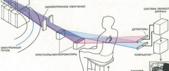
In the second option, as well as in the implementation of another prototype method, a scanning X-ray radiation flux is formed, passed through a filter that passes predominantly the high-energy part of the radiation spectrum of the X-ray tube, which is installed in front of the volume under study, and the radiation flux passing through the volume under study is directed to a line of sensors-recorders of X-ray quanta, read information from the sensors, and use a computer to obtain an X-ray image of the volume under study. In contrast to the prototype method, the X-ray radiation flux is additionally passed through a filter that passes predominantly the low-energy part of the radiation spectrum of the X-ray tube, which is installed in front of the volume under study, and the radiation flux passing through the volume under study is directed to an additional line of sensor-recorders of X-ray quanta, installed parallel to the existing line . Due to the fact that with this version of the method, the study is carried out using X-ray radiation of different energies, a computer is used to determine what part of the radiation is retained by calcium, fat and soft tissues contained in the volume under study, and the corresponding X-ray images are obtained.
The proposed methods make it possible to diagnose osteoporosis based on the determination of calcium content in bones, determine the calcium content in the aorta and large vessels - atherosclerosis, determine the calcium content in tuberculous foci - their potential activity. The ability to determine the content of adipose tissue makes it possible to diagnose lipomas.
1. A method for obtaining an x-ray image, in which an x-ray radiation flux is formed, passed through a filter that passes predominantly the high-energy part of the radiation spectrum of the x-ray tube, installed in front of the volume under study, sent to sensor-recorders of x-ray quanta, information is read from them and images are obtained using a computer. , characterized in that additionally the X-ray radiation flux is passed through a filter that transmits predominantly the low-energy part of the X-ray tube radiation spectrum, installed in front of the volume under study.
2. A method for obtaining an X-ray image, in which a scanning X-ray flux of radiation is formed, passed through a filter that passes predominantly the high-energy part of the radiation spectrum of the X-ray tube, installed in front of the volume under study, directed to a line of sensor-recorders of X-ray quanta, information is read from them and obtained through An image computer, characterized in that the additional X-ray radiation flux is passed through a filter that passes predominantly the low-energy part of the X-ray tube radiation spectrum, installed in front of the volume under study, and is directed to an additional line of sensor-recorders of X-ray quanta installed parallel to the existing line.
