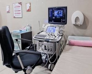
Ultrasound examination or ultrasound has long been firmly established in medical practice and is widely used to diagnose a wide variety of pathological changes, including in blood vessels. It is highly informative and absolutely safe. Therefore, the procedure can be carried out as many times as necessary without fear of harming the body, including children and pregnant women. One of the areas of ultrasound diagnostics of blood vessels is the study of blood vessels of the neck and head - brachiocephalic vessels.

Technical side of the process
The Doppler effect is a physical phenomenon. It lies in the fact that when the medium that reflects the ultrasonic waves or the ultrasound source moves, changes in the emitted ultrasonic waves occur. If we transfer this effect to the medical plane, then the moving medium being studied is the blood flow in various vessels. The change in ultrasonic waves directly depends on the speed of movement of blood particles.
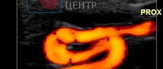
Doppler examination in its classical form does not provide an image on a screen. With its help, only the movement of blood flow and its direction are determined. Ultrasound can provide visualization. Modern ultrasound machines make it possible to perform two studies at once and see on the screen an image of a specific organ, the vessels inside it, and the blood flow inside these vessels.
Doppler ultrasound is also called Doppler ultrasound, Doppler ultrasound, or duplex scanning. The information read by the ultrasonic sensor is processed by a computer, and a two-dimensional color image is displayed on the screen, from which the speed and direction of blood flow and areas of its blockage can be determined.
FAQ
- Is it painful to have an ultrasound examination?
No, this is a non-invasive and painless diagnostic method.
- Can all prescribed treatment be completed with you?
Yes, we have all treatment methods and all equipment. Moreover, you can start treatment on the first day of contacting us.
- Will ultrasound be enough to make a diagnosis?
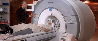
It all depends on the disease; for some diseases, only an ultrasound is sufficient, while others will require a more in-depth examination, such as magnetic resonance imaging or blood tests.
- How often can an ultrasound be done?
The ultrasound diagnostic method is absolutely harmless; if necessary, it can be done as many times as necessary.
- Is it possible to consult a specialist after an ultrasound?
Yes, you can get advice from a specialized specialist - a traumatologist, orthopedist, physiotherapist or sports doctor. You can also undergo treatment with us, and you can start on the same day you contact us.
What body functions does ultrasound study?
By performing an ultrasound scan of the vessels of the neck, one can diagnose pathologies of the carotid and vertebral arteries, and scanning the vessels of the head allows one to assess the condition of the carotid, subclavian, vertebral arteries and the main arteries of the brain. Such examinations are usually carried out simultaneously. This allows you to obtain the most complete and reliable information about the state of the vessels supplying the brain. There are several parameters by which the condition of the vessels of the neck and head, as well as the blood flow in them, is assessed.
How elastic are vascular walls?
A decrease in the elasticity of vascular walls can lead to strokes, heart attacks, and varicose veins. The reasons for decreased elasticity are: poor diet, high blood cholesterol, bad habits, age factor. Timely detection of the fact of decreased elasticity of the walls of blood vessels will allow the doctor to prescribe therapy in a timely manner and prevent serious problems with the patient’s health.
What condition is the inner surface of the vessel in?
The inner surface of the vessel is in direct contact with the blood and releases special substances that prevent the clotting process. In other words, the main function of the inner surface is to prevent the formation of blood clots. Violation of the structure of the inner layer of the vessel and its integrity can lead to thrombus formation.
The presence or absence of changes in the integrity of the vessel walls
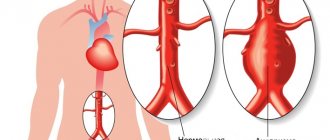
It is logical that a violation of the integrity of the walls of blood vessels leads to a disruption of blood flow, a change in its movement, and therefore a disruption of the blood supply to the brain. The doctor’s task when conducting an examination using ultrasound dopplerography ( ultrasound Dopplerography of the vessels of the neck and head
) identify ruptures in the vascular walls, assess the complexity of the situation.
Intraluminal formations of arteries or veins
Formations in the lumens of veins or arteries are blood clots that impede blood flow. Accordingly, the blood supply to the brain becomes more difficult, which can result from a stroke, cerebral infarction and other pathological conditions.
Ultrasound of the carotid arteries
Ultrasound of the carotid arteries reliably shows the condition of the blood vessels and is one of the safest and most painless technologies. Ultrasound waves do not harm the body, and the non-invasiveness of the method negates the risk of vessel damage. Therefore, this type of diagnosis of diseases and pathologies of the carotid arteries is the most common. There are practically no obstacles in the path of ultrasonic waves when examining the carotid arteries using ultrasound, which makes it possible to obtain reliable readings about the condition of the vessel. A pleasant advantage of this research method is its low cost.
What does the study clarify?
After ultrasound with Doppler, the specialist is able to determine a number of factors.
Vessel patency
It is important to assess how freely blood flows through the vessel, whether there are any blood clots or blood clots in the lumens of the veins and arteries.
To what extent does the course of the vessel correspond to the normal trajectory?

The existing direction of blood flow during scanning is compared with the norm along the trajectory of the anatomical course of the vessel. An increase in the tortuosity of blood vessels leads to disruption and slowing of blood flow.
Diameter and location of the vessel lumen
Vasomotor function is a change in the diameter of the lumen of a vessel under the influence of certain factors. This function is necessary to regulate blood pressure in blood vessels, heat exchange, and metabolism. Pathological narrowing of the lumen of the vessel leads to increased pressure in the spasmodic area, and local blood supply is disrupted.
Length of visibility of the changed lumen
When identifying pathological changes in the lumen of the vessels of the neck and head, it is important to identify the area of localization of the pathology, as well as the length of visibility of the changed lumen. This will allow you to accurately diagnose and prescribe treatment.
The main parameters that are examined during ultrasound of the BCA
- Detailing the thickness and structure of the vascular wall, its uniformity;
- Patency of blood vessels, as well as assessment of the lumen of the vessel (narrowing or dilation);
- The nature and symmetry of the speed characteristics of blood flow along the BCA.
- Turbulence (turbulence in the blood flow relative to the longitudinal axis of the vessel stack) of the blood flow is also assessed;
- The presence of signs of atherosclerosis and the degree of its severity in%, an assumption is made about the significance of the identified changes for hemodynamics (blood circulation);
- The presence of anatomical features of the BCA (bends, deformations, hypoplasia and small diameter of the vertebral artery, taking into account the speed characteristics of the blood flow, high entry of the vertebral artery into the canal of the transverse processes of the vertebrae).
- By the bends of the vertebral artery in the canal of the transverse processes of the vertebrae, one can indirectly judge whether the patient has vertebral instability and disc herniation at the specified level.
Indications for use of the procedure
Doppler ultrasound is prescribed to patients for a number of indications.
Causeless migraine and dizziness
If a patient suffers from migraines quite often, but does not associate it with fatigue, stress, or physical exertion, he should undergo an ultrasound scan.
to identify the true causes of pathology. The same can be said about causeless dizziness. Perhaps this is caused by impaired cerebral circulation.
Noise in the ears and head
Noise in the ears and head can be a symptom of atherosclerosis, vegetative-vascular dystonia, hypertension and other diseases. Doppler ultrasound will allow you to accurately make a diagnosis.
Poor health, which is accompanied by attacks of weakness and a feeling of shortness of breath
The feeling of weakness and lack of air cannot always be associated with poor physical fitness of the patient or problems with the breathing apparatus. Perhaps the reason lies in impaired blood supply to the brain, anemia or other pathologies that ultrasound scanning will help identify.
VSD (vegetative-vascular dysfunction)
VSD is manifested by paroxysmal or constant palpitations, increased sweating, headache, tingling in the heart area, redness or paleness of the face, chilliness, and fainting. This pathology is not considered as an independent disease - it always accompanies some organic pathology, which may be associated with impaired blood supply to the brain.
Hypertension
Hypertension, or a sustained increase in blood pressure, can cause many diseases, including those associated with poor circulation in the vessels of the head and neck. Doppler ultrasound of the vessels of the head and neck is the best way to confirm or refute the presence of just such pathologies.
Ultrasound ultrasound: progress of the procedure
An ultrasound scan of the vessels of the head and neck lasts, as practice shows, approximately 45-50 minutes.
The patient lies on his back and throws his head back. To facilitate the movement of the sensor, the doctor applies a special gel to the skin. During the examination, the sensor is constantly moved to assess blood flow in different parts of the head and neck. During the procedure , ultrasound examination of the vessels of the head and neck
It may be necessary to perform some functional tests. To do this, the doctor asks the patient to breathe deeply, and he can press the vessels with his fingers or a transducer.
Operating principle of ultrasonic equipment
Ultrasound is sound waves whose frequency exceeds 20,000 hertz. Human hearing organs are not able to catch this sound, but whales, dolphins, bats and other representatives of the animal world communicate freely on it. Ultrasound waves propagate perfectly in the soft tissues of the human body without disrupting their functionality or provoking pathogenic changes.
The human body is heterogeneous. It contains air, water, hard and soft tissue. All of these structures impede the passage of sound waves in different ways, which scientists call acoustic impedance. This indicator depends on the density and elasticity of the medium, which prevents the propagation of sound. This is exactly the principle that an ultrasound machine works on. It performs several tasks at once - it creates ultrasound, records its propagation, acoustic resistance of different parts of the body and converts it into a picture. A three-dimensional image of the scanned area is displayed on the computer screen, and the doctor, using a special sensor, can “move” around the patient’s body. Ultrasound comes precisely from the sensor, which is moved over the surface of the skin, so the picture largely depends on its power and size.
Human skin reflects about 100% of sound vibrations. For the free passage of ultrasound it is necessary to use a transition medium. Liquid gel plays this role. It allows the sensor to slide freely over any surface and provides a high-quality signal due to its specific viscosity.
The information recorded by the ultrasound device sensor enters the reconstruction system. There it is processed and turned into a three-dimensional image in black and white. An interesting fact is that 64 different shades of a black and white scale are used to create an image. The color changes depending on the intensity of the sound wave, the degree of its reflection and the density of the area of the body from which the sound was reflected. The most intense waves are displayed in white, and the minimum ones are recorded in black.
If it is necessary to assess the condition of the vessels, echo contrast is performed. This is an ultrasound examination with intravenous administration of a contrast agent. The information content of the procedure may well compete with computed tomography. But ultrasound examination has an advantage - complete safety and comfort of the patient. Contrast improves visualization of organs and blood flow, details the final image and increases diagnostic accuracy.
Decoding the results
In order to understand what an ultrasound scan of the vessels of the head and neck shows, and to decipher the results of an ultrasound and Doppler examination, the doctor must compare the following actual blood flow indicators with the norm:
Features of blood flow in the vessels of the subject;
During the examination, it is necessary to assess the direction of blood flow, identify areas of blood penetration through vessels into adjacent cavities, and find out whether there is pathological tortuosity of the vascular system.
Systole size (highest speed of blood movement);
To assess the quality of blood flow, its maximum speed must be determined. In this case, the width of the channel and the diameter of the vessel are taken into account.
Minimum blood flow velocity (diastolic);
The main reason for the decrease in the level of blood supply to organs is the drop in blood flow speed. During ultrasound, it is determined in which area of the vascular system blood moves at the slowest speed and what reasons contribute to this.