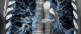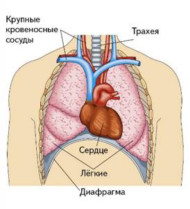
Organs of the chest schematically Magnetic resonance imaging allows you to study the structure of the internal organs. The method provides comprehensive information about the condition of soft tissues and is less indicative of bones. MRI diagnostics are prescribed to detect diseases of the chest organs. Scanning in most cases is not the method of choice for examining the lungs, but it allows for good visualization of the trachea, bronchi, pleura, heart, thymus, large blood and lymphatic vessels. What a chest MRI shows often determines the final diagnosis. The method is used as an alternative and addition to CT, radiography or ultrasound examination of the heart.
Do they do chest MRI?
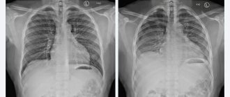
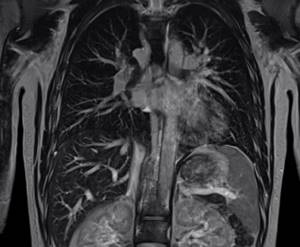
The low information content of MRI scans regarding the lungs is due to the large amount of air contained in their structure. Due to the low concentration of hydrogen and constant movement, the pictures are blurry. However, magnetic resonance imaging can detect tumors in the respiratory system, foci of inflammation, fibrous degeneration or necrosis. With its help, the mediastinal organs are examined.
Most often, an MRI scan is recommended as a follow-up examination when any abnormalities are detected by other methods (in case of unclear darkening on an X-ray of the lungs, etc.). Indications for chest MRI are:
- suspicions of heart pathologies (congenital and acquired valve defects, structural anomalies of the muscular part of the organ, diagnosis of post-infarction complications, etc.);
- hemodynamic disturbances in large veins and arteries;
- control of blood circulation after operations on blood vessels, including the heart;
- questionable results of X-ray and/or CT scan of the lungs (detailed study of the pleura, suspicion of abscesses, tumors, etc.);
- diagnostics of the state of the lymphatic system;
- suspicion of tumors of the chest organs and control of the spread of metastases to this area;
- clarification of the nature of inflammatory changes in the studied area;
- suspicion of fluid accumulation in the pleural cavity;
- the need to examine the organs of the chest cavity if CT is contraindicated, etc.
MRI scan of the chest organs
The procedure cannot be performed if you have a pacemaker or defibrillator, or an insulin pump. The presence of any metal implants in the body is considered a contraindication. If there are any, all patients at the Magnit diagnostic clinic must provide a document from the medical institution where the operation was performed, with a detailed description of the material. Some people are wondering: “Do they do a chest MRI after stenting?” This issue is resolved on an individual basis. Typically, diagnosis can be carried out six months after stent placement. The following are recognized as relative contraindications for MRI:
- first trimester of pregnancy;
- fear of closed spaces (claustrophobia);
- the patient’s age is under 5 years due to the child’s inability to remain stationary for a long time;
- the patient's weight is more than 120 kg;
- mental illness;
- neurological impairment or acute pain.
Women who are pregnant in the first trimester should consult a gynecologist and, if possible, reschedule the examination to a later date. For claustrophobia and excess body weight, diagnostics are carried out using open-type devices. For children and patients with acute pain, the examination is done under sedation or anesthesia in a hospital setting.
Advantages of MRI in the hospital
- The duration of the procedure is about 15 - 20 minutes;
- MRI with the introduction of a contrast agent allows you to most accurately assess the condition of blood vessels;
- in our hospital the procedure is performed using modern Ingenia 1.5T equipment (Philips), which produces high-quality images;
- maximum possible tunnel diameter (73 cm) - patient’s waist circumference (circumference) up to 225 cm
- a full range of studies due to the maximum configuration;
- additional information through advanced software;
- there is an external environment control system (music from the patient’s playlist, choice of lighting, personal climate control);
- automated system for quality control of studies and descriptions;
- IT platform with triple control of research results - with the support of professors, leading specialists from Russia, Europe and Israel (“second opinion”);
What is included in a chest MRI?
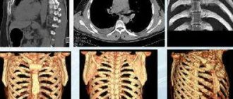
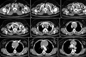
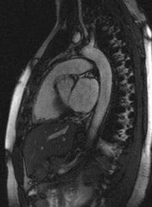
The result of MR scanning is layer-by-layer images (slices) of the area under study in three mutually perpendicular planes. The presence of special software helps to construct three-dimensional images based on them. MRI of the chest and mediastinum allows you to study:
- pleura and pleural cavity;
- lungs;
- lower part of the trachea;
- thymus gland (thymus);
- heart and pericardium;
- large lymphatic vessels;
- ribs;
- thoracic spine and spinal cord at this level;
- diaphragm.
Contrast scanning provides information about the speed of blood flow, the condition of the vascular walls, and the patency of arteries and veins. The method is indispensable for diagnosis, monitoring the development and effectiveness of treatment of tumor diseases.
MRI of the mediastinum and lungs
Indications
The costosternal joints connect the ribs to the sternum. Their ligaments go to the anterior and posterior surfaces of the sternum and connect to the opposite and neighboring ligaments. The cause of pain in the chest is often costosternal syndrome (found under the names costochondritis, cartilage dystrophy, arthrosis), “sliding” rib syndrome, Tietze syndrome.
Costochondritis is an inflammation of the cartilage tissue at the junction of the ribs and sternum. Muscular, osteochondral structures are involved in the pathological process, and its main symptoms are pain in the sternum, which intensifies with coughing, movements, deep breaths, and, less commonly, swelling. Added to these are psychopathological factors - increased anxiety with disruption of normal lung function, depression. Symptoms of costochondritis are similar to angina pectoris, ischemic heart disease and other cardiovascular diseases.
To differentiate diseases, to understand what exactly causes pain and what treatment method to prescribe, an examination is carried out. Therefore, if alarming symptoms appear, you cannot postpone a visit to the doctor.
Preparing for a chest MRI
Native magnetic resonance imaging does not require specific preparation. If contrast is required, you should have a light snack 45 minutes before the scan.
At the Magnit diagnostic clinic, the procedure is carried out by appointment. The patient should arrive 5-10 minutes earlier than the appointed time in order to fill out the documents without haste.
Preparation involves consultation with a radiologist. The doctor asks questions regarding the purpose of the study and whether the patient has any contraindications. If none are identified, the person is taken to the locker room. Here he must remove jewelry (hairpins, glasses, rings, earrings, chains, piercings), clothes with metal inserts or fittings, and leave electronic devices (phone, watch, player, etc.).
Anxious patients may experience fear or discomfort during the test or feel unwell after the procedure is completed. To avoid unpleasant phenomena, they practice the use of sedatives before the study, but only on the recommendation and prescription of a doctor. The patient should inform the radiologist about anxiety in advance.
How is the examination carried out?
MRI of the costosternal joints does not require preparation in the form of diet or other measures. It is enough to make sure there are no contraindications and get a doctor’s referral. Before the procedure, you will need to remove clothing to the waist, jewelry, and accessories with metal components.
The results of the study are analyzed together with data obtained using other methods: radiography of the sternum, ultrasound of the mediastinum, examination of the clavicular joint.
If you have any questions, need advice, or want to choose a time convenient for your visit, fill out an application on the website or call the clinic administrator on duty yourself.
How is a chest MRI done?
After preparation, the patient is taken to the diagnostic room. The following is done here:
- a person lies on the tomograph platform. The conveyor moves so that the study area is in the center of the apparatus frame;
- The x-ray technician records the patient’s position, places cushions if necessary, offers to use headphones, explains the rules of behavior during the procedure, and gives a button for emergency communication;
- The radiologist leaves the office and checks that the speakerphone is working properly. Reminds the patient not to move and turns on the tomograph. The study lasts 20-30 minutes. The introduction of contrast prolongs the procedure by 10-15 minutes;
- At the end of the study, the patient is helped to stand up. The man goes to the locker room and takes his personal belongings.
You can wait for the results in the rest room. Their preparation lasts 15-60 minutes. The patient receives a report on paper and a digital medium with images. During a personal consultation, the radiologist gives explanations to the patient. If you have left your email address in advance, the research results will be sent there as well.
MRI of the chest
How is it carried out?
MRI does not require special preparation. Before starting the study, the patient must remove all metal objects.
The research algorithm is as follows:
- the patient lies down on a special table, which is part of the tomograph, the genitals are protected with special pads;
- during the procedure, the device makes characteristic clicking sounds, which is absolutely normal; if these sounds bother the patient, you can wear special headphones;
- You cannot move during the procedure; you can take good pictures only in conditions of complete immobility;
- it is necessary to strictly follow all commands of the specialist who conducts the research.
If your general condition worsens, dizziness, breathing problems, or nausea occur, you should immediately inform your doctor.
Indications for diagnosis
The reason for the research is:

- Pain syndrome radiating to the sternum, under the shoulder blade, shoulder, kidneys, abdominal region, ribs; Radiculopathy;
- Compressive myelopathy; Paraparesis of a spastic nature; Loss of sensitivity;
- Functional disorders of visceral organs: bronchopulmonary system, trachea, duodenum;
- Paresthesia; Thoracalgia; Intestinal motility disorder; Angina pectoris of unknown origin; Intercostal neuralgia;
- Difficulty breathing; Local swelling; Hyperemia; Restriction of natural mobility; Deformations, asymmetry of the musculoskeletal system, violation of statics;
- Muscle spasms in the spinal region; Hump, pathological kyphosis and lordosis.
Scanning is required during the period of preoperative preparation and after radical treatment to assess its quality and the degree of recovery of the body. It is indicated to prevent complications. Allows timely adjustment of therapy.
Contraindications
There are some contraindications for MRI that limit the use of this method for certain patients. This is the presence in the body of sewn-in:
- metal orthopedic prostheses;
- metal heart valves;
- neurostimulants;
- insulin pumps;
- pacemakers;
- intrauterine devices;
- metal dentures;
- metal fragments in the body;
- ear implants.
There are also conditional restrictions for patients with claustrophobia, pregnant women and children under 7 years old.
How much does the examination cost?
The price varies from 2200 to 11000 rubles. It depends on the policy of the medical center, the tomograph model, and the need for additional manipulations.
The easiest way to see how much an examination at a selected institution will cost is in the price list on the main page. There are two amounts indicated there: the first is native, that is, standard tomography, the second is a study with paramagnetic preparations.
Here you can find information about benefits and current promotions. And by booking a ticket through the service, our visitors will receive a discount of up to 1000 rubles.
What other research may be needed?
In addition to imaging tests, laboratory tests and lumbar puncture are often required. It involves taking cerebrospinal fluid from the subarachnoid space for its subsequent study. It allows you to identify neuroinfections and cancer.
Myelography shows the condition of the subarachnoid space of the spinal cord and its roots. Discography helps to study the integrity of intervertebral discs. Requires administration of contrast. For a comprehensive study of the neuromuscular system of the back, electroneuromyography and EMG are prescribed.
Which parts of the thoracic spine are visualized?
The technique shows:
- Injuries and damage to bone elements;
- Position of fragments;
- Severity of lordosis;
- The presence of pathological bends;
- The relative position of the vertebral bodies, their height;
- Protrusion of the fibrous ring;
- Diameter of the spinal canal;
- Circulation of cerebrovascular cerebrospinal fluid;
- Compression of surrounding structures;
- Condition of the facet joints;
- Swelling;
- Rupture of muscle fibers, fascia, ligaments;
- Pinched roots;
- Areas of necrosis, ischemia;
- Localization, stage and size of space-occupying formations.
Advantages of this technology
She happens to be:
- Non-radiological, that is, it does not exert radiation ionizing load on the body. Therefore, unlike CT, it does not require a recovery period and can be performed as often as necessary.
- Non-invasive - does not require damage to the skin and mucous membranes. This technique does not cause discomfort and eliminates iatrogenic injuries.
- Painless.
- Highly informative;
- Available for use in pregnant women and young children.