Ultrasound scanning of the carotid arteries is a safe and informative method for diagnosing many diseases of these vessels. In patients over 55 years of age, this study should be performed annually to identify risk factors for ischemic stroke.
Ultrasound of the main arteries of the head is a non-invasive imaging method that, by recording reflected ultrasound waves, allows one to obtain information about individual anatomical features, the extent and degree of damage to the arteries, as well as assess the influence of these factors on blood flow. The examination is absolutely painless and does not cause significant discomfort to the patient. The high diagnostic value of the study is due to the fact that the specialist gets a real-time understanding of the anatomical formations and the nature of blood flow in the carotid arteries.
Ultrasound examination of the carotid and vertebral arteries (MAG ultrasound) is performed to identify risk factors for ischemic stroke. Today it has already been proven that atherosclerotic narrowing of the internal carotid arteries significantly increases the risk of stroke due to thrombosis of the carotid artery or embolism - transfer of a piece of plaque to the cerebral vessels. This statement is also true for the vertebral arteries.
Great vessels supplying the brain with blood
In order to understand the principle of ultrasound scanning, let’s consider what great vessels are – the main subject of research.
The brain is fed by two vertebral and two carotid arteries; they are considered paired and form a circle at the base of the brain. These are the main blood supplying arteries. And if pathologies of these vessels occur, the brain tissue will be subject to significant changes, which will necessarily affect the entire body. With severe atherosclerosis and lipid metabolism disorders, there is also a risk of severe conditions due to insufficient blood supply to brain tissue.
So, the main vessels to study are the large arteries of the head and neck. In turn, the main vessels should be divided into areas of their location:
- large arteries located when entering the skull - extracranial area or section;
- vessels located directly in the area of the skull and its bone canals.
Indications
Our clinic offers a service - duplex scanning in Moscow. In what cases can this study be prescribed:
- tinnitus, clearly manifested when bending over or exercising;
- frequent fainting, epileptic seizures not related to epilepsy;
- loss of coordination and balance, which leads to severe dizziness and even falling;
- pain in the head area;
- vision problems;
- tremor;
- increased intracranial pressure.
These are the main reasons why duplex scanning of blood vessels may be performed.
What is Doppler ultrasound of blood vessels?
Any changes affecting at least one artery entail disruption of normal blood circulation. The most accessible and accurate method in this situation is considered to be the method of vascular Doppler ultrasound.
In simple terms, MAG ultrasound is a duplex ultrasound examination of the largest arteries of the head, features of blood flow and its speed. Overall, the procedure is a combination of the familiar ultrasound and Doppler effect.
The principle of operation of the device is quite simple from a physical and mathematical point of view. During the procedure, changes in the frequency of the signal reflected from bodies in motion are recorded. The equipment is a kind of device with location characteristics, which, when scanning, captures any slightest changes and, thus, reveals the violations for which they are characteristic.
The main popular and extremely effective methods of ultrasound diagnostics of the vessels of the neck and head today are Doppler ultrasound (USD), duplex scanning, color scanning, also called triplex mapping.
Typically, ultrasound mag and ultrasound mag procedures are prescribed for the following symptoms and diseases:
- hypertension, high blood pressure;
- acute cerebrovascular accidents and brain necrosis with severe paresis and loss of sensitivity;
- suffered acute coronary circulatory disorders, stroke;
- narrowing of the cerebral lumens of the cerebral arteries, atherosclerosis of cerebral vessels;
- arteriovenous malformation with the manifestation of hemorrhages;
- stroke with temporary neurological deficit;
- visual impairment;
- osteochondrosis of the spinal column;
- dizziness of unknown origin;
- preparation for surgery with a diagnosis of ischemia;
- dysfunction of the vestibular apparatus;
- vegetative-vascular dystonia;
- traumatic brain injuries, bruises of the head, cervical spine, soft tissues of the skull, concussion;
- encephalopathy;
- surgical interventions in the neck area;
- hereditary defects of the spinal column and cervical region;
- parallel chronic diseases that aggravate cerebral vascular diseases, obesity, smoking addiction, diabetes mellitus.
Are your legs bothering you? Check your blood vessels! Who can benefit from ultrasound scanning of lower extremity vessels?
Spider veins, swelling, a feeling of heaviness in the legs - these signs can be, in particular, a manifestation of vascular diseases of the lower extremities. Ruslan Akhyaevich Bidzhiev, ultrasound diagnostics doctor at Clinic Expert Stavropol, talks about in which cases ultrasound scanning of the vessels of the legs should be performed and how this study is performed.
— Ruslan Akhyaevich, Ultrasound scanning of the vessels of the lower extremities - what kind of study is this?
- This is a modern, highly informative method for identifying pathological changes in the vessels of the legs - arteries and veins. The abbreviation “UZDS” stands for “ultrasonic duplex scanning”.
I would like to note that until recently, Doppler ultrasound (USDG) was considered one of the widely used diagnostic procedures for assessing the condition of the vessels of the extremities. However, with its help it was possible to evaluate only one indicator - the patency of the vessel.
Unlike ultrasound doppler scanning, duplex ultrasound scanning of leg vessels allows you to visualize in detail the condition of the vascular wall of both large arteries and veins and small ones, identify the presence of blood clots and atherosclerotic formations, and determine the speed, direction and intensity of blood flow in the vessels. That is, we have the opportunity to obtain more detailed information.
— For what symptoms is ultrasound scanning of the vessels of the lower extremities performed?
— The main complaints of patients for which ultrasound scanning may be prescribed are pain and swelling of the legs. Redness of the skin, spider veins, various compactions (especially in the popliteal region), nodules in the projection of the vessels of the legs can also serve as a reason for prescribing this study.
Typically, ultrasound duplex scanning of the arteries and veins of the lower extremities is prescribed to the patient by the attending physician - a vascular surgeon, cardiologist, neurologist, therapist, traumatologist.
— In the diagnosis of what diseases can ultrasound of the arteries and veins of the lower extremities help?
— Vascular surgeons and phlebologists prescribe it to detect varicose veins, phlebitis (inflammation of a vein), superficial and deep thrombosis (phlebothrombosis and thrombophlebitis).
After operations on the lower extremities, ultrasound scanning is used to monitor the consistency and quality of the performed manipulations. Traumatologists prescribe duplex scanning to prevent thrombosis of the vessels of the lower extremities, associated either with compression of the vessels by a plaster cast or with a blood clotting disorder.
Ultrasound scanning will also be informative in case of some heart pathologies, after chemotherapy, and will also help to distinguish some infectious diseases from vascular pathology (in particular, erysipelas from thrombosis). In addition, this research method makes it possible to detect traumatic injuries and anomalies in the structure of blood vessels, intervascular malformations (connections).
— Do I need any preparation for ultrasound duplex scanning of the vessels of the lower extremities?
- No.
— How is ultrasound scanning of the vessels of the lower extremities performed?
— Traditionally, this study is performed in various projections. Despite the fact that, according to generally accepted recommendations, the patient is examined in a standing position during ultrasound scanning, practice shows that sometimes this is not enough, so our clinic provides a more expanded and thorough approach to this issue.
I would like to emphasize that we carry out diagnostics step by step, relying in our work on the laws of physics. First, the patient is placed on the couch. After normalizing the blood pressure, we perform an ultrasound scan of the femoral vessels, then the patient turns over on his side and the back of the thigh and lower leg are examined.
The next stage is to examine the patient’s limbs in a standing position, that is, under load. If necessary, we perform the examination in a sitting position.
— Does it happen that to clarify a patient’s diagnosis, ultrasound duplex scanning of the vessels of the legs is not enough, and some other research methods are required? And if so, which ones?
— Today, ultrasound scanning is one of the most informative diagnostic methods, which, by the way, is considered the gold standard for studying the blood vessels of the legs. In my opinion, the best method for detecting and assessing pathologies of the veins of the extremities has not yet been invented. To confirm this, I would like to add that, based on our conclusions, traumatologists, vascular surgeons, and cardiologists prescribe treatment and successfully perform operations.
Traditional angiography or CT angiography may also be prescribed to assess the condition of the arteries.
If necessary, the attending physician may additionally recommend that the patient undergo laboratory blood tests. Among them, in particular, assessment of blood clotting indicators (including prothrombin index) and a number of other studies.
You can sign up for ultrasound duplex scanning (USDS) of blood vessels here. ATTENTION: the service is not available in all cities
Interviewed by Sevilya Ibraimova
The editors recommend:
Doppler, duplex, triplex... What types of vascular ultrasound are there? Ultrasound of neck vessels: when is it prescribed? When is ultrasound of cerebral vessels prescribed? When is CT angiography needed?
For reference:
Bidzhiev Ruslan Akhyaevich
Graduate of the Faculty of Medicine of Stavropol State Medical University in 2015.
In 2021, he completed an internship in the specialty “Therapy with a course in dietetics.”
He completed training and advanced training courses in ultrasound diagnostics at the FMBA research center (Moscow).
Currently holds the position of ultrasound diagnostics doctor at the Expert Clinic, Stavropol. Receives at the address: Dovatortsev St., 39A.
Main goals of duplex ultrasound
The ultrasound scanning method is aimed at obtaining the necessary information to identify the extent of damage to the main main arteries of the head. Hypodynamic changes recorded by MAG ultrasound, which occur during low activity and mobility of a person, usually relate to cholesterol deposits. As a result, deformation of the arterial walls occurs in the form of their narrowing. This process can continue until the lumen is completely blocked by cholesterol, which will inevitably lead to impaired blood supply and coronary heart disease. This is a very dangerous condition that threatens heart failure and myocardial infarction.
Thus, through duplex Doppler mode, highly accurate information about the stage of the lesion can be obtained.
Combining the study of the condition of the arteries and the assessment of the speed and direction of blood flow, the ultrasound method makes it possible to diagnose not only the very fact of the presence of pathologies, but also the history of their origin, that is, the cause.
The main purpose of cerebral Doppler is to identify pathologies of the main functions of the great vessels of the brain and neck caused by various reasons. This can be thrombosis and embolism, growth of the vessel wall, compression of the lumen between the arteries and disruption of their patency.
The main indicators revealed during the MAG ultrasound procedure
Scanning helps identify:
- quantitative and qualitative characteristics of cholesterol deposits and blood clots;
- the degree of damage to the arteries, deformation of their shape;
- the quality and degree of thickening of the walls of blood vessels and their loss of plasticity;
- parameters of imbalance between vasoconstriction and vasodilation of the endothelium (motor function of arteries);
- damage and integrity of vascular walls;
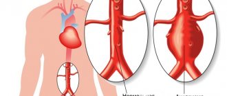
- the presence of aneurysms and their parameters;
- the degree of disturbance and the size of the gaps in the walls of the arteries;
- any conditions that cause brain hypoxia due to circulatory disorders.
This procedure can also be prescribed to monitor the quality of medical therapy for disorders of the blood supply to the brain.
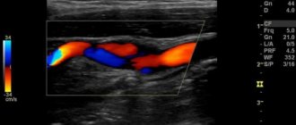
The most complete picture of the extent of pathologies can be given by color (triplex) scanning. In this case, with changes in blood flow and its speed characteristics, the colors of the image also change. Thus, color Doppler sonography provides the optimal number of changes and a better result regarding the state of blood circulation.
How to prepare for the MAG ultrasound procedure
Patients who are prescribed an ultrasound diagnostic method should already 24 hours before the procedure avoid such dishes and products as natural coffee, energy drinks, tea, alcoholic drinks, and pickles. It is better, in general, not to eat anything salty, as this can affect the overall tone of the vascular system and give incorrect diagnostic indicators.
In addition, you should avoid smoking and swimming in hot water 2-3 hours before the examination.
Before the study, it is necessary to stop taking certain medications, but, as a rule, the doctor warns about this at the previous consultation.
If these simple rules are followed, the probing procedure will be the most accurate and will provide the maximum information.
How is Dopplerography of the vessels of a child’s neck and examination of the vessels of the head performed?
To get tested, contact Human Health. The medical center is located in Moscow, in the North-Eastern Administrative District. You can drive up to it by car or take the metro. You need to get off at Otradnoe, Bibirevo or Vladykino stations.
During the examination, the doctor:
- Ask the child to lie down on the couch.
- He will apply a special conductive gel.
- Attach sensors to the skin and examine the necessary vessels.
The doctor will then fill out the form and give it to you. The attending physician deciphers the examination results.
If your child is taking any medications, tell the doctor about it. They may affect blood pressure. In this case, you will have to interrupt the course of treatment for several days.
How does an ultrasound scan work?
The examination procedure itself takes from several minutes to one hour. The patient needs to undress to the waist and take a horizontal position. The doctor uses a special acoustic gel for the procedure. This is done to make it easier to glide over the skin, as well as for greater sound conductivity.
During the scan, in order to scan all the blood vessels of the head and neck as much as possible, the patient must roll over onto his side and stomach as directed by the specialist. In this way, external and internal large main vessels are scanned - vertebral and carotid arteries, veins of the spinal column and jugular vessels.
Sometimes the doctor asks the patient to change his position to a vertical one, that is, to sit up, hold his breath, or take some medicine. Such tests are needed to identify a more complete picture of disorders and are used, most often, when there are certain hereditary pathologies of blood vessels and their structure, for example, the absence of the internal carotid artery.
In practice, after the end of the probing, a qualified doctor can make a diagnosis and write a medical report based on the results of the examination.
The document will indicate the presence and description of the anomalies found, the direction and speed of blood movement, the condition of the vessels and their walls.
Since during normal blood circulation the vessels are not narrowed, there are no deposits or blood clots in them, the blood movement is not subject to turbulence, and the examination result is compared with the normal characteristics of the vessels, the doctor can immediately see all pathologies and disorders.
This study is considered safe and does not harm patients, so this diagnosis can be used by people of any age category. In some cases, ultrasound probing is even used for newborns and infants who need an accurate diagnosis.
Advantages
For duplex scanning of head vessels, the price in our clinic is one of the most reasonable in Moscow. The study is complex and requires expensive, high-precision ultrasound equipment and qualified doctors. We have both. When using DSMAG you can get the following advantages:
- Maximum level of information content.
- No pain during the procedure.
- No side effects.
- There are no contraindications.
- It is possible to repeat the session and you do not have to wait a certain amount of time.
- No special preparation required.
- The duration of the event is no more than half an hour.
Duplex scanning of the vessels of the head is performed in the same way as any other ultrasound.
Ultrasound diagnostics in children
The Doppler ultrasound method is widely used in childhood diseases, especially in the field of vascular diseases of the nervous system. In addition, disruption of normal blood circulation often occurs in newborns with postpartum hypoxia.
Ultra-precise diagnostics helps to detect pathologies at the earliest stages, and avoid the development of serious conditions when a burst vessel can cause disaster.
It is with the help of ultrasound that it is possible to recognize developmental defects and the subsequent occurrence of problems with the blood supply to the body based on symptoms such as headache in a child, decreased memory, attention and learning ability, chronic hypoxia due to congenital pathologies, and vegetative-vascular dystonia.
When diagnosing childhood diseases, a painless and safe ultrasound scanning procedure is especially relevant.