The gallbladder is classified as an auxiliary organ of digestion. Its main task is the accumulation of bile, which is produced by the liver. Our body needs bile to digest animal fats. Therefore, disturbances in the functioning of the bladder are accompanied by digestive disorders and pain in the right hypochondrium. In this case, the patient undergoes an ultrasound of all abdominal organs, during which the gallbladder is examined.
- What it is?
- Who is prescribed an ultrasound of the gallbladder?
- How should you prepare for an ultrasound?
- How is the procedure done?
- What are normal indicators for the gallbladder?
- What pathologies of the gallbladder can be detected on ultrasound?
What is an ultrasound examination of the gallbladder?
The gallbladder is located just below the liver, approximately at the level of the last rib, on the right side of the abdominal cavity. In normal condition, this organ cannot be palpated. Instrumental and laboratory methods (ultrasound, radiography, computed tomography) are used to diagnose it. The simplest and most accessible way to examine an organ is ultrasound.
Ultrasound, or echography, is a fast, informative and safe diagnostic method for the patient’s health. It is based on the ability of ultrasonic waves to penetrate the structures of our body and be reflected from them. The denser the tissue or organ is in the path of ultrasound, the more the characteristics of the reflected wave change. The ultrasound machine detects these changes, processes them and displays an image of the organ on the monitor.
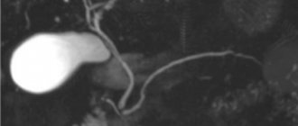
Echography of the gallbladder allows you to determine the shape and location of the organ, its size, examine the ducts and identify stones and tumors. For ultrasound examination of the gallbladder, the transabdominal method is used, that is, scanning through the anterior wall of the abdomen. The procedure is carried out using ultrasound sensors with a frequency of 2.5-3.5 MHz. With this scanning mode, ultrasound waves can penetrate only to a depth of 23-25 cm. Therefore, for very obese people, ultrasound is a little informative method.
In addition to the standard procedure for ultrasound scanning of the gallbladder, echography is also prescribed to determine the function of the gallbladder (ultrasound with a choleretic breakfast). The essence of such an ultrasound is to monitor how much the organ contracts when eating food. During the study, the patient is asked to eat choleretic food (eggs or sour cream), then the release of bile is observed. Diagnosis of bladder function takes place in several stages. This ultrasound takes about an hour.
Advantages of examination in our clinic
- The most accurate result. Our clinic employs experienced staff and uses modern equipment, which allows us to guarantee the accuracy of our diagnoses.
- Real time saving. We carry out all procedures by appointment. Thanks to this, we save your time - you come at the appointed time, spend ten minutes on the examination and receive the result five minutes later.
- Detection of the disease at an early stage. Detection of diseases at an early stage facilitates quick and easy treatment, most often without hospitalization and complex operations.
- Safety and painlessness. Ultrasound does not cause any harm to a person, unlike radiography; this examination method will not harm even pregnant women. Ultrasound is performed several times in a row without any consequences.
- Study at home. If you are unable to get to our clinic, we can perform an ultrasound at your home.
Who is prescribed an ultrasound of the gallbladder?
The gallbladder is examined in cases of liver dysfunction, as well as during ultrasound diagnostics of the abdominal organs. In addition, the reasons for prescribing an ultrasound are:
- pain on the right side of the body,
- discomfort or heaviness in the liver area,
- icteric syndrome,
- injuries and damages,
- monitoring of the treatment performed,
- suspicion of stones or tumors formed.
The only contraindication for ultrasound is an open wound in the scanning area. Using an ultrasound sensor in such a situation can lead to infection in the wound.
Indications
An ultrasound of the gallbladder is prescribed by a gastroenterologist, therapist or surgeon in the following cases:
- frequent pain in the right hypochondrium that is not relieved by painkillers;
- a feeling of heaviness or discomfort in the liver area;
- feeling of bitterness in the mouth;
- yellowness of the skin and visible mucous membranes;
- severe nutritional disorder: abuse of spicy, fatty, fried, smoked foods;
- irregular meals;
- excessive passion for low-calorie diets;
Note: patients with a removed gallbladder undergo a specialized ultrasound - dynamic echo-choledochography (ultrasound examination of the ducts with a food load).
Contraindications
Apart from severe damage to the skin in the area of study (open wounds, burns, infections), there are no contraindications to the procedure.
How should you prepare for an ultrasound?
In order for the echography of the gallbladder to be accurate and informative, patients need to go on a diet. The main goal of the diet is to reduce the process of gas formation in the intestines. Gas bubbles interfere with the passage of ultrasound waves, which affects the quality of the ultrasound image.
A few days before the ultrasound scan you should avoid:
- carbonated and alcoholic drinks,
- dairy products,
- fatty and spicy foods,
- fresh vegetables,
- sweet fruits,
- legumes,
- black bread and yeast baked goods.
If patients are prone to flatulence, they are recommended to take adsorbents (activated carbon) and enzyme preparations (pancreatin). If you need to take any medications, you should tell your doctor about them. Taking medications may affect the accuracy of the results. The procedure is carried out on an empty stomach. The last meal is allowed 8-10 hours before the ultrasound. During this period of time, the gallbladder has time to fill with bile again, so it will be easier to examine it.
Dietary restrictions also apply to children. Remove sweets and fruits from your child's diet a few days before the ultrasound. Children, like adults, undergo the procedure on an empty stomach. Children under one year old should not be fed 3 hours before an ultrasound scan, children under 4 years old should not be fed 4 hours before, and children under 8 years old should not be fed 6 hours before. Preparation for an ultrasound examination for children over 8 years of age is the same as for adults.
If you have previously undergone an ultrasound examination of the gallbladder, then take with you copies of the protocols; by comparison, you can evaluate the dynamics of recovery or worsening of the organ’s condition. Bring a small amount of food to the clinic before your ultrasound function test. This could be boiled chicken egg yolks, cottage cheese or sour cream. You will be told exactly what to take and in what quantities when you schedule an ultrasound.
Contraindications
There are no absolute contraindications to ultrasound of the biliary tract, except for the reasons why the examination will be uninformative. Such conditional contraindications include:
- a patient with skin lesions at the time of exacerbation;
- the presence of wounds, purulent skin lesions;
- presence of scars in the abdominal area.
With such lesions, the ultrasound probe will not be in close contact with the skin, which will prevent the penetration of ultrasonic waves to the required depth, and it will be difficult to obtain complete information about the condition of the area under study.
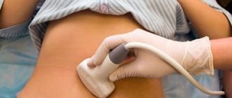
How is the procedure done?
As a rule, ultrasound is performed in the first half of the day, always on an empty stomach. Under no circumstances should you drink or eat before the procedure. Even a small amount of water or food provokes the release of bile from the bladder. The organ decreases in size, which makes it difficult to diagnose. You should also refrain from chewing gum before visiting the clinic; it also provokes the secretion of gastric juice and bile. If you are scheduled for an ultrasound examination in the afternoon, then a light breakfast is allowed 6-8 hours before the ultrasound.
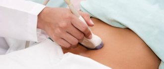
The duration of the procedure is 10-15 minutes. The patient is asked to remove his outer clothing and lie on his back on the couch. Before the ultrasound, the sonologist applies a small amount of gel to the patient's skin in the area being examined. The gel ensures continuous contact between the ultrasound transducer and the skin and improves the transmission of ultrasound waves.
During the scan, the doctor moves the sensor over the patient's skin at the location of the organ. To view the gallbladder from a different angle, the patient is asked to change position (sit down or roll over on his left side). The monitor displays the organ and surrounding tissues. The data obtained is included in the study protocol, which is given to the patient. Your attending physician will decipher it and make a preliminary diagnosis.
Carrying out an ultrasound with a choleretic breakfast differs from the standard procedure. During the study, the patient needs to eat several chicken eggs, a glass of full-fat sour cream or cream. Instead of food, a sorbitol solution can be used. First, the organ is scanned at rest. Then the patient must eat a choleretic breakfast, after which 4 ultrasounds are performed at intervals of 15 minutes. On each of them, the ultrasound specialist notes how much the gallbladder has shrunk. Ultrasound with determination of function allows you to determine which type of biliary dyskinesia the patient has (hypomotor or hypermotor disorder).
What are normal indicators for the gallbladder?
During an ultrasound, first of all, the shape, contours and size of the organ are determined. The gallbladder has the shape of a hollow pear, with smooth and clear edges. Its length ranges from 7-14 cm, and its width from 3 to 5 cm. The thickness of the bubble walls is 2-3 mm. The volume of the bile is 40-70 ml. These parameters are considered normal ultrasound for an adult. Normal sizes for the gallbladder of children depend on the height and weight of the child; they are determined using specially compiled tables.
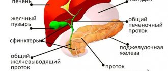
In addition to determining the size of the organ, during the scanning process its ductal system (common bile and lobar bile ducts) is studied. Their diameter, permeability and the presence of concretions (stones) in them are determined. The diameter of the lobar ducts is 2-3 mm, and the common bile duct is 6-8 mm. The common bile duct unites with the Wirsung (pancreatic) duct and flows into the duodenum. Bile enters the gastrointestinal tract along this route.
In normal condition, the organ and its ducts do not have stones or other formations. If any deviations from normal values are detected, the patient is prescribed additional diagnostics.
The norm for ultrasound to determine function is considered to be a reduction of 70% from the initial level, which suggests the absence of dyskinesia.
Decoding the results
During the procedure, the doctor evaluates the following indicators:
- organ location and mobility;
- shape, size, thickness of the walls of the gallbladder;
- diameter of the bile ducts;
- contractile function of the organ;
- the presence of stones, polyps, neoplasms.
The size of the gallbladder is normal
- length 7-10cm;
- width 3-5cm;
- transverse size 3-3.5 cm;
- volume from 30 to 70 cubic meters. cm;
- wall thickness up to 4mm;
- common bile duct with a diameter of 6-8 mm;
- the internal diameter of the lobar bile ducts is up to 3 mm.
The shape of the gallbladder is pear-shaped or oval, the contours are clear, the bottom of the bladder can protrude from under the lower edge of the liver by 1-1.5 cm. However, it is worth noting that the size of the organ largely depends on the build of the patient himself. Therefore, a competent doctor pays attention to the overall picture, rather than simply measuring individual structures.
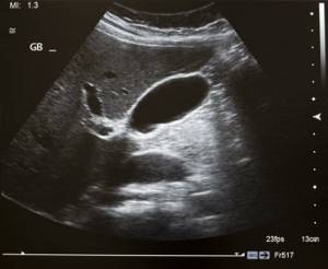
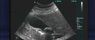
Photo: one of the projections of the gallbladder on ultrasound
What pathologies of the gallbladder can be detected on ultrasound?
Ultrasound can detect the following pathologies:
- cholecystitis (acute or chronic),
- organ dyskinesia,
- cholelithiasis (presence of stones),
- tumors
- congenital pathologies (absence or atypical position of the gallbladder, protrusion of its walls).
Cholecystitis is inflammation of an organ. Inflammation of the bladder can occur in chronic or acute form. The acute form is accompanied by intense pain in the right side, the chronic form is accompanied by nausea, heaviness, discomfort and dull pain in the area where the organ is located. Acute cholecystitis on an ultrasound image is determined by thickening of the walls and an increase in the size of the organ, while blood flow increases in the arteries of the bladder. In the chronic form, thickening and deformation of the walls, as well as a decrease in the size of the organ, are visualized.
Dyskinesia is a disorder of motor function in which there is insufficient contraction of the muscles of the organ. The ultrasound image shows an inflection of the neck of the organ and an increase in the tone of its muscles.
Cholelithiasis, or cholelithiasis, is the formation of calculi (stones) in the bile duct or its ducts. On echography, stones are displayed as areas with a dense echo structure, which can shift depending on the position of the body. Since stones reflect the signal well, an acoustic shadow from them can be seen on the echogram. In case of tumors, ultrasound shows formations on the walls of the organ that are larger than 2 cm in size. There is also a thickening of its walls and deformation of the contours.
The ultrasound procedure is sometimes not enough to make an accurate diagnosis, so patients are prescribed additional tests. Ultrasound allows you to quickly identify any abnormalities that have arisen, which makes it an indispensable method for diagnosing gallbladder diseases.
What does an ultrasound of the liver show?
As a result of the examination based on the results of ultrasound of the liver
The following diseases can be seen:
- hepatitis, both acute and chronic;
- cirrhosis;
- hepatoma;
- hemangioma;
- calcification;
- cyst;
- fatty degeneration.
Decoding the results
The patient receives the results of the examination at the end of the procedure. They include an examination protocol with printed images of the liver attached to it. The protocol describes the size of the liver, its structure, contours, and large vessels. As a rule, the ultrasound specialist who conducted the study does not make a diagnosis. It indicates deviations or the presence of any formations or inclusions. Based on the data obtained, the attending physician decides whether any additional research is necessary or makes a diagnosis and prescribes treatment.
Normal liver sizes according to ultrasound in adults
Ultrasound of the liver: normal values in adults in the table (for a child these numbers will be slightly different)
| Parameter | Size, mm |
| Whole liver length | 140-180 |
| Diameter | 200-225 |
| Sagittal size | 90-120 |
| Left lobe | |
| Thickness | 70 or less |
| Height | 100 or less |
| Right lobe | |
| Thickness | 112-126 |
| Length | 110-150 |
| Oblique-vertical size | up to 150 |
| Vessels | |
| Common bile duct | 6-8 |
| Portal vein | 13 or less |
| Vena cava diameter | 15 or less |
| Hepatic veins (distance from mouths up to 2 cm) | 6-10 |
| Hepatic artery at the porta hepatis | 4-7 |
Liver formation
The most common changes on liver ultrasound are various formations. They come in several types: focal benign, focal malignant and infectious.
Benign formations include adenoma, lipoma, polycystic disease, hemangioma, focal nodular hyperplasia, bile duct hamartoma. Over time, under the influence of negative factors, benign formations can progress to the malignant stage. Therefore, it is advisable to undergo a follow-up examination every few months.
Malignant liver tumors are divided into metastatic and primary. When primary malignant tumors (hepatoblastoma, sarcoma, peripheral cholangicarcinoma, hemangiosarcoma, hepatocellular carcinoma, etc.) are detected, treatment is prescribed after all the necessary examinations, but the outcome of the disease is often unpredictable.
Metastatic ones occur due to the manifestation of cancer in nearby organs: the gastrointestinal tract, lungs, ovaries and mammary glands in women. These are the most dangerous types of formations. They are usually resistant to drug treatment, and there is no point in removing them from the liver through surgery while there is a tumor in another organ.
Formations in the liver can also appear as a result of certain infections: viral hepatitis (both acute and chronic), candidiasis, tuberculosis, echinococosis and toxocariasis, abscess.