Due to the anatomical features of the genitourinary system, bladder problems are rare in men. In most cases, they are associated with age-related features or a malignant tumor. Therefore, performing an ultrasound of the bladder
will help to diagnose possible diseases in a timely manner.
Indications for ultrasound
Ultrasound of the bladder is prescribed along with blood and urine tests and, upon receipt of the examination results, can be supplemented with more in-depth diagnostic methods. Indications for ultrasound of the bladder in men are any abnormalities in the condition of the genitourinary system: frequent and painful urination, the urge to urinate without urination, pain in the penis and above the pubis, purulent and bloody discharge, redness and swelling of the genitals. All these symptoms may be accompanied by fever.
What problems can ultrasound of the genitourinary system help detect?
Ultrasound diagnostics helps to identify many diseases of the genitourinary organs. Among them:
- inflammatory kidney diseases (pyelonephritis, glomerulonephritis);
- urolithiasis (kidney stones are visualized very well on ultrasound, as are stones in the ureter; ultrasound in general is one of the preferred methods for diagnosing urolithiasis);
- inflammation of the bladder (cystitis);
- inflammatory process in the urinary tract (urethritis, etc.);
- prostate diseases (prostatitis, prostate adenoma, prostate stones);
- vascular pathologies;
- cysts, nodes and other neoplasms, etc.
How to prepare for a bladder ultrasound for men?
Since the bladder is a hollow organ, scanning it when empty is difficult. What you need to undergo an ultrasound of the bladder is always discussed when you sign up for the procedure. An hour before the procedure, you will need to drink at least 1-1.5 liters of water so that the bladder expands. This way the echostructure of the walls, as well as the adjacent prostate, will be clearly visible.
Is it possible to eat before an ultrasound of the bladder?
Many patients are concerned about whether they can eat anything before an ultrasound of the bladder. Regardless of which method of scanning the bladder will be used, three days before the ultrasound you will need to go on a diet: give up fatty foods, sweets, high-fiber foods and carbonated drinks. Gases in the intestines can distort the results of a bladder test. Before the transrectal ultrasound of the bladder, you will need to do an enema and take a laxative to cleanse the intestines. What results will be obtained as a result of an ultrasound of the bladder in men is determined to a large extent by preparation.
Ultrasound of the pelvis in men: what is included, how to prepare for the study at MEDSI
Table of contents
- How is the procedure performed?
- What is included in the examination?
- Indications for examination
- Types of research
- What diseases can be detected
- Preparation for the procedure
- Survey results
- Advantages and disadvantages of ultrasound
- Advantages of carrying out the procedure at MEDSI
Ultrasound diagnostics
is a type of examination of the patient’s body using sound waves of a certain frequency. The basis for the analysis is the difference in the reflection of such waves from tissues of different structures.
This procedure allows you to identify diseases and pathologies and determine compactions in organs. It is accurate and completely safe, so it can be used repeatedly even for a short period of time. Such a study does not require surgical intervention, and the result is immediately displayed on the monitor of the doctor who performs the examination.
How is the procedure performed?
Ultrasound of the pelvic organs in men does not take much time and does not require recovery procedures afterwards. The procedure for conducting the examination depends on its type and on the organs that need to be examined.
When ordering an abdominal test or Doppler ultrasound, the following steps must be taken:
- The patient lies on his back on the couch and removes clothing from the lower abdomen
- The doctor applies a special agent to the area being examined that improves the passage of sound waves through the body tissues
- Then he moves a special sensor over the patient’s abdomen, and the results are displayed on the monitor and recorded in the protocol
With this type of pelvic ultrasound, a man must first undergo the procedure with a full bladder and then with an empty one.
For transrectal examination, the procedure is slightly different:
- The patient should remove clothing from the lower part of the body, sit on the couch in a position lying on his side and bend his knees
- The doctor puts a protective device (condom) on the sensor and lubricates it with gel for better ultrasound transmission
- The sensor is inserted into the patient's rectum, the data is transmitted to the screen and noted in papers
Doppler examination differs in results - in this case, vessels are displayed, marked in different colors, which allows you to determine the quality and speed of blood flow. It can be done using one of the above methods.
The time spent on the procedure is no more than 20 minutes.
What is included in the examination?
Ultrasound of the pelvic organs in men includes a comprehensive analysis of several organs. This:
- The prostate is an endocrine gland that protects the bladder from infection and also produces a number of hormones and secretions that support reproductive health.
- Seminal vesicles are glands with a cell-like structure in which seminal fluid is formed.
- The bladder is an organ that is necessary for storing and removing urine from the body.
- Adjacent structures and lymph nodes
When examining the condition of the seminal vesicles and ducts, abdominal and transrectal examinations are used. They determine the presence in an organ of elements uncharacteristic of its structure. The transrectal method allows you to identify the smallest formations and compactions.
An ultrasound of the bladder is performed abdominally, first with the organ filled with urine, and then a second time with the organ empty. This is necessary to determine how completely this fluid is removed from the body. The study helps determine the presence of stones and other pathologies or neoplasms.
When examining the prostate, its volume is measured, its shape and structure are determined, as well as the presence or absence of any pathologies. In men over forty years old, it often increases in size more than the body needs for proper functioning.
Also, studies make it possible to determine the patency of the vessels of the pelvic organs and the speed of blood flow in them, which is also important for the proper functioning of the structural elements of the genitourinary system.
Ultrasound allows you to examine the tissues and lymph nodes adjacent to the bladder and prostate. This possibility is especially important when inflammation is diagnosed, but its nature is unclear or undefined.
The pelvic organs are located quite close to each other. There is a high probability that an infection that gets into one of them will quickly affect all the others. That is why it is necessary to undergo a comprehensive examination regularly.
Indications for examination
Doctors prescribe pelvic ultrasound for men with the following symptoms:
- Pain when urinating
- Inability or excessively frequent urine output (especially at night)
- Discomfort and pain in the lower abdomen
- The appearance of blood or pus in the urine
- Problems with potency
- Impossibility of conception
- Presence of sexually transmitted diseases
- There are abnormalities in urine tests
- Unexplained pain in the groin
Also, men from 40 years of age need to regularly undergo a comprehensive ultrasound of the pelvic organs, since it is at this age that the likelihood of problems and pathologies in the genitourinary system increases.
Types of research
Three types of ultrasound examinations are used to diagnose the male pelvic organs:
- Abdominal
- Transrectal
- Dopplerographic
The first type of examination is carried out using a sensor that allows you to examine organs through the wall of the abdominal cavity - the doctor moves the device over the patient’s abdomen, and an image is projected onto the monitor. This is a precise and painless procedure. It allows you to identify many pathologies and diseases, including detecting stones (calculi) in the bladder, changes in the size of walls in organs, and the presence of neoplasms.
Transrectal ultrasound is necessary when an abdominal examination cannot be used (due to an abdominal wound or the inability to completely fill or empty the bladder) or does not provide sufficiently detailed results. At the same time, inserting a sensor into the patient’s anus can be quite painful, so they try not to use this method unless absolutely necessary. It can be used to look for very small tumors or cysts.
Dopplerography allows you to detect disorders and pathologies in blood vessels and evaluate their ability to transfer blood to the cells of the body: diameter, patency, blood flow speed, wall thickness. The study visualizes them in different colors on the screen. It represents an additional scan when using either of the two methods described above. This type of ultrasound with Doppler is called duplex.
What diseases can be detected
Ultrasound of the pelvic organs in men can help in diagnosing a number of disorders and diseases. This:
- Inflammation of the prostate gland (prostatitis) in acute or chronic form
- Urolithiasis and the presence of stones (stones and sand)
- Seminal duct disease (vesiculitis)
- Bladder problems (cystitis)
- Oncological formations (malignant tumors)
- BPH
- Cysts, polyps, benign tumors
- Vascular diseases of the testes and testicles
- Problems with blood circulation in organ tissues
To identify such ailments at an early stage, it is recommended to undergo a preventive examination from time to time, and if the slightest symptoms appear, immediately consult a doctor.
Preparation for the procedure
For different types of pelvic ultrasound in men, preparation is somewhat different.
Before performing an abdominal examination, you must:
- Stick to the diet for two or three days. This is necessary to eliminate the possibility of excessive gas formation in the intestines, since it interferes with a high-quality view during the examination:
- On preparation days, food should be low-fat (fish, meat and poultry are acceptable), you can eat cereals, hard cheeses and drink weak tea
- Dairy products, fatty meat and fish dishes, vegetables, fruits, coffee, alcohol should be excluded from the diet.
Transrectal examination requires different preparation:
- The day before it is necessary to use drugs to clean the rectum. When choosing a product, it is recommended to consult with your doctor
- Another preparation option is to use an enema on the day of the examination (2-3 hours before it)
- During the standard procedure, a full bladder is not required, but occasionally (when it is necessary to identify the causes of infertility, erectile disorders, etc.), the doctor may prescribe an examination with a full bladder. In this case, you need to drink 0.5-1 liter of water
Doppler ultrasound requires preparation similar to that for the examination method that will be used.
If an urgent examination is necessary due to injury or other reasons, it is carried out without preparation.
Survey results
During an ultrasound, the data is displayed on the monitor and recorded by the doctor in a special protocol, which contains a comparison of the obtained indicators with the normative ones. The diagnostician evaluates:
- The size and shape of the pelvic organs: they should not be pathologically enlarged or reduced, and their shape should remain clear, uniform and clearly distinguishable. There should be no cysts or tumors
- Their location: organs should not be displaced
- Tissue echogenicity (their ability to reflect ultrasound). There are normal, reduced and increased types of echogenicity, and it is also possible to have it completely absent
In addition to the above data, the compliance of the actual sizes of organs with normal indicators is assessed:
- Seminal vesicles – no more than 1 cm
- Prostate volume – up to 30 cm3
- Prostate sizes:
- Transverse – 27-43 mm
- Anteroposterior – 16-23 mm
- Upper anterior – 24-41 mm
Advantages and disadvantages of ultrasound
The use of pelvic ultrasound as a diagnostic method in men has a number of pros and cons.
Advantages:
- Painlessness (in the case of the transrectal method - low pain)
- No abdominal incisions required
- High accuracy and information content
- Safety (no x-rays used)
- Results are immediately visible on the monitor
- Gives a complete picture of both the structure and structure of organs and their localization
- Displays neoplasms and vascular dysfunctions
- Allows you to diagnose disorders at an early stage
- The procedure is short - takes up to twenty minutes
The disadvantages of ultrasound examination include:
- Discomfort during transrectal ultrasound
- Difficulty in performing an abdominal examination in the presence of wounds or traumatic rashes in the abdominal area
- In case of severe pain, it may be difficult for the patient to take the required position for examination
Advantages of carrying out the procedure at MEDSI
- The latest devices models Pro Focus 2202, Philips iU22 for performing pelvic ultrasound in men allow diagnosing diseases most accurately
- Leading doctors (candidates of medical sciences) will expertly interpret the results and give recommendations for the prevention of diseases of the genitourinary system
- You can consult with a professional at a convenient time and in a convenient place, since there are more than 20 MEDSI clinics in Moscow, and you can make an appointment by phone
- Ultrasound machines are suitable for adults and children
- Possibility of urgent research
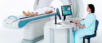
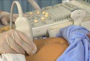
How is the procedure performed?
Transabdominal ultrasound of the bladder in men is carried out as in women: the patient lies on his back, undressed from the waist down, and the sonologist examines the lower abdomen, moving a transducer lubricated with gel over it. If necessary, the sonologist asks the patient to empty the bladder and scans it again with residual urine. For transrectal ultrasound, the patient lies on his side, is asked to bend his knees towards his stomach, after which a special thin scanner is inserted into the rectum. The quality of the image depends on how correctly the patient prepares for an ultrasound of the bladder. In rare cases, intracavitary ultrasound may be necessary, which is performed by inserting the thinnest sensor into the urethra.
How it's going
The patient undresses to the waist and lies on his back.
Sometimes the doctor may ask you to turn on your side or stomach. To diagnose kidney prolapse, the patient must be in an upright position. A special gel is applied to the skin. The doctor then moves the sensor over the body, pressing it lightly. In this case, an image of the organs is obtained on the screen. The examination lasts about 30 minutes. The patient does not experience any discomfort. The results can be recorded on any digital media.
Registration at the Novomed clinic is carried out by telephone. Remember that an accurate diagnosis is the basis for effective treatment. Call us at the first alarming symptoms!
What does the study show?
If the patient is aware of how to prepare for an ultrasound of the bladder, the scan will show the echostructure, shape and size of the organ. Whether bladder cancer is visible on an ultrasound depends on the size of the tumor. Small tumors may not be visible, and the structure of large tumors is difficult to decipher - this will require a more in-depth examination.
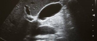
Normal bladder
The norm in form is that longitudinal ultrasound of the bladder
shows an oval shape, and the transverse one shows a round shape. An ultrasound of the bladder, which shows the smooth and clear outlines of the organ and its symmetry, is characterized as positive. The thickness of the walls of the bladder in a healthy state is from 0.3 to 0.4 cm. The bladder is a hollow organ, there should be nothing inside it.
Signs of pathology on ultrasound of the bladder in men
Based on what an ultrasound of the bladder in men shows, one can judge the presence of various types of pathologies. Cystitis on an ultrasound of the bladder is visible as an accumulation of cells, epithelium and salts - this is a sediment inside the bladder. In advanced stages of cystitis, thickening of the walls is observed. Masses on the walls of the bladder can be kidney stones or polyps. Movable formations in the bladder in a man: blood clots, stones, foreign bodies or air trapped in the organ as a result of inflammation or fistula. As a result of prostatic hyperplasia or swelling, the bladder may become enlarged.
Advantages of conducting research in our clinic
- Experienced doctors. Our specialists not only perform urological ultrasounds, but also treat identified pathologies. To do this, they have all the necessary skills and knowledge.
- Modern equipment. It allows you to perform the study with high accuracy and in the shortest possible time, with minimal discomfort for the patient.
- Optimal cost of ultrasound. We will tell you the prices for all services in advance, which will allow you to plan your expenses.
- Maximum comfort of procedures and no queues.
If you want to undergo diagnostics in our clinic and receive quality medical care, call the number provided to make an appointment. Specialists will answer all your questions.
Bladder norms according to ultrasound:
- The bubble can take different shapes, depending on the degree of fullness and the location of nearby organs. In women, the form is closely related to the number of pregnancies and births, the course of pregnancy, and the location of the uterus.
- The structural structure in a normal state should be echo-negative. This characteristic is influenced by the patient’s age; the greater the age and the presence of inflammatory processes, the greater the echogenicity.
- The volume of the organ differs between men and women. For men, this value ranges from 350-750 ml, for women it varies from 250 to 550 ml. This parameter can be affected by pregnancy, previous surgeries, and the presence of tumors on nearby organs.
- The wall thickness on average can reach 2-4 millimeters. Any violation of the local type of parameter usually suggests the occurrence of pathology.
- The normal filling rate is within 50 ml/hour. The first calls for emptying occur in an adult with a volume of 100 ml, while the norm at one time is 150-250 ml. urine.
- The remaining urine in the bladder should not exceed 50 ml.
Pathologies that ultrasound can detect
As a result of an ultrasound of the kidneys and bladder, the doctor can identify a tumor and perform a biopsy under the control of imaging equipment. In addition, thanks to the scanning device, pathologies such as the presence of cysts, stones, and obstacles to the normal outflow of urine become visible.
You can identify the size of the kidneys and urinary tract, their shape. If necessary, the study can be expanded; in addition, other visualization methods are used to provide more complete information about the detected changes. This may be MRI, CT and other types of examination.
How is an ultrasound of the prostate gland (prostate) performed?
Ultrasound of the prostate gland can be performed in two ways:
The first method is a standard ultrasound through the abdominal wall, the so-called transabdominant method of examination. This method is no different from ultrasound of other abdominal organs. The procedure time is 15-20 minutes and is absolutely safe for the patient.
The second method is transrectal ultrasound (TRUS) of the prostate gland. With this method, the sensor is inserted through the patient's rectum, which in turn gives more accurate examination results.
It is worth noting that you will receive the results of the study on the spot, after the first examination. Thus, if necessary, the urologist will immediately prescribe you the necessary course of prevention or treatment.
Is ultrasound diagnostics necessary?
How to determine that your man needs to visit an ultrasound diagnostic room? Of course, it would not be superfluous to undergo such an examination periodically to exclude any problems, but such consciousness, you see, is not characteristic of all representatives of the stronger sex.
But if your companion is approaching his fortieth birthday, a visit to an ultrasound specialist should be included in the plan of mandatory activities. After all, this particular age limit is characterized by an increase in problems with prostate tumors. And as you know, preventing a disease is always easier than treating it.
Cost of ultrasound of the prostate gland (prostate)
| Name of service | Price, rub.) |
| Primary appointment with a general practitioner | 1800 rub. |
| Repeated appointment with a general practitioner | 1300 rub. |
| Primary appointment with a urologist-andrologist | 1800 rub. |
| Repeated appointment with a urologist-andrologist | 1300 rub. |
| Ultrasound of the prostate gland (transrectal + transabdominal) | 2000 rub. |
| Ultrasound of the prostate gland (transabdominal) | 1400 rub. |
| Ultrasound of the scrotum with colorectal dosage | 2000 rub. |
| Ultrasound of the penis (Doppler examination) | 3000 rub. |
All our services and prices
Structure of the bladder
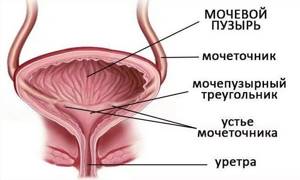
The organ is tilted at an angle.
Its tip “looks” forward and up, and the bottom position looks down and back. The body of the bubble itself is located between them. Tapering downwards it forms the urethra. In this place is the cervix, which ends with the urethra. The urinary duct connects the front of the apex and the navel. On the surface, the bubble is divided into anterior, posterior, superior and lateral surfaces. At the back, in the superficial layer there are two lumens into which the orifice of the ureter enters. The function of holding urine is performed by two valves - sphincters:
- Involuntary. It is formed by a muscle group twisted in the form of a spiral, which passes through the muscles into the small pelvis. Their design is similar to an inverted umbrella.
- Arbitrary. The sphincter is covered by the pelvic muscles, ligaments that form the obturator muscle.
The structure of the bladder has a special structure. Its shell consists of four layers:
- Interior. It is lined with transitional epithelium, which forms a reliable barrier and folds that smooth out when filled with urine. The mucous layer that covers the inside of the organ is combined with muscles. This area is called triangle triangle. The interurinary fold, located near the mouths of the ureters, protects urine from flowing backwards.
- Connective . The submucosa consists of auxiliary tissue of low density. It contains the lymphatic and circulatory systems, as well as nerve endings.
- Muscular. Muscle tissue is the basis of the organ septum. The ligaments of smooth muscles, which run in three layers, combine to form the main muscle, which performs the function of pushing urine out. Near the orifices of the ureters, circular fibers create sphincters. If muscle function is disrupted, the result may be pathological changes in the process.
- Serous. It lines the back and sides, as well as the bubble itself. On all other surfaces it is reduced to a fairly dense connecting layer, which performs a supporting function.
The bladder is designed to perform two functions: reservoir or urine collection and evacuation or urine removal. The physiological volume at which the call comes from the brain to empty it is on average about 200 - 400 ml, and for the fair sex the figure is slightly lower than for men. With age, muscle strength weakens, so older people have a larger bladder volume.
From the ureters, urine enters the bladder approximately every 30 seconds. The rhythm of filling is influenced by factors: the amount and nature of fluid consumed, the presence of stressful situations, and ambient air temperature.
Nerve endings that are located in the wall of the bladder quickly respond to the pressure caused; they are also called mechanoreceptors; they control the maintenance of a certain volume of urine in the bladder.
The evacuation function of the bladder is carried out by contracting it if the walls are significantly stretched, when the mechanoreceptors of the urethra are exposed to urea, and by relaxing the muscles of the urethra when the walls of the bladder are irritated.
The bladder plays an important role in maintaining a normal internal environment in the body, removing and cleansing the body of waste products. The characteristics of the urine of a healthy person are unchanged. However, due to certain diseases in which stagnation of urine occurs, it changes its chemical composition. And this leads to inflammatory processes and the occurrence of urolithiasis.
Preparing for a prostate ultrasound
If ultrasound of the prostate gland is performed through the abdominal wall (transabdominal examination method), then you need to know:
- The patient's clothing should be comfortable, not tight and not put pressure on the abdominal cavity.
- Fill your bladder with fluid: drink 1-2 liters of water an hour or two before the examination, or do not visit the toilet 3-4 hours before the appointment.
- If you were examined with the use of a contrast agent immediately before the ultrasound of the prostate gland, then inform your doctor about this, he will correct the results.
Are there any contraindications to transrectal ultrasound?
Transrectal ultrasound of the pelvic organs in men is harmless and does not harm the man’s body. When performing TRUS, ionizing radiation is not used, which allows the procedure to be performed the required number of times.
Contraindications to transrectal pelvic ultrasound in men include:
- exacerbation of hemorrhoids;
- anal fissures;
- recovery period after rectal surgery;
- severe mental disorders that may interfere with diagnosis.
Decoding the results
The results of the procedure will be issued in writing on average within 15 minutes after the study. The doctor will draw up a conclusion that reflects the condition, structure of the organ, characteristics and size, as well as the volume of residual urine. In general, such a study allows you to obtain the same information as a standard ultrasound - detect inflammatory diseases (signs of acute or chronic cystitis), suspect urolithiasis, see tumors and neoplasms. Additionally, the doctor will determine how much residual urine remains in the bladder after emptying.
If during the examination a residue of more than 30 ml is detected, the specialist will give recommendations on further actions. If prostate adenoma is suspected, additional diagnostic methods, consultation with a urologist, and laboratory tests may be required.
Who is prescribed a prostate ultrasound? Symptoms for undergoing a prostate ultrasound?
Regardless of the symptoms, you first need to see a doctor, and only after an initial examination, a urologist, if necessary, will prescribe an ultrasound of the gallbladder if he recognizes the following symptoms or diseases:
Make an appointment with a urologist
Make an appointment
Sign up for a study at the Dr. AkNer
Clinic specialists Dr. AkNer offers to conduct a comprehensive ultrasound of the urinary system at a time convenient for you. Our ultrasound diagnostic doctor has extensive experience in conducting ultrasound examinations, which guarantees the most accurate and reliable results. The specialist conducts an ultrasound in accordance with the protocol, provides a competent interpretation and explains in detail everything that was discovered.
Sign up for an ultrasound using the online form or call the contact numbers.
June 09, 2021
Ultrasound diagnostics doctor, candidate of medical sciences, Evseeva Ekaterina Leonidovna.