Lymph nodes are part of the body's immune system and help fight infection by producing special white blood cells called leukocytes. Lymph nodes trap bacteria, viruses and even cancer cells. An enlarged lymph node can be associated either with its inflammation or indicate the occurrence of pathological processes in neighboring organs and throughout the body.
A biopsy is performed to determine the cause of enlarged lymph nodes, as well as if the presence of cancer cells in the lymph nodes is suspected. It can be performed in the groin area, armpits, neck, lower jaw, and also for nodes behind the ears.
There are two types of biopsy - puncture and open.
A puncture biopsy is performed using local anesthesia and comes in two types:
-Fine needle biopsy (a thin hollow needle is used to obtain tissue samples);
-Trephine biopsy (a special large needle with barbs is used to cut out a piece of tissue)
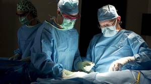
An open biopsy is performed using general anesthesia. The lymph nodes are removed through an incision.
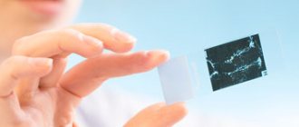
Materials obtained during the biopsy are sent for examination to the histological laboratory. The duration of the study depends on the equipment and qualifications of specialists. In UNIM laboratories, the period for histological examination is 3 days.
Based on the results of histological examination, a diagnosis is made and treatment is prescribed, or additional studies (immunohistochemical) are prescribed.
Lymph node biopsy
Posted at 18:56h in Services by doctor
Lymph nodes are organs of the immune system that are responsible for protecting our body from attacks by infectious agents. This is where pathogenic microorganisms are destroyed. When they enter the body, they enter the lymph nodes along with the lymph flow. It is worth noting that these organs also fight cancer cells. There are about 600 lymph nodes in the human body.
However, such organs also suffer from a variety of ailments. It is not always possible to make an accurate diagnosis. Therefore, a lymph node biopsy is often performed. Often this is simply necessary. Such a study is prescribed to clarify the etiology of the disease or lymphostasis and lymphodema.
About lymph
The lymphatic system consists of lymph nodes and the vessels that feed them. It is multifunctional, but its main function is to protect the body from various pathogens.
For this purpose, immune cells exist in the lymph nodes - T-lymphocytes and B-lymphocytes.
The passage of blood through small capillaries creates a plasma filtration effect. Some of the blood enters the surrounding tissues, and some goes back into the bloodstream. Another one - into the vessels. The result is the formation of lymph. It is a clear, watery liquid containing immune cells.
Lymphatic vessels, uniting, enter the thoracic lymphatic ducts, right and left, the same, in turn, flow into the veins. This results in the return of fluid to the bloodstream.
Lymph nodes, as part of the body's immune system, are located in different parts of the body. Their task is to be filters, a kind of barriers for infectious carriers and pathogenic cells.
In what cases is it prescribed?
A lymph node biopsy allows for timely detection of disorders occurring in the body. Therefore, similar studies are carried out for many indications:
- the procedure is prescribed to determine the etiology and malignancy of lymphadenopathy, if it is not possible to determine this using non-invasive diagnostic methods;
- a lymph node biopsy is performed for lymphadenopathy that lasts a long time, even when the patient receives adequate therapy;
- if the patient has signs of a tumor etiology of lymphadenopathy, for example, proliferative or metastatic lesions of lymph node structures;
- when during the initial examination, greatly enlarged, dense, but painless lymph nodes are detected, accompanied by signs of general intoxication of the body.
Indications
There are many indications for the procedure:
- Node over 1 cm;
- Soreness;
- Inflammatory process for no apparent reason;
- Suspicion of a malignant tumor;
- The lymph node does not change, although treatment has been carried out for a long time;
- Sarcoidosis or lymphogranulomatosis, etc.
This procedure provides an understanding of:
- What is the specificity of the course of the disease;
- What is the level of infection;
- If a patient with cancer has metastases.
Who should not have a biopsy?
Biopsy of a lymph node in the armpit, groin area or neck has a number of contraindications, including:
- cervical kyphosis of the spine, which does not allow a biopsy of a lymph node located in the neck;
- if there is suppuration in the lymph node itself or the tissues surrounding it;
- with hypocoagulation syndrome, which is a blood clotting disorder.
Lymph node biopsy requires professional intervention. After all, these organs are elements of the defense of the human body. Their damage in the presence of certain disorders can only aggravate the patient's condition. Therefore, you should choose a clinic for testing and a specialist with special attention.
3.How to prepare for a lymph node biopsy?
Tell your doctor:
- About a possible or confirmed pregnancy;
- If you are taking any medications;
- If you are allergic to any medications or latex;
- If you have problems with the circulatory system or are taking blood thinning medications.
A lymph node biopsy can be performed under local or general anesthesia. If your doctor chooses the second option, you will be given instructions on how to prepare for anesthesia. A biopsy under local anesthesia does not require any preparation.
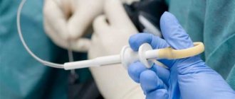
Aspiration biopsy
This biopsy method is performed using a thin needle. The instrument is carefully inserted into the subcutaneous lymph node structure located under the jaw or above the collarbone, and material is removed for further research. The procedure is performed exclusively on an outpatient basis. It is worth noting that during the biopsy, the patient experiences virtually no pain. Therefore, the aspiration method is used very often in modern medicine.
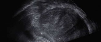
The tool used to extract the material is made in the form of a thin hollow needle. If the lymph node is not palpable, then an ultrasound device can determine its location. This technique is often used for infectious or metastatic processes in tissues.
Localization of lymph nodes in the neck
Lymph nodes are located throughout the body. Now we are interested in the neck, where there are such groups of lymph nodes:
- Chin;
- Submandibular;
- Preglottic;
- Jugular;
- Parotid;
- BTE;
- Occipital;
- Posterior cervical;
- Supraclavicular.

Bone biopsy
A medical examination involves feeling them. An enlarged lymph node means certain pathological processes occurring in it. This can be an inflammatory, purulent process, the occurrence of infection, as well as oncology.
Needle biopsy
How is a lymph node biopsy performed in this case? The puncture method involves obtaining a column of biological material for various histological studies.
It is worth noting that this procedure is carried out in almost the same way as aspiration. The difference lies in the tools. For a puncture biopsy, a needle equipped with madrain is used, which ensures excision and retention of biological material.
However, this method is not recommended for use in cancer pathologies, as this can lead to the spread of affected cells. In addition, puncture and aspiration biopsies often show incorrect results.
Varieties
Performing a lymph node biopsy is not technically particularly difficult. There are several methods for taking material. Depending on the size and location of the lymph node in the neck - deep or superficial, choose:
- open biopsy - cutting off a piece of tissue. It is carried out exclusively under anesthesia. The formation can be completely removed;
- puncture biopsy - removal of material using a needle and syringe. Typically used for large nodes. There is a distinction between fine-needle aspiration - the contents of the node are sucked out, and trephine biopsy, when a thin section of tissue is taken.
Excision method
An open lymph node biopsy involves obtaining material intended for further research through surgery. The procedure lasts at least an hour. During the intervention, the doctor makes a small incision through which the lymph node and a small piece of connective tissue are excised with a scalpel.
In this case, the patient is placed on the operating table and then general anesthesia is administered. The place where the incision will be made is treated with a disinfectant. After this, the doctor begins to remove the lymph node. Finally, the incision is carefully sutured and a bandage is applied over it.
This biopsy method is used more often than other methods. This is due to the greatest information content, as well as the reliability of the results obtained. In some cases, an open biopsy is performed intraoperatively. This allows for rapid diagnostics. If the tissue turns out to be cancerous, the doctor performs extensive surgery.

How to do it
The patient needs to prepare for the procedure in advance.
To do this, he must pass urine tests prescribed by the doctor. When examining the cervical lymph nodes, an X-ray examination may be prescribed. It is necessary to exclude kyphosis of the spine.
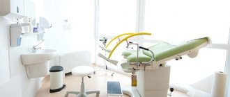
Excisional biopsy is otherwise called open: it requires access to the lymph node. The manipulation is carried out in the operating room.
Before the intervention, the required area is treated and numbed. As we have already noted, local anesthesia is more often used; in some cases, intravenous anesthesia may be indicated.
In the projection of the lymph node, the doctor makes an incision in the skin, after which he separates the tissue and, having reached the lymph node, removes it.
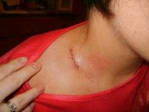
After completion of the manipulation, the incision is sutured. The duration of the entire procedure can range from ten minutes to half an hour. It all depends on the complexity.
Sentinel lymph nodes
A biopsy of sentinel lymph nodes is performed to determine the extent of malignant processes, as well as to remove several lymph nodes, rather than the entire group.
Thanks to this procedure, it is possible to preserve part of the organs of the structure. Sentinel lymph nodes are those that are first affected by malignant cells. The tendency of tumors to metastasize to these organs is the main problem in the treatment of oncology. Today, sentinel lymph node biopsy has almost become a standard procedure in the presence of a tumor.

Lymphatic system of the mammary gland
As is known, malignant tumors have the ability to metastasize. The mammary gland is distinguished by an abundance of lymphatic vessels and a variety of pathways for the possible outflow of lymph, which is one of the main routes for the spread of tumor cells.
For a long time, the main way to prevent the occurrence of secondary tumors spreading through the lymph flow was considered to be the removal of nearby nodes - in the armpit, under the collarbone, under the scapula.
How can we determine which nodes are affected by metastases and really need to be removed? In the armpit alone there can be from 14 to 45.
What should be done before the biopsy?
Before taking such a test, you must visit a doctor and clearly describe your condition. In this case, you should talk about all pathologies and health problems, allergic reactions. It is also worth mentioning blood clotting disorders and pregnancy if the woman is pregnant.
If the patient has been prescribed medication therapy, the specialist should be told about this before the operation. A week before the scheduled procedure, you must stop taking medications that cause blood irritation. Such drugs include Heparin, Cardiomagnyl, Aspirin, Warfarin, Aspercard.
If general anesthesia will be used during the biopsy, then you should stop eating and drinking 10-12 hours before.
References
- Federal clinical guidelines for the diagnosis and treatment of Hodgkin lymphoma (lymphogranulomatosis), 2014.
- Bit-Sava E.M. Biopsy of sentinel lymph nodes in the surgical treatment of patients with breast cancer. // Bit-Sava E.M., Egorenkov V.V., Anchabadze M.G., Damenia A.O., Melnikova O.A., Akhmedov R.M., Monogarova M.A. et al. – Research and practice in medicine, 2021 No. S2. P.29.
- Dvoretsky L.I. A patient with lymphadenopathy. Diagnostic search algorithm. – Clinician, 2007 No. 2. P.66-72.
- H. L. Liu, J. C. Chung. The Lymph Node Content of Supraclavicular Lymph Node Flap: A Histological Study on Fresh Human Specimens – Lymphatic research and biology, 2021. Oct;17(5):537-542. Doi: 10.1089/lrb.2018.0056.
After the procedure
A lymph node biopsy does not cause pain, as it is performed either under general anesthesia or with the use of local painkillers. The duration of the operation is from 30 to 50 minutes.
After the procedure, the patient will have to refrain from taking a shower or bath, visiting a bathhouse or sauna for several days, since it is not recommended to wet the place where the puncture was made. You should also avoid excessive physical activity. The research result will be ready within 7-10 days.
How to detect metastases in lymph nodes
The problem is not simple, since if there are a small number of mutated cells in the lymph nodes, there are usually no symptoms. If there is any suspicion, the doctor must make an accurate diagnosis.
This is where lymph node biopsy comes to the rescue. It is carried out in different ways:
- Biopsy with needle insertion into a questionable node. Carry out under local anesthesia. The procedure takes up to 30 minutes.
- Open biopsy. This is a surgical procedure performed under anesthesia, local or general, with the removal of part or all of the lymph node.
- Sentinel lymph node biopsy. A radiopharmaceutical or fluorescent dye is injected into the tumor. Penetrating the lymph vessels, it reveals the problem node, the so-called sentinel node.
After the biopsy procedure, all materials are sent to the laboratory for study.
What complications can there be?
After a lymph node biopsy, complications develop in some cases. First of all, do not forget that the procedure is, in fact, a surgical intervention. Although the operation is considered minimally invasive, in any case penetration into the body occurs. It is for this reason that such surgical intervention increases the risk of complications. Such phenomena often occur:
- infectious complications;
- dizziness or fainting;
- damage to lymphovascular and nerve tissues;
- numbness of the part of the body where the lymph node being examined was located;
- bleeding, which, in the absence of serious damage, goes away on its own after a few hours.
After the biopsy, doctors warn patients that if any complication occurs, they must immediately seek help from specialists. It is also worth visiting the clinic if swelling occurs in the area of the lymph node to be examined, if there is a fever, or an increase in body temperature. If pain does not disappear even 7 days after surgery, you should consult a doctor. The reason for visiting a specialist may be the occurrence of grainy or purulent discharge from the wound through which the puncture was performed.
Why is a percutaneous biopsy of the abdominal organs and retroperitoneal space performed?
This study helps solve important diagnostic problems:
- confirm the malignant nature of the tumor and establish its type;
- distinguish benign from malignant neoplasms;
- correctly determine the indications for surgery and plan treatment tactics;
- conduct molecular genetic and immunohistochemical analyzes of tumor tissue and correctly select targeted drugs and immunotherapy drugs;
- plan treatment for tumors with unknown primary localization;
- establish an accurate diagnosis for diffuse lesions of parenchymal organs, such as the liver, kidneys.
Using percutaneous puncture, a biopsy of the liver and pancreas, kidneys, ovaries, adrenal glands, prostate gland, lymph nodes, and soft tissue formations can be performed. This procedure is not performed for tumors in hollow organs, for example, the stomach, colon, because it is much easier and safer to obtain a tissue sample during an endoscopic examination.