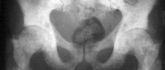
Quite often, just examining a small patient, palpation and test results are not enough for the doctor to make a final diagnosis. In this case, additional studies are prescribed. An illustrative example is cystography of the bladder in a child. This type of diagnosis is a picture taken using an x-ray
with a contrast agent previously introduced into the body.
general information
Despite the fact that X-rays have gained a reputation as not the most favorable type of effect on the human body, and the method itself has a number of contraindications, cystography in children should be carried out without fail in cases where the health risks as a result of the disease are more obvious. rather than the risk of exposure. In addition to X-rays, there is another method of examination, this is MRI diagnostics. However, for some patients, tomography is quite expensive. In addition, it requires the child to remain motionless inside the tomograph for at least half an hour, and such a requirement cannot always be met.
Cystography of the bladder is done both in children under one year old and in older children. Parents should not have any doubts if a doctor recommends this procedure. Of course, a thorough examination must first be carried out, aimed at identifying absolute contraindications.
Contraindications to bladder cystography
Contraindications to contrast cystography are due to radiation exposure, the use of contrast agents, as well as the procedure technique (the need to insert a catheter).
| Radiation exposure | Applying Contrast | Method of implementation |
|
|
|
Indications for the procedure for a child
Cystography in children under one year of age, infants and older children is prescribed as part of a set of studies aimed at identifying pathologies and abnormalities in the development of the genitourinary system in several cases at once.
Pathologies of the bean-shaped organs and bladder
Such diseases can be congenital or acquired; in most cases they require either surgical intervention or conservative (medicinal) treatment. Pathological processes are usually the result of injuries and abnormal intrauterine development.
Urinary incontinence
Cystography of the bladder can be performed in cases of urinary incontinence in a child caused by structural features of the genital organs and bladder, and weakness of the pelvic floor muscles. In some cases, research is necessary for enuresis (involuntary urination at night).
Bladder ruptures and reflux
Here, the examination (usually followed shortly by surgery) is aimed at identifying the area of ruptures and the total area of injured tissue. In the case of reflux (backflow of urine from the bladder into the ureters), the diagnostician finds out what caused such a malfunction in the genitourinary system.
Detection of a tumor of unknown etiology
In some cases, cystography in children is aimed at confirming or excluding the most tragic consequences, namely various malignant tumors. The earlier the diagnosis is made, the higher the chance of achieving a favorable treatment outcome.
Kidney tuberculosis; accumulation of sand and salt stones
By analogy with the previous case, kidney tuberculosis, sand and salts (the last two can subsequently turn into stones) require medical intervention as soon as possible. To assess the scale of the disease, understand how quickly it progresses, and create the most adequate treatment regimen, several sessions of cystographic examination will be required.
Anomalies in the development of the genitourinary system
Bladder cystography in a child will help assess the severity of congenital or acquired abnormal changes in the genitourinary system. Parents should remember that in almost half of the cases, anomalies affect not only the kidneys, bladder and adjacent organs, but also the reproductive system.
Cystoscopy: what it is, preparation, how it’s done, contraindications
Table of contents
- When is it carried out?
- Indications
- Contraindications
- Treatment with cystoscopy
- How to prepare
- How does the procedure work?
- Cystoscopy for different patients
- Consequences of the procedure
- Advantages of carrying out the procedure at MEDSI
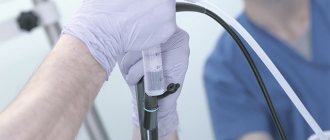
Cystoscopy of the bladder is an endoscopic examination, that is, a visual examination of the walls of the urethra, bladder and ureteric outlets using optics to identify pathology.
It is a diagnostic procedure, but it allows for targeted sampling of biomaterial (simultaneous biopsy) when pathological foci are detected and medication is administered. The study allows not only to examine the bladder cavity, but also to evaluate the functions of each kidney separately by the nature of the ureter separated from the right and left MOUTH, since they open into the bladder and are clearly visualized during the procedure; can be used as an auxiliary method for diagnosing adenoma and malignant neoplasm of the prostate - the medical indications for CYSTOSCOPY are quite wide.
When is it carried out?
The procedure can be prescribed at any age and is the main diagnostic method for MANY diseases of the genitourinary system, when safer research methods (ultrasound, radiation, magnetic resonance) do not provide the necessary information, as well as when it is necessary to take biomaterial from foci of inflammation for histological examination. neoplasms, foreign bodies (obtaining samples of urinary stones in order to clarify their composition in order to prescribe the correct treatment and diet). If calculi (stones) are detected, small formations can be destroyed and removed using an endoscope, polyps can be removed and sent for analysis.
In the presence of ulcerative lesions of the mucous membrane, electrocoagulation (cauterization) of the damaged areas can be done.
For tumors and inflammatory diseases of the prostate, CYSTOSCOPY in men will help determine the extent of the process to surrounding tissues, the degree and nature of the involvement of the bladder and urethra in the pathological process.
Indications:
- Cystitis and urethritis - pain, burning and cramping when urinating, pain in the lumbosacral spine, frequent urge to go to the toilet with small portions of urine excreted
- Tumors of the bladder - symptoms are similar to cystitis, however, when examining a smear from the urethra or urine, atypical cells were found
- Prostatitis, adenoma and cancer of the prostate - frequent urge to urinate, feeling of incomplete emptying of the bladder, urinary incontinence/retention, nocturia (frequent urge to urinate at night)
- Sexual disorders in men (male infertility) – to assess the condition of the seminal tubercle
- Suspicion of urolithiasis with localization of stones in the bladder - pain and cramping in the lower abdomen, difficult painful urination in small portions, a feeling of insufficient emptying of the bladder, turbidity of the urine to a whitish tint, the appearance of salt crystals in the urine (crystalluria)
- Enuresis – bedwetting (urination occurs during sleep) in the absence of mental and neurological pathologies
- Pyuria – discharge of pus in the urine (appearance of foreign light clots in the fluid, turbidity)
- Hematuria – blood in the urine (change in color and transparency of the liquid, the appearance of bloody clots) in the absence of injury
- Anomalies of the development of the genitourinary system or suspicion of them - to assess the volume and shape of the reservoir and urinary tract
- Evaluation of treatment effectiveness
Contraindications
Contraindications mean that cystoscopy in these cases is indicated only if other methods are not informative.
- Acute inflammatory processes in the bladder (acute cystitis), urethra (acute urethritis), prostate (acute prostatitis), testicles (acute orchitis) - in men, in the uterus and appendages - in women, during fever
- Bleeding from the urethra of unknown etiology
- Injuries to the urethra and bladder
- Disturbances in the hemostasis system (hemophilia)
Therapeutic manipulations during cystoscopy
Despite the fact that cystoscopy is a diagnostic procedure, with its help, as with almost all endoscopic examinations, some therapeutic manipulations can be performed:
- Crushing and removing small stones
- Removal of polyps and small tumors with their further examination with simultaneous coagulation of wound surfaces
- Coagulation of erosions and ulcers of the urethra and bladder
- Removal of clots or foreign bodies and restoration of patency of the urinary tract when obstructed by blood, pus or small stones
- Administration of medicinal solutions, lavage of the bladder and urethra (wash water is also collected for research)
How to prepare?
Cystoscopy
under anesthesia
will require advance (10–12 hours before) refusal of food and liquid intake (3–4 hours), after the procedure you will need time to recover, so it is not recommended to use personal transport and engage in potentially dangerous activities that require concentration attention.
Cystoscopy
without anesthesia
does not require any special preparation: it is enough to arrive on an empty stomach, having toileted the genitals before leaving home. Before the procedure, you should empty your bladder.
The choice of type of anesthesia will depend on the indications: cystoscopy under anesthesia or “during sleep” is indicated for easily excitable or mentally unstable patients. Anesthesia can be either general or spinal (only the lower half of the body, from the lower back, loses sensitivity, consciousness is preserved).
Since the structure of the male urethra is somewhat more complex (it can be 6 times longer than the female one), spinal or general anesthesia is often recommended for cystoscopy in men to eliminate pain. Also, anesthesia may be recommended if a long-term examination is expected, removal of multiple tumors, or if the patient’s bladder has a small capacity (150 ml or less).
How does the procedure work?
- Before the diagnosis begins, the examinee is given a sterile gown, he is asked to undress and lie on the couch on his back with his knees bent, they explain how the examination will be carried out and what sensations will arise during this process.
- The external genitalia are treated with antiseptic solutions, the endoscope is lubricated with glycerin to improve gliding. For men, an anesthetic is injected into the urethra using a syringe with a rubber tube and held with a clamp until the anesthetic takes effect (about 10 minutes)
The procedure technique will vary depending on the type of instrument. There are rigid and flexible endoscopy.
- Rigid endoscopy
of the bladder is performed using a rigid endoscope on a long (30 cm) metal tube. Such an endoscope straightens tissue well, simplifying examination, but is more traumatic and causes more discomfort to the subject, especially men. A rigid endoscope is not used in the presence of large tumors of the pelvic organs or pregnancy. During rigid cystoscopy, an endoscope tube is inserted into the urethra and liquid is supplied to the bladder, which simultaneously washes it and straightens the folds of the mucosa, improving visualization. To supply and drain fluid, a two-way valve is connected to the endoscope tube, since if there is pus or blood in the cavity that clouds the environment, the organ will need to be washed before examination. Wash water is collected for analysis
- Flexible endoscopy
uses a flexible endoscope - a movable thin tube made of polymer material with optics and a lamp at the end. The device follows the curves of the body and therefore can easily penetrate hard-to-reach places, which makes the examination quite informative. This method allows you to minimize injuries and eliminate pain during the procedure. In modern diagnostics, flexible cystoscopy is gradually replacing rigid cystoscopy.
Cystoscopy for different patients
Cystoscopy of the bladder in women.
As a rule, cystoscopy in women does not cause difficulties and does not require general anesthesia, since the female urethra is straight and short (up to 5 cm). For pain relief, a local anesthetic is applied to the endoscope tube. Difficulties arise in the presence of large tumors of the uterus or in late pregnancy, when the uterus compresses the bladder and changes its configuration. In this case, the use of flexible endoscopy is indicated. Examination during pregnancy is performed only for vital reasons, since any interventions on the pelvic organs can provoke spontaneous abortion.
Cystoscopy of the bladder in men.
The male urethra measures from 17 to 22 cm in length, so examination requires special care and experience from the endoscopist, especially at the stage of insertion of the instrument. During the procedure, an anesthesiologist must be present in the operating room at all times, who can anesthetize the patient if he experiences severe pain during the examination.
For children, cystoscopy of the bladder is performed only with a flexible pediatric endoscope, which is much thinner than an adult, and only by an experienced pediatric diagnostician.
Consequences of the procedure
After the anesthetic wears off, patients usually experience slight discomfort and burning in the urinary tract, aggravated by urination (especially after cystoscopy in men), and frequent urge to go to the toilet. After using a rigid endoscope, light pink mucus may be discharged. To reduce pain, it is recommended to increase the amount of fluid consumed (which, in turn, will reduce the concentration of urine), and use a one-time painkiller.
If symptoms do not go away within three days or are accompanied by fresh blood discharge, chills, or fever, you must immediately return to the clinic or call a doctor.
Advantages of carrying out the procedure at MEDSI:
- Given the high reputation of the clinic’s doctors, the price of cystoscopy is at the level of the average cost of the service in private clinics in Moscow
- Territorial accessibility
- Opportunity to conduct an examination and receive specialist advice in the same branch before and after the procedure
- Prevention and control of complications, hospitalization at will or according to indications in the clinic’s hospital
- Experienced diagnosticians with extensive experience, availability of pediatric specialists
- Tactful staff, technical and friendly service
To make an appointment, call 24/7 8 (495) 7-800-500.
Preparation for the procedure
One of the main questions of parents who are faced with the need to carry out a procedure such as cystography in children is how is this examination done? Other problems are no less important. Is advance preparation necessary? Will another x-ray be needed? How effective does the attending physician consider the method?
Diet
Approximately two days before the visit to the diagnostician, it is necessary to transfer the child to dietary nutrition. However, this does not mean that you need to exclude all familiar foods from your diet. It is enough to temporarily stop giving legumes, as well as other foods that cause gas formation: bread, raw vegetables and fruits, milk. It is recommended to give preference to porridge and boiled meat. For drinks, unsweetened teas and herbal infusions, such as chamomile or fennel, are preferred. They not only normalize intestinal function, but also calm the baby’s nervous system, because the procedure is stressful for him.
Cleansing enema
Preparation for cystography in children does not end with a diet. On the day of the procedure, you need to give a cleansing enema. If a child is worried before the diagnosis, have a friendly conversation with him, try to put him in a positive mood, explain that cystography does not pose anything terrible or dangerous to his health.
What does cystography show?
Depending on the methodology, cystographic diagnostics makes it possible to determine such parameters of the bladder as its shape, size and position, existing damage, and the presence of pathological fillings in it in the form of stones or neoplasms. In order to obtain more detailed information, double contrast is used. Thanks to it, it is possible to identify even minimal defects in the mucous membrane of the bladder, which may be a sign of inflammatory processes, tuberculosis or erosion. Pathological reverse flow of fluids is detected when the contrast penetrates into the ureters.
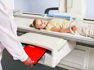
How is cystography done for children?
How is cystography done in boys and girls? The first thing you need to do is lie on your side, bending one leg and extending the other. Girls, on the contrary, lie on their backs. The doctor should gradually insert a catheter into the urethra; local anesthesia can be administered first (if there are no contraindications). A contrast agent is injected through a catheter into the bladder. Then x-rays are taken in several projections. At the end of the procedure, the contrast is released through the urinary tract, this usually takes no more than 2-3 days.
Types of bladder cystography
Cystography involves the use of liquids or gas. Depending on the form of the substance used, different types of procedures are distinguished.
| Shape of Contrast | View | How is it carried out? |
| Liquid (“Triomblast”, “Iodamide”, laquo;Urografin”) | Ascending cystography (retrograde) | The procedure involves the use of a catheter through which a substance is injected into the patient’s bladder, after which an image is taken. On average, about 200 ml of contrast is administered. |
| Descending cystography (excretory) | Contrast is administered by injection into a vein. Together with the bloodstream, it penetrates the kidneys, after which it enters the bladder. Once he has completely filled it, they take a picture. | |
| Vaccine cystography | The procedure differs in that the filming is carried out at the moment of urination. Before it is performed, a contrast agent is injected into the bladder through a catheter. | |
| Gas (nitric oxide, oxygen, carbon dioxide) | Pneumocystography | The MP is filled with gas in an amount of about 150 ml. |
| Lacunar cystography | Double contrast is performed with the introduction of 20 ml of liquid contrast and 200 ml of gas through the catheter. | |
| Sedimentary pneumocystography | It involves the use of a 15% solution of barium sulfate, to distribute it evenly on the walls of the organ, the patient is asked to take a lying position and, after a certain period of time, turn over not only on different sides, but also on the stomach and back. After half an hour, the suspension is removed by urination and nitric oxide is administered. |
Since ascending and descending cystography cannot detect small tumors and non-contrast stones, gas or double contrast is used.
Contraindications
Cystography can be done for a child as often as your doctor recommends, especially in cases where it is necessary to monitor the course of the disease over time. However, there is a certain limit - no more than 5 or 6 times during the calendar year. More radiation may harm a child, especially since children are twice as susceptible to X-ray radiation.
Inflammation in the scrotum, urethra, bladder
Carrying out diagnostics in the presence of inflammation, especially in acute form, can worsen the course of the disease and paint an incorrect picture. This will interfere with making a definitive diagnosis.
Excretion of blood clots in the urine or gross hematuria
On an x-ray, a diagnostician will not always be able to distinguish clots from neoplasms, no matter how experienced he or she is.
Indications for bladder cystography
Cystographic diagnosis is performed in adult patients and children in order to confirm the diagnosis. It is prescribed by specialists such as a urologist or nephrologist, and sometimes by a surgeon or traumatologist.
| When is it performed on infants? | When is it performed in adults? |
MP anomalies:
|
|
Cystography in CELT - painless and effective
Our clinic has been providing services on the Moscow paid medicine market for more than 25 years, and this says a lot. All those who want to be sure that they will eliminate the risk of complications after the procedure and the need to repeat it due to a lack of quality images turn to us.
We employ radiologists with many years of experience, and we also have a powerful diagnostic base that allows us to carry out the procedure safely and as comfortably as possible for the patient.
You can schedule a consultation with us online or by phone.
Results of the procedure
Cystography can detect the presence of urethral diverticulum, vesico-vaginal fistulas, vesicoureteral reflux, and will also demonstrate dye leakage both inside and outside the peritoneum. The results of cystography will help the attending physician exclude or diagnose the disease, as well as prescribe the necessary treatment.
After examining the bladder, it is recommended to consume increased amounts of fluid for 24 hours. This promotes rapid removal of the contrast agent. There may be slight pain when urinating and a pink tint to the urine for 1-2 days.
What can cystography show?
Cystography is highly informative, making it possible to detect pathological changes in organs at an early stage.
This is especially important for diagnosing cancer. In the early stages, neoplasms take the form of polyps. The polypous structure and thickening of the bladder walls are clearly visualized by contrast radiography. In addition, cystography allows you to identify:
- pathological narrowing of the bladder;
- chronic cystitis;
- sand and stones;
- urolithiasis;
- diverticula;
- enterovesical fistulas;
- reflux of the vesicoureteral type;
- urethritis;
- complications after infectious or inflammatory diseases;
- benign neoplasms.