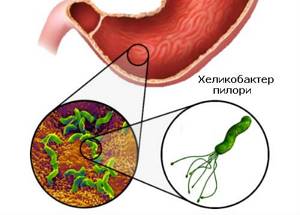
Helicobacter pylori (Helicobacter pylori) is a bacterium that is the main cause of gastrointestinal diseases such as gastritis, stomach and duodenal ulcers. Infection occurs through dirty hands, poorly washed vegetables and fruits, and common household items with an infected person. The bacterium affects the mucous membranes of the gastrointestinal tract and increases the production of hydrochloric acid. The result of infection is inflammation and ulcers on the mucous membranes.
Determining the presence of bacteria is important for choosing a treatment method. An express test for Helicobacter will help with this - a quick and accurate test to determine infection in the gastric mucosa.
Detailed description of the study
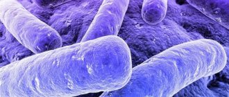
Helicobacter pylori (H. pylori) is a spiral-shaped bacterium that affects up to 50% of the population worldwide, with a higher prevalence in developing countries. H. pylori is the most significant cause of chronic gastritis, peptic ulcers, lymphoma and gastric cancer.
Infection with H. pylori occurs in early childhood, and in most cases the bacterium continues to remain in the body of the infected person throughout life. Transmission of H. pylori occurs primarily through the fecal-oral route (usually through dirty hands due to poor hygiene), and less commonly through the oral-oral route - through contact with infected saliva, for example, when an infected parent or relative kisses a child during childhood. Among children under 10 years of age, on average, 5%-10% are infected with this infection. The prevalence of H. pylori varies around the world, with higher rates of infection observed in developing countries. In Russia, about 60% of the adult population is infected with H. pylori.
There are four important components that lead to the formation of diseases caused by H. pylori. Firstly, this bacterium has the enzyme urease, which neutralizes chemically aggressive gastric juice. Secondly, the presence of flagella helps H. pylori actively move towards the epithelial cells of the host's stomach. The third advantage is the release of special adhesin proteins that interact with stomach cells, which leads to the successful establishment of the pathogen in the host body. Fourth, the bacterium produces toxins that allow it to survive and multiply in the stomach. All of these factors ultimately lead to damage to the stomach tissue and the development of chronic inflammation.
Most people have no symptoms of H. pylori infection. Clinical manifestations, if present, are usually associated with symptoms such as abdominal pain, predominantly on an empty stomach, nausea or heaviness after eating. Examination often reveals a violation of the integrity of the gastric mucosa - ulcers or erosions. There are various extragastric manifestations associated with H. pylori infection, such as anemia and chronic immune thrombocytopenia. The long-term presence of this bacterium in the body increases the risk of developing lymphoma and stomach cancer. This risk is higher among those whose close relatives suffered from these diseases.
When infected with H. pylori, microscopic inflammation of the stomach is always observed. Changes in the mucous membrane of organs are often not visible during endoscopic examination. The “gold standard” for detecting H. pylori infection is taking a biopsy from several parts of the stomach and then examining the resulting samples under a microscope.
Taking a small fragment of stomach tissue, followed by staining and fixing the resulting micropreparation on glass, allows you to study in detail the changes in the cells of this organ under a microscope. Imaging of H. pylori in such a study has a sensitivity and specificity of 90% or more.
Indications for testing
A Helicobacter pylori test with material collection (Helpil test) is recommended for:
- Feeling of heaviness and pain in the stomach (more often occurs in the morning and on an empty stomach).
- Frequent heartburn, belching with an unpleasant aftertaste.
- Bloating, flatulence.
- Suspected or already diagnosed gastritis, peptic ulcer, stomach cancer or other gastrointestinal diseases.
- Iron deficiency anemia with unknown cause.
- Detection of Helicobacter pylori in other family members.
Testing is also necessary to monitor the effectiveness of the course of treatment, and can also be recommended during a preventive health examination.
How the test works
There are several methods to detect H. pylori:
- Invasive methods – high-precision diagnosis of mucosal tissue, which is taken using endoscopy.
- Non-invasive methods - breath test or blood and stool tests. Less accurate, but often used.
The principle of operation of test helpil (helic test) is based on the ability of bacteria to produce urease - a special enzyme that decomposes urea into carbon dioxide and ammonia. In the urease breath test, the analyzer evaluates the presence of ammonia in the exhaled air, and in the rapid test for Helicobacter pylori, an analysis is performed based on the reaction on a disk soaked in urea and an indicator. If the test result is positive, the disc turns blue. The reaction occurs in 3 minutes, and the entire rapid test for Helicobacter pylori with material collection and esophagogastroduodenoscopy (EGD) will take about 15-20 minutes.
Other studies can be carried out with a sample of the mucous membrane: histological, cytological and others.
HELICOBACTER PYLORI ACCURATE DIAGNOSIS
Cytology for gastric biopsy is a study to determine quantitative and qualitative changes in cells, the presence of intra- and extracellular inclusions and microorganisms in biomaterial, which is used to diagnose a wide range of oncological, infectious, autoimmune and idiopathic diseases of various organ systems.
How do you obtain biomaterial for research?
When performing gastroscopy, the endoscopist performs a forceps biopsy of the mucous membrane in the most and least changed lesion, which is not felt by the patient, and then places this biomaterial in a special container for sending to a morphologist.
General information about the study
Cytological examination is a laboratory diagnostic method that allows one to evaluate qualitative and quantitative changes in cells, the presence of intra- and extracellular inclusions and microorganisms. The method has found wide application in the diagnosis of a whole range of diseases of various organ systems. To diagnose malignant neoplasms, infectious diseases, autoimmune and idiopathic conditions, washings, brush biopsies, lavage, punctures of fine-needle aspiration biopsy (FNA) and any other biomaterial obtained during endoscopic examination can be subjected to cytological examination.
Depending on the method by which the biomaterial was obtained during endoscopy and for what purpose the cytological examination is carried out, the following parameters are assessed in the sample: cellular composition and its quantitative changes (macrophages, neutrophils, lymphocytes, eosinophils, erythrocytes, epithelium, Langerhans cells), qualitative cell changes (presence of multinucleated cells, syncytium, atypical cells of epithelial and non-epithelial origin), inclusions (sulfur granules, dust particles, asbestos bodies, PAS-positive material in alveolar proteinosis, amyloid), microorganisms (bacteria, fungi, viruses).
Cytological examination is indispensable in the diagnosis of many diseases of the gastrointestinal tract. The digestive tract can be examined almost throughout its entire length using fibrogastroduodenoscopy (FGDS), sigmoidoscopy and colonoscopy. The resulting swabs, brush biopsies and FNA can be subjected to cytological examination. This method is especially useful when excisional biopsy is difficult (eg, in the presence of strictures), less informative (patients with Barrett's metaplasia and ulcerative colitis) or risky (risk of bleeding, perforation, tumor dissemination). If it is planned to perform both a brush biopsy for cytological examination and an excisional biopsy for histological analysis from the same suspicious area of the mucosa, it is recommended to first collect biomaterial for cytological examination, and only then for histological examination. Cytological examination is used to identify dysplastic changes in the epithelium of various parts of the gastrointestinal mucosa and diagnose malignant neoplasms. In addition, using this method, esophagitis caused by herpes simplex virus and cytomegalovirus can be diagnosed, and pseudomycelium of Candida albicans can be detected. The cytological method is used to diagnose H. pylori infection (sensitivity 98%). The distal part of the duodenum, jejunum and ileum cannot be visualized by endoscopy and therefore are not subjected to cytological examination.
Cytological examination is an additional method for diagnosing malignant neoplasms of the larynx. Its sensitivity is inferior to that of the histological method.
The detection of any pathological signs in a smear does not always indicate the presence of a particular disease, since the finding can sometimes be due to contamination of the sample during an endoscopic examination. Also, a normal smear does not completely exclude the presence of the disease. The result of the cytological examination is assessed taking into account additional clinical, instrumental and laboratory data.
What is the research used for?
- For diagnosis and control of treatment of oncological, infectious, autoimmune and idiopathic diseases of various organ systems.
After gastroscopy
The gastroscopy procedure with biopsy lasts 30-60 minutes. After its completion, the patient remains under the supervision of doctors until the effect of the sedatives wears off. The patient will be able to eat after the numbness of the tongue and throat caused by the local anesthetic has completely passed. Due to taking sedatives and also taking a biopsy within 12-14 hours, it is better not to drive, drink alcohol, take irritating foods (hot, spicy) and blood thinners (aspirin, etc.).
Fill out an application on the website and you will receive a FREE consultation with a medical specialist from a clinic operating under a license from the Israeli Ministry of Health. Personal information left on the site is strictly confidential and is not transferred to third parties.
What diseases are caused by Helicobacter pylori?
Helicobacter pylori can remain in the body for several years without manifesting itself in any way. As soon as favorable conditions arise (for example, immunity decreases), the bacterium begins to rapidly divide, releasing enzymes that destroy the mucous membrane of the duodenum and stomach. This contributes to the penetration of bacteria deep into the organ and the development of certain diseases:
- gastritis;
- inflammation of the duodenal mucosa (duodenitis);
- peptic ulcer;
- lymphoma of the stomach;
- cancerous tumors.
Malignant tumors appear in the digestive organs if left untreated for a long time.
Preparing for the examination
All kinds of inflammatory diseases of the digestive tract can be called an epidemic of the 21st century. Esophagitis, gastritis, ulcers, cholecystitis, pancreatitis - all these pathologies are diagnosed in millions of patients every year. Previously, the occurrence of these diseases was explained by the influence of various factors: stress, poor diet, alcohol consumption, smoking. Of course, all these unfavorable factors significantly increase the risk of developing an inflammatory process in the gastrointestinal tract, but today it is reliably known that all of the above diseases are of a bacterial nature, namely, they arise due to the presence in the body of Helicobacter Pylori, a bacterium that infects various parts of the stomach and intestines.
- Before starting a gastroscopy with taking a biopsy for an HP test, the clinic’s doctors collect a complete history of the patient’s life and illness: facts such as radiation or surgical treatment of the esophagus and stomach in the past, allergies, bleeding disorders, heart pathology, pregnancy is mandatory must be known to the doctor.
- Information that is very important for Top Ichilov specialists is a list of medications that the patient has taken and/or continues to take over the past two weeks. Some medications, such as antibiotics, acid-lowering drugs, can cause a false result. Be sure to tell your doctor about the medications you are taking; you may need to stop taking them for a while.
- The same applies to drugs that reduce clotting and thin the blood: in order to prevent bleeding at the site of taking the biopsy, it is better to stop them for a while.
- 8 hours before the start, you should limit your food and liquid intake.
How else can Helicobacter pylori be detected?
- Study of material obtained during gastric endoscopy (FGDS , gastroscopy). The study provides a precise and unambiguous answer. The disadvantage is the need to insert an endoscope into the stomach .
- Blood test for Helicobacter. Determination of antibodies to Helicobacter is not always informative. We see a lot of false positive results from these tests. Antibodies in the blood can persist for many years after helicobacteriosis and this does not require treatment.
- Analysis of stool for Helicobacter antigen. We see many false negative results from such tests (H. pylori is obtained during endoscopy of the stomach, but the antigen is not detected in stool).
Why do we recommend performing the respiratory urease Helik test to determine Helicobacteriosis? It's reliable, fast, cheap, painless
Gastroscopy with biopsy: procedure
An endoscopic examination that also includes a biopsy is an unpleasant procedure, so at Top Ichilov it is performed under “light” sedative anesthesia (medical anesthesia), thanks to which the patient does not feel any pain or discomfort.
The doctor inserts an endoscope into the patient’s stomach through the mouth and esophagus and examines the gastric mucosa. Then biopsy forceps are inserted into the instrumental channel of the endoscope, with the help of which two pieces of tissue are taken from the antrum and body of the stomach.
The extracted samples are placed in a special reagent, with which doctors determine the presence/absence of H. Pylori on the gastric mucosa.
- Helicobacter
- 5
- 4
- 3
- 2
- 1
(5 votes, average: 5 out of 5)
Ways and symptoms of Helicobacter pylori infection

It is very easy to become infected with the Helicobacter bacteria. The most common ways:
- through dirty water;
- by contact.
The microorganism is easily transmitted through touch, coughing and sneezing. The everyday route of entry of bacteria into the body is very common in families where at least one person is a carrier.
The peculiarity of the microorganism is that it remains dormant until the body’s defenses resist. As soon as immunity decreases, Helicobacter pylori manifests itself. The bacterium begins to rapidly multiply, its waste products poison the body and cause inflammation of the mucous membranes of the duodenum and stomach. This may be due to illness or stress.
In most cases, doctors and people themselves do not connect the appearance of gastric pathologies with the action of the body and begin long but unsuccessful treatment.
Signs of Helicobacter pylori infection are:
- frequent heartburn ending with an attack of nausea;
- systematic diarrhea or diarrhea;
- attacks of stomach pain;
- belching after eating;
- allergic reactions that are associated with aversion to certain products;
- Change in breath odor to very untidy.
The vital activity of the microorganism also affects the condition of nails and hair. The hair noticeably thins and loses its healthy appearance. Nails become dry and brittle, and spots may appear on them.