History of the discovery of X-rays
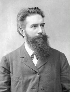
Wilhelm Conrad Roentgen. The science of radiology got its name in honor of Wilhelm Conrad Roentgen, a professor at the University of Würzburg, who discovered X-ray radiation on November 8, 1895. Roentgen made the discovery unexpectedly for himself: late in the evening, leaving the laboratory, the scientist turned off the light in the room and noticed a greenish glow in the darkness, fluorescence emanating from a screen covered with crystals of platinum-barium bluehydride. As it turned out, the crystals reacted to the influence of a nearby electric vacuum (Crookes) tube on them, which at that moment was under high voltage. When the current was turned off, the screen glow stopped, and when turned on again it resumed. The tube was wrapped in black opaque paper, so Roentgen suggested that when an electric current passes through it, it emits some kind of invisible rays that can penetrate opaque media and excite barium crystals. Roentgen called these unknown rays X-rays.
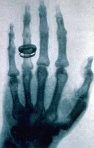
After 50 days, the scientist presented a 17-page manuscript containing a description of the rays he discovered to the chairman of the Würzburg Physico-Medical Society. This day, December 28, 1895, went down in history as the official date of the discovery of X-rays. Along with the manuscript, the scientist also presented the first X-ray photograph taken earlier, on December 22, which showed the hand of his wife Bertha Roentgen. After a woman saw an X-ray of her hand, she, not understanding the intricacies of physics, was so impressed that she exclaimed: “I saw my death.”
On the evening of January 23, Dr. Roentgen gave a lecture to a packed auditorium of the Würzburg Physico-Medical Society. After a discussion about his experiments, Roentgen invited the society's chairman, Albert von Kölliker, a renowned anatomist, to take an image of his hand using the new X-rays. When the finished image was shown to the audience, they burst into deafening applause. Dr. von Kölliker, impressed by the discovery, proposed calling the new rays X-rays - his proposal was met with applause by the audience.
The discovery of X-rays caused a wide resonance among scientists around the world, including among Russian scientists. In early January 1896, Roentgen's pamphlet was published. Within a few weeks, it was translated into Russian, English, French and Italian, and already at the end of January, A. S. Popov produced the first X-ray machine in our country, with the help of which Russian scientists repeated Roentgen’s experiment, making the first X-ray photograph in Russia. A photograph of the resulting image was placed in the Russian translation of Roentgen’s brochure, published that same month in St. Petersburg under the title “A New Kind of Rays.”
Wilhelm Roentgen continued to study his discovery, and by May 1897 he finally formulated all the basic properties of X-rays, publishing two more scientific papers. The most valuable practical property of X-ray radiation, which has found wide application in science and medicine, turned out to be its ability to penetrate through opaque bodies. In 1901, Wilhelm Roentgen was awarded the first Nobel Prize in physics for his discovery. Subsequently, the science that studies the effects of x-rays on the body was called radiology.
The first X-ray photograph showing the hand of the scientist's wife, Bertha Roentgen, and her wedding ring.
The year of birth of veterinary radiology in Russia can be considered 1896, when S.S. Lisovsky was the first to use X-rays to scan a dog. In 1899 M.A. In addition to X-raying, Maltsev also took photographs of the head, neck and limbs of a dog, the metatarsus and fetlock of a horse, and the metacarpus of a cow; The scientist used anesthesia to secure the animals during the study. Three years later, an X-ray unit was assembled in the laboratory of the Kharkov Veterinary Institute, with the help of which they diagnosed bone fractures and dislocations, identified foreign bodies, and also carried out studies of fetuses in small domestic animals.
However, these studies were isolated; they were carried out on primitive devices assembled on their own. Only by 1924 did the production of X-ray machines begin in the workshops of the former USSR, and thanks to G.V. Domrachev and A.I. Vishnyakov from the Kazan and Leningrad Veterinary Institutes, this type of research has been widely used in veterinary medicine.
Subsequently, the workshops for the production of X-ray machines turned into X-ray factories, which by 1931 began to produce devices suitable for studying not only small animals, but also large ones, thanks to which in 1932 the first X-ray rooms.
X-ray of the hand of anatomist Albert von Kölliker, taken on January 23, 1896 by V.K. X-ray during his public lecture at a meeting of the Physico-Medical Society.
From this moment on, the intensive development of veterinary radiology began in the former USSR, to which many Soviet veterinary radiologists made a significant contribution. Among the most significant discoveries are the following:
- In 1931, A. I. Vishnyakov wrote the first book on X-ray diagnostics of animal diseases, “Fundamentals of Veterinary Radiology”
- In 1935, a book by Prof. A. V. Sineva “Clinical diagnosis of internal diseases of domestic animals”
- In 1939, A. Yu. Tarasevich’s book “Lameness of Farm Animals” appeared.
- In 1940, A. I. Vishnyakov’s voluminous textbook “Veterinary Radiology” was published, which describes the principles of X-ray physics, X-ray engineering, and also provides extensive and systematized material on X-ray diagnostics of various animal diseases and X-ray therapy
- A.A. Weller has published articles on the use of X-rays in military settings. Weller also studied the possibility of diagnosing diseases of the limbs, withers and intestines in horses
- G. G. Wokken published a number of works on age-related and comparative x-ray anatomy of animals, x-ray osteology, anthropology and angiology
Veterinary radiologists of Russia and the former USSR made a great contribution to veterinary science on such issues as the determination of mineral metabolism in farm animals and birds, the diagnosis of respiratory diseases of large and small animals, the diagnosis of diseases of the digestive system, comparative x-ray anatomical studies in farm animals, the determination of the location and depth of foreign bodies.
Due to the advent of even more advanced X-ray machines, the possibilities for studying animals have increased significantly. Digital radiography is actively developing, which, thanks to multiple improvements in image quality, is gradually replacing classical, analog radiography.
Invention of the first X-ray machine
It all started with the discovery of the X-ray beam. This happened in 1895. The author of the discovery was the German scientist Wilhelm Roentgen, after whom the phenomenon, unusual for those times, was named. A year later, special radiation formed the basis for the operation of special medical equipment. The first steps in the development of X-ray diagnostics were also taken by Wilhelm Roentgen. The scientist himself did not consider his discovery grandiose, and throughout his life he never patented it.
The first X-ray machine was very different in design and operating principle from the equipment used by modern doctors. Due to the insufficient sensitivity of the film and the low level of equipment at the beginning of the twentieth century, it took several hours to create an x-ray image. To solve the problem over time, research began to use special intensifying screens located on both sides of the film.
Achievements in the field of X-ray production in the USSR and the Russian Federation
In 60-70, some of the best domestically produced X-ray machines were produced: RUM-15, RUM-18, RUM-22 and other analogues. There were no foreign parts or spare parts for this equipment.
RUM-20
RUM-20 is considered one of the most successful domestically produced radiological diagnostic systems. The best specialists worked on this model.
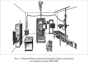
Flexible high-voltage cables, silicon rectifiers, an electronic image intensifier, and X-ray tubes with a rotating anode were used especially for RUM-20.
Electroradiography
In the 70s, Soviet scientists proposed an innovation that would subsequently allow significant savings on consumables. Experts developed a special tactic for forming an X-ray image: they used light-sensitive selenium plates on plain paper. The technology is called the “electroradiography method”.
Mammographs for diagnosing cancer
Later, on the eve of the 90s, another revolutionary project was organized, thanks to which the first microfocus mammography complex “Electronics-M” appeared.
In 1989, the developers received an award for this outstanding invention. This device became a breakthrough in the field of domestic production of medical equipment. "Electronics-M" was used to diagnose breast cancer.
Switch to imported parts
If during the Soviet era all the parts and elements used were strictly domestically produced, then already at the end of the twentieth century there were more parts of imported production (approximately 20-70%).
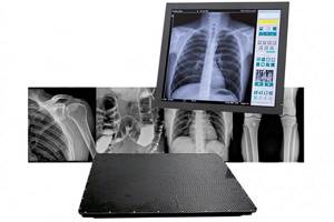
Digital and analog X-rays for radiology diagnostics
Today, many X-ray rooms are not equipped with special equipment for darkrooms and developing film images. The fact is that most modern clinics use digital X-ray machines.
Currently, analog X-rays are still relevant and remain on the global market. They are still quite popular in Russia due to their affordability.
However, in recent years, digital equipment has become more numerous. According to forecasts of leading experts, one day digital X-ray machines will completely replace film technology.
Further development of X-ray machines
Already at the beginning of the twentieth century, cumbersome installations were replaced by mobile units created specifically for the navy and army. The main contribution to the modernization of equipment was made by N.A. Velyaminov. Thanks to the great Soviet leader, compact devices quickly complemented the medical equipment of military field hospitals.
A century ago, with the help of X-ray machines, doctors could diagnose tuberculosis and cancer in the early stages. X-ray therapy became increasingly widespread, and at some points became a fashionable whim. History knows many cases when the inappropriate and unnecessary use of X-ray irradiation became the cause of high-profile scandals. But this is an inevitable part of the development of almost all discoveries that today help doctors save millions of lives.
Cathode ray research
In the middle of the 19th century. many physicists studied electrical phenomena. One of these phenomena was a gas discharge. Gas at low pressure began to glow under the influence of high voltage.
At the same time, it was discovered that the cathode in the balloon (cathode tube) emits invisible rays, which were called cathode rays. Cathode rays caused some chemical compounds to glow, and they were identified by this glow. At the end of the 19th century. It has been proven that cathode rays are a stream of negatively charged particles (electrons).

Rice. 1. Cathode rays.
Properties of X-rays
It was immediately assumed that X-rays were electromagnetic radiation. In this case, they must demonstrate wave properties and, in particular, the ability to diffraction. However, no diffraction at narrow slits could be detected. Consequently, X-rays either had a nature other than electromagnetic, or had such a short wavelength that the distance between the slits used was too large.
The assumption of a short wavelength was confirmed when crystals were taken as a diffraction grating. Narrow beams of rays passing through the crystal lattice showed a clear diffraction pattern on the screen. It turned out that the wavelength of X-rays is much shorter and comparable to the size of atoms.
Currently, the boundaries of the X-ray range are taken to be $0.005...10$nm (radiation frequency - $3×10^{16}...6×10^{19}$Hz). In the long-wavelength part, X-rays border on ultraviolet radiation, in the short-wavelength part - on gamma rays.
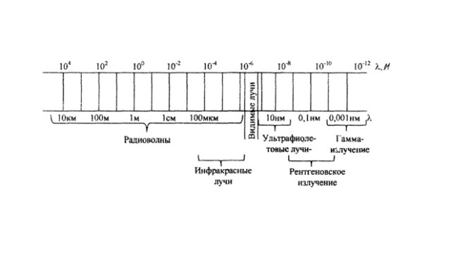
Rice. 2. X-ray scale.
Due to their shorter wavelength, X-rays have higher energy than UV radiation, so it has a high penetrating power, which has led to its use in medicine and scientific research.
There are two types of X-ray spectrum: continuous and line. The continuous spectrum is also called the stopping spectrum, since it is formed when fast electrons are sharply slowed down by a substance. The line spectrum is formed during the transition of electrons in atoms from level to level and characterizes the properties of the substance itself.