X-rays are a very important imaging tool used throughout the world. Originally used to take pictures of bones, X-rays have saved countless lives and led to many discoveries.
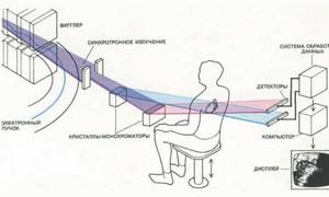
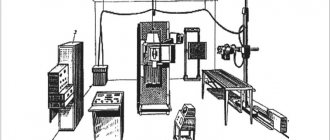
Diagram of the passage of X-rays through the human body when examining the heart
X-rays are a type of electromagnetic radiation; they are produced when charged particles (electrons or ions) collide with a material with sufficient energy.
For many years, scientists have expressed concern about the health effects of X-rays; After all, these rays expose the patient to radiation. But do the benefits they provide justify the risks of their use?
In this article, we will discuss what X-rays are, how they are used in medical science, and the risks they pose.
Briefly about X-rays
Here are some key facts about X-rays.
- X-rays are a type of radiation.
- We are exposed to cosmic rays and radiation from other sources every day.
- X-ray radiation is considered carcinogenic.
- Different x-ray procedures produce different levels of radiation.
- The benefits of using x-rays in medical diagnosis of body pathologies outweigh the potential negative factors.
- The discovery of this type of radiation is attributed to Wilhelm Roentgen.
- For the first time for medical purposes, X-rays were tested on a little boy who had a broken wrist.
- Computed tomography (CT) produces a higher level of radiation exposure than all other types of fluoroscopy.
Wilhelm Roentgen (1845-1923)
Wilhelm Roentgen is considered the discoverer of this type of rays. Just a few weeks after the discovery of the ability to image bones, X-rays began to be used for medical purposes.
The first person to use X-rays for medical purposes was young Eddie McCarthy from Hanover, who fell and broke his left hand while skating on the Connecticut River in the winter of 1896. Everyone living on our planet is exposed to some dose of radiation in their daily lives. Radioactive substances are naturally present in air, soil, water, rocks and plants. The largest source of natural radiation for most people is radon gas.
In addition, the Earth is constantly bombarded by cosmic radiation (cosmic ionizing radiation), which includes X-rays. These rays are not harmless, but it is impossible to hide from them, and the level of their radiation is so small that it can be neglected.
Pilots, flight attendants and astronauts are at greater risk of exposure to higher doses of radiation because as altitude increases, so does the power of cosmic radiation. But there are only a few studies that track the link between aviation and aerospace jobs with increased cancer exposure.
Applications of X-ray tubes in industry, science and security
To determine defects and inhomogeneities in materials, X-ray transmission is used - X-ray flaw . X-ray devices for flaw detection are stationary or mobile and have high power. For example, in the production of metal structures, X-ray tubes with a voltage of 150-400 kV or more are used. Increasing the voltage makes it possible to reduce the exposure time, which is important when illuminating structures of considerable thickness.
To determine the structure and composition of substances, X-ray diffraction analysis . Tubes for such analysis produce, in addition to bremsstrahlung radiation, intense characteristic radiation. Their power is usually low.
The scientific application of X-rays is to determine the atomic-molecular structure of substances and decipher the structure of molecules.
X-rays are used to ensure security at airports and customs – cargo and luggage are scanned to determine their contents.
Types of X-ray diagnostics
To take an X-ray, the patient himself (or his body part being examined) is positioned in front of the X-ray receiver and is illuminated with short pulses of X-ray radiation. Because bones contain a lot of calcium, a chemical element with a high atomic number, X-rays are absorbed by them, appearing white in the resulting image. Any gases in tissue, such as the lungs, appear as dark areas on the image because the absorption of rays by gases is relatively low.
Radiography
The most common type of X-ray imaging for us. Used to take pictures of broken bones, teeth and chest. X-rays use low levels of radiation. To analyze photographs printed on film, a X-ray viewer is usually used.
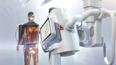
X-ray complex General Electric
X-ray
It can be compared to the “cinematic equivalent” of radiography. The radiologist can view the X-ray image of the patient in motion in real time, and, if necessary, take static images. This type of diagnosis can be used to study intestinal activity, for which it is necessary to take food containing barium. Fluoroscopy uses higher levels of radiation than radiography, but the level of radiation remains relatively low.
Philips X-ray complex
Computed tomography (CT)
The patient lies on a table that slides into a ring-shaped scanner. A fan-shaped beam of rays passes through the patient's body and hits a variety of sensors. The patient slowly moves through the scanner, so that the scan takes a series of cross-sectional images of his body ("slices"), from which a three-dimensional image is formed. This procedure uses the highest level of radiation compared to other x-ray imaging options because many images are taken at once.
Computer tomograph Siemens
Use of X-ray tubes in medicine
In medicine, X-rays are used for the following purposes:
- diagnosis of various diseases;
- radiation therapy.
X-ray diagnostics
The very first diagnostic method was fluoroscopy . X-ray radiation, passing through the human body, hit a special fluorescent screen on which an image was projected in real time. This method was accompanied by a large radiation dose to the patient and did not allow the doctor to subsequently review the diagnostic results again.
To solve this problem, radiography - the image was projected onto photographic film, which was then developed and made it possible to carefully study the patient’s organs and tissues through the light. But due to the fact that X-rays cannot be focused with lenses, the image was large, and a large area of photographic film was required.
fluorography method was developed - radiation was projected on a fluorescent screen, as in fluoroscopy, and then photographed with a conventional camera. Due to this, it is possible to take smaller X-ray images, which has reduced the cost of examination and carried out mass preventive examinations of the lungs among the population.
After the widespread use of computers, they were used in medicine. Now the image is formed without photographic film - in the form of a digital image that can be processed programmatically and obtain clearer data.
Thus, computed tomography allows you to obtain layer-by-layer images of the human body due to the movement of the X-ray tube around the patient, then process them, reconstruct them in 3D and analyze them. At the same time, the radiation dose on modern CT scanners is very low and allows examinations to be carried out as often as necessary for medical reasons.
Modern fluoroscopy has also undergone changes - a fluorescent screen is no longer used. Instead, an X-ray image intensifier and electron-optical converter are used, and the image is displayed on a monitor. The data can be saved on a computer and used for later analysis.
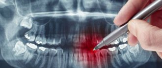
X-ray tubes are used for mammography , a non-invasive examination of the breast for tumors.
All of these diagnostic methods require a different approach to X-ray tubes. For example, CT tubes must be resistant to high centrifugal force, increased heat capacity up to 8 million thermal units and increased filament current up to 400 mA - to extend the service life of the tube and the ability to conduct studies on patients with large body weights.
Standard X-ray tubes are not suitable for mammography. Mammography tubes have peak voltages of 20-35 kVp (kilovolts peak), 0.1 mm/0.3 mm focal values and a molybdenum or rhodium target, while conventional tubes have 40-150 kVp, 0 focal spot, 6 mm/1.0 mm/1.2 mm, and the target is made of tungsten.
X-ray therapy
X-ray radiation, due to its damaging effect on biological tissues, is used to treat malignant tumors. There are two types of X-ray therapy devices, the tubes for which will differ accordingly:
- short-focus - radiation energy ranges from 10 to 60 kV, penetration depth up to 6-7.5 cm;
- devices for deep X-ray therapy - radiation energy in the range from 100 to 250 kV, the irradiation distance is from 30 to 60 cm.
To limit radiation from the tubes, casings with cylindrical or rectangular tubes and different sizes of the output window are used.
Risks associated with X-rays
X-ray radiation can cause mutations in our DNA and thus can lead to cancer in the future. For this reason, X-ray radiation is classified as carcinogenic by the World Health Organization (WHO). However, the undeniable advantages of x-ray diagnostics outweigh the possible negative consequences of its use.
According to some estimates, about 0.4% of cancer cases were caused by CT scans. Some scientists believe that the incidence rate for this reason will increase along with the increasing popularity of the use of this type of diagnosis.
According to studies, by the age of 75, the risk of cancer increases slightly due to X-ray procedures, by only 0.6 - 1.8%. In other words, the risks are minimal compared to the benefits of using X-rays for medical imaging.
All X-ray procedures have varying degrees of risk associated with them, which depends on the type of procedure as well as what damaged part of the body is being examined. The list below shows some x-ray procedures and compares radiation doses relative to normal everyday radiation exposure.
- Chest examination - 2.4 daily doses of natural background.
- Examination of the skull – 12 daily doses of natural background.
- Examination of the lumbar spine - 182 daily doses of natural background.
- Intravenous urogram – 1-year dose of natural background.
- Upper gastrointestinal examination - 2-year dose of natural background.
- Irrigoscopy – dose of natural background for 2.7 years.
- CT scan of the head – 243 daily doses of natural background.
- Computed tomogram of the abdominal cavity - natural background dose over 2.7 years.
Note:
Here are the radiation levels for adults; Children are more sensitive to radiation!
Application of X-ray computed tomography in a multidisciplinary clinic
Grigorieva E.V., Khutornoy N.V., Solodov A.A., Krylov V.V.
Radiation diagnostics is an integral stage of patient examination both at the diagnostic stage and in preparation for surgery, as well as when assessing the effectiveness of the prescribed treatment. Modern diagnostic techniques make it possible to obtain reliable data on the structure and function of almost any human organ in a short time.
Since all radiological techniques used today involve wave radiation, the term “radiology” is used to describe them. November 8, 1895 can be considered the beginning of the era of radiation diagnostics. On this day, X-rays were discovered; later, in 1901, Roentgen would be awarded the first ever Nobel Prize in Physics for this.
The first X-ray images resembled photographs and were received with enthusiasm by the public. Six months after the publication of X-ray, images of Egyptian mummies from Vienna museums and photographs of limb bones had already appeared, and authoritative doctors advised everyone to see an image of their brain. Contemporary X-ray surgeons quickly found use of new radiation to detect foreign bodies in the patient’s body and in the diagnosis of urinary tract stones. In 1896, Dr. Henry W. Cattel of the University of Pennsylvania wrote: “The surgical imagination may easily be lost in the endless possibilities of application of this remarkable method.”
In Russia, X-rays were already called X-rays in 1896, possibly on the initiative of one of Roentgen’s students, A.F. Ioffe. The first X-ray machine was designed by A.S. Popov in 1896, and in March of the same year, Professor N.V. Sklifosovsky began regularly using X-rays to diagnose skeletal bone fractures.
Fig.1. X-ray of the hand of anatomist Albert von Köliker, taken by Roentgen during a report on the discovery on December 28, 1895 at the Physics Institute of the University of Würzburg
In 1897, the first reports of illnesses associated with uncontrolled use of X-rays appeared. Lead protective suits have become an integral part of any X-ray room equipment over time.
According to standards, today the total effective radiation dose for a healthy person undergoing preventive examinations should not exceed 1 mSv per year.
Fig.2. Protective suit for an X-ray room assistant, circa 1918.
As in the days of X-ray, today, according to X-ray data, fractures of the bones of the skull, spine and skeleton, divergence of cranial sutures, foreign bodies, primary calcified formations, accumulation of air in the cranial cavity, in the pleural and abdominal cavities, pathology of the paranasal sinuses, and urinary tract calculi are verified. and many other diseases. However, despite the low cost and availability of the x-ray method, it fades into the background due to low contrast and the inability to visualize changes in soft tissues. Classic radiography is being replaced by X-ray computed tomography.
X-ray computed tomography (CT) entered clinical practice in the 70s of the last century. The method is based on layer-by-layer transverse scanning of an object with a thin beam of X-rays. Body tissues absorb radiation differently, and images can be constructed based on these differences.
The theoretical basis of computed tomography was developed by A. McCormak in 1956-1973, and an employee of the central scientific laboratory EMI Ltd GNHounsfield designed the first working version of the scanner. In 1979 both scientists were awarded the Nobel Prize in Physiology or Medicine.
Fig.3. The first X-ray computed tomograph EMI Mark I and the first axial section of the brain in a woman with an intracerebral tumor, obtained on October 1, 1971. (tumor indicated by arrow)
Today there are 5 generations of computed tomographs, which differ in the nature of the movement of the scanning device, the type of radiation beam and the number of detectors. Since the third generation, CT has used spiral scanning technology, in which, while the table is moving, the X-ray tube and detectors (or just the tube) rotate 360° around the patient’s body. One rotation of the tube can produce from 16 to 360 slices, allowing large volumes to be scanned in a short time and with high spatial resolution. For example, it is possible to obtain simultaneous images of the brachiocephalic and intracranial arteries against the background of intravenous bolus contrast enhancement with high quality visualization of vessels with a diameter of up to 1-2 mm, which is used to assess the functioning of vascular anastomoses or for widespread stenoses of the internal carotid artery and its branches.
With spiral scanning, depending on the area of study, the effective radiation dose is reduced to 88%, and the maximum scanning volume is increased to 80-160 cm. This allows for onco check-up - scanning the brain, neck, chest, abdominal and pelvic organs in one study in patients with tumors and suspected metastatic lesions. Intravenous bolus administration of a contrast agent at high speed helps to obtain separate images of the arterial, venous and delayed phases of contrast and to identify the feeding vessels of the tumor, differentiate different types of tumor formations, and assess the extent and extent of invasion. Based on the results of the study, you can get a consultation with an oncologist at the clinic to determine treatment tactics, prescribe and administer chemotherapy.
Rice. 4. CT examination (onco check-up) of a patient with rhabdomyosarcoma of the left shoulder girdle. From left to right, top to bottom: 2D reconstruction helps determine the volume of tumor nodes (arrows) and metastases to regional lymph nodes (short arrow); 3D reconstruction of the same volume allows us to evaluate the lumen of the brachiocephalic and subclavian arteries at the level of the tumor; 3D reconstruction using the onco check-up program is needed to identify foci of destruction of skeletal bones; high-volume scanning with contrast allows the diagnosis of distant metastases during one study (short arrows)
A modern technique for studying the vascular bed is CT angiography. This method allows you to obtain high-quality images of arteries and veins at the examination level due to preliminary contrast. According to Russian and foreign clinical guidelines, CT angiography is one of the mandatory preoperative examination methods for patients with stenosis and occlusion of the carotid and vertebral arteries, pathological deformation of the arteries of the head and neck, Takayasu and moyamoya disease, steel syndrome and arterial dissection. The indications for surgery are not only to determine the degree of arterial stenosis, but also the extent of the stenosis, which can be difficult to determine with duplex scanning, especially with stenosis and tortuosity at the level of the craniovertebral junction.
Rice. 5. CT angiography, various post-processing methods. From left to right: MPR, sagittal plane reformation, partially calcified plaque in the ampullary portion of the internal carotid artery (arrow); the same patient, 3D reconstruction in the transparent vessel program; 3D reconstruction of CT angiography in a patient with occlusion of the internal carotid artery (indicated by a long arrow)
One of the areas of application of CT angiography is the diagnosis of intracranial vascular formations, primarily aneurysms and arteriovenous malformations, including against the background of non-traumatic intracranial hemorrhages. According to various authors, the capabilities of CT angiography in the diagnosis of intracranial aneurysms are almost equal to digital subtraction cerebral angiography, and the sensitivity and specificity are 97.1% and 98.5%, respectively. Multiplanar reformation (MPR) and maximum intensity projection (MIP) reliably convey the relationship between the aneurysm and the supporting vessel, and volumetric (3D) reconstructions serve to depict the overall anatomical structure of the arterial circle and the relationship of the aneurysm with the bones of the base of the skull.
Modern tomographs provide the ability to create volumetric reconstructions. This takes preoperative preparation to a whole new level. This is of particular value in patients with head and neck tumors and intracranial formations, as they allow one to clearly show the relative position of the tumor and the great vessels and bone structures, which facilitates access planning and reduces the risk of complications.
Fig.6. CT angiography in patients with intracranial aneurysms. From left to right: aneurysm of the cavernous part of the internal carotid artery, 3D reconstruction with skull bones; aneurysm of the bifurcation of the internal carotid artery, 3D reconstruction after subtraction of the skull bones (aneurysms are indicated by arrows)
Computed tomography technologies are successfully used in the Department of Radiation Diagnostics of the A.I. Evdokimov Moscow State Medical University. The clinic has an Aquillion computed tomograph (Toshiba), which allows obtaining 64 slices per rotation of the tube with a minimum slice thickness of 0.5 mm and a radiation dose reduction program. As a result of close work with the neurosurgical department of the clinic, headed by the chief freelance specialist of the Ministry of Health of the Russian Federation, Academician V.V. Krylov, CT angiography of intra- and extracranial arteries and veins became a routine examination in the department.
Active work in the clinic of maxillofacial surgeons made it possible to develop techniques that are rare for routine work. CT is actively used to diagnose diseases of the salivary glands. With CT sialography, in addition to visualizing the ducts of the affected salivary gland, it is possible to assess the condition of the parenchyma of the gland itself, regional lymph nodes, as well as diagnose pathology of other salivary glands at the research level.
Radiation methods are also actively used in ophthalmic surgery. For the period 2017-2018. The clinic's ophthalmologists and neurosurgeons performed more than 20 orbital decompression operations in patients with endocrine ophthalmopathy and exophthalmos. The purpose of this operation is to reduce compression of the optic nerve due to increased extraocular muscles and fiber volume. In preparation for surgery, examination of the orbits with intravenous contrast enhancement makes it possible to assess the volume of extraocular muscles and retrobulbar tissue, visualize the condition of the orbital arteries and veins and the degree of compression of the optic nerve.
Fig.7. CT examination of the orbits in a patient with endocrine ophthalmopathy. From top to bottom: 2D reconstruction in the coronal plane with assessment of the thickness and shape of the extraocular muscles and the degree of compression of the optic nerve; axial section with assessment of exophthalmos; sagittal 3D reconstruction allows you to determine the course of the ophthalmic artery and the condition of bone structures
In addition to the described methods, the department widely uses standard examinations of the brain, spine, large and small joints. The main indications for primary CT scanning are complex fractures, arthrosis and tumors, both in primary bone formations and in metastatic lesions.
The use of computed tomography in a multidisciplinary clinic allows you to receive specialist advice on the day of the examination and immediately determine further treatment tactics.
Benefits of X-ray examination
The fact that X-rays have been used in medicine for such a long time proves how important they are. Although x-rays alone are not always sufficient to diagnose problems, they are an important part of solving such puzzles.
Here are just some of the benefits of using x-rays:
Non-invasive.
X-rays allow you to diagnose medical problems and monitor the progress of treatment without physically interfering with the patient's body.
Directionality.
X-rays help doctors accurately insert medical instruments – catheters, stents, etc. – into the patient’s body. X-rays can also help treat tumors, remove blood clots and other similar formations.
Shows the unexpected.
Sometimes an x-ray can reveal features or pathologies of organs that were not the original purpose of the examination. For example, infections in bone tissue, accumulations of gas or fluid in tissues where they should not be, or tumors and neoplasms.
X-ray safety debate continues
It is necessary to keep the risks in mind for the future. On average, due to the use of CT scans, the risk of fatal cancer is 1 in 2000. If you compare this figure with the average global cancer mortality rate of 1 in 5, this risk is clearly negligible.
Moreover, there is debate about whether x-rays can in principle cause cancer. The latest study on this topic, published just recently by the American Journal of Clinical Oncology, notes that X-ray procedures do not pose any risks at all.
The document states that the radiation power used for scanning is not sufficient to cause any amount of serious long-term damage to the body. The authors state that any damage caused in the body by X-ray examination is repaired by the body itself, and no mutations occur. True, only if a certain threshold at which irreversible damage occurs is not exceeded; this threshold, according to the authors, is significantly higher than that used in any type of x-ray studies.
Experts note that humanity is bombarded daily by cosmic rays and natural background radiation, and is living longer than ever, thanks in part to advances in modern medical technologies such as computed tomography and other X-ray technology.
Overall, the importance of making the correct diagnosis and prescribing the right type of treatment makes x-ray examinations much more beneficial than dangerous. X-rays will remain in medical practice, regardless of whether there is even a small risk of its use, or if there is no risk at all.