Various types of tumors in the brain account for approximately 6% of all tumor formations in the human body. They occur in approximately 10 - 15 cases per 100 thousand population.
ALENA PARETSKAYA
Pathophysiologist, immunologist, member of the St. Petersburg Society of Pathophysiologists ALEXANDER SERYAKOV MD, professor, oncologist, hematologist, radiologist (radiation therapist) of the highest category, leading oncologist of the SM-Clinic holding »
What is a brain tumor
“Brain tumors,” says oncologist Alexander Seryakov, “are intracranial neoplasms, including both tumor lesions of the brain tissue itself, as well as nerves, membranes, blood vessels, and endocrine structures of the brain.
Such formations can be benign or malignant in nature. Benign tumors put pressure on certain areas of the brain directly adjacent to the tumor and can lead to disruption of the functioning of individual structures in the brain itself.
A malignant tumor is distinguished by very rapid growth and the ability to grow into the surrounding tissues. Glioma is one of the most common types of malignant brain tumor. Its most aggressive type is glioblastoma. It grows quickly and has no clear edges. It is difficult to treat, with a high relapse rate.
Other malignant brain tumors include:
- meningiomas – tumors of the meninges;
- neuroepithelial tumors (gangliomas and astrocytomas);
- Neuromas are special tumors from the membranes of nerve cells.
Methods for diagnosing brain tumors
Doctors at the neurology and oncology clinics of the Yusupov Hospital use non-invasive and invasive methods for diagnosing brain tumors:
- Neurological, pathopsychological, neuro-ophthalmological and otoneurological examination;
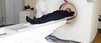
- Computed and magnetic resonance tomography;
- Echoencephalography (ultrasound examination);
- Electroencephalography;
- Study of cerebrospinal fluid (CSF pressure, protein content, isoenzyme composition, B-glucuronidase activity);
- Computed tomography with intravenous contrast;
- Scintigraphy;
- Immunohistochemical diagnostics.
Immediately on the eve of surgery, a puncture biopsy is performed to finalize the diagnosis.
During a neurological examination, the doctor evaluates the patient's eye movements, hearing, sensitivity, muscle activity, smell, balance and coordination. Using special tests, a neurologist evaluates memory and intelligence.
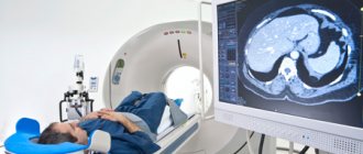
In order to identify a space-occupying lesion, its location and type, computed tomography is performed. The method allows you to identify cerebral edema, hemorrhages and other associated conditions. Computed tomography is used to evaluate the effectiveness of therapy and early diagnosis of tumor relapses.
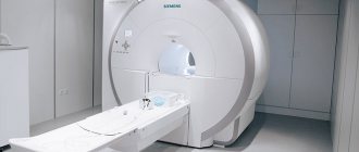
The essence of magnetic resonance imaging is to create a detailed image of the complex structures of the brain using a magnetic field. The resulting three-dimensional image of the brain is necessary for specialists to determine the location of the tumor as accurately as possible.
Magnetic resonance imaging (MRI) is the gold standard for diagnosing brain tumors. Modern devices that the Yusupov Hospital is equipped with allow doctors to obtain a three-dimensional image of the tumor. Using MRI, you can visualize space-occupying formations located near bones, small tumors, and tumors of the brain stem. MRI is also performed if necessary during surgery.
Doctors at the Yusupov Hospital determine brain activity using PET-CT (positron emission tomography). The method helps to distinguish newly appeared cancer cells from those killed as a result of radiation therapy. Thanks to the data obtained with PE-CTT, the accuracy of modern radiosurgical technologies increases. Often PET-CT is performed in conjunction with computed tomography.
Also, oncologists at the Yusupov Hospital use the following instrumental studies to diagnose brain tumors:
- Emission computed tomography – allows you to determine the degree of malignancy of the tumor;
- Magnetoencephalography – helps to evaluate the functioning of different parts of the brain;
- MRI angiography – used to assess cerebral blood flow before surgical removal of space-occupying lesions with abundant blood supply.
A brain tumor biopsy is a surgical procedure during which the doctor takes biological material from a suspicious area. Tumor tissue samples are sent to a histology laboratory to be examined for signs of atypia, a type of tumor cell. This study is carried out as an independent diagnostic procedure or during surgery to remove a tumor. If a standard biopsy is fraught with disruption of vital functions, doctors at the Oncology Clinic of the Yusupov Hospital use alternative techniques: fine-needle or stereotactic biopsy.
To identify abnormal cells or tumor markers in the cerebrospinal fluid, samples of cerebrospinal fluid are obtained using a lumbar puncture. Before performing the procedure, patients at the Oncology Clinic of the Yusupov Hospital undergo computed tomography or magnetic resonance imaging to ensure the safety of the procedure.
Causes of brain tumors in adults
Scientists have not yet determined the exact cause of tumors, but there are assumptions about the connection of their growth with the effects of radioactive radiation, toxins penetrating the body, and environmental pollution.
Children may develop congenital neoplasms - one of the reasons is considered to be intrauterine development disorders. A possible factor may be traumatic brain injury, which can also intensify an already existing process.
There is evidence that some brain tumors can develop after radiation therapy prescribed for the treatment of other pathologies, immunosuppressive treatment, and HIV infection. There is a hereditary predisposition to certain types of brain cancer. But for many people the cause remains unknown.
About 10 - 30% of brain tumors are of metastatic origin - these are cells that are carried by the blood flow (less often lymph) into the brain vessels, tissues, and membranes. About 60% of such tumors are multiple.
Most often it metastasizes to the brain:
- in men – lung damage, colorectal cancer, kidney damage;
- in women - breast cancer, melanoma, colorectal and lung cancer.
Intracerebral metastases occur with cancer of the uterus, digestive tract or prostate.
Causes of brain tumors
The exact mechanism of formation of malignant tumors has not been established. Risk factors include age over 70 years and being male. More often, tumor pathologies of the brain are found in patients who have:
- family history;
- contacts with carcinogenic substances;
- radiation exposure;
- history of traumatic brain injury;
- past inflammatory pathologies of the brain.
In some patients with detected tumors, doctors are unable to identify a single provoking factor, while in people who are regularly exposed to them, there are no brain tumors. Such information casts doubt on existing hypotheses about the origin of oncopathologies of the central nervous system (CNS).
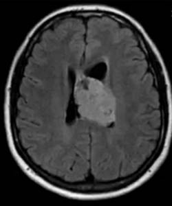
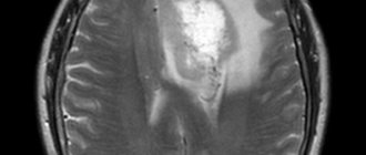
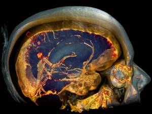
Ependymoma on MRI scan of the brain
Head tumors are often secondary in nature, i.e. metastases. According to localization, formations can be located in the bones of the skull, soft and dura maters, in gray and white matter. Melanomas, cancers of the kidney, prostate, lung, breast, and stomach most often metastasize to the structures of the head.
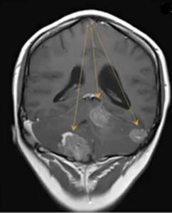
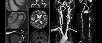
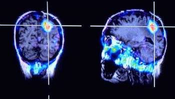
Brain metastases on post-contrast images (indicated by arrows)
Signs of a brain tumor in adults
Focal symptoms are often among the first to appear.
They arise due to the pressure of the tumor on the surrounding tissues and chemical reactions to foreign cells, hemorrhages, blockage of blood vessels with a tumor embolus, compression of tissues and nerves. As the tumor grows, symptoms from neighboring areas appear, then general brain symptoms. If the tumor is large, a so-called mass effect may occur (the main structures of the brain are displaced, causing the cerebellar region to become wedged into the opening of the skull). One of the early signs is headache. It is usually local and occurs due to irritation of blood vessels, nerves, and meninges. There may be diffuse pain throughout the head, which is typical of meningeal lesions. The nature of the pain is paroxysmal, deep, intense or bursting.
Another sign is vomiting, which is not associated with food intake. It may or may not be at the peak of the headache.
Dizziness occurs in the form of dips, rotation of the body or objects around.
Possible muscle weakness, muscle hypertonicity, uneven on one and the other side of the body, changes in tendon reflexes. Muscle-joint sensations may also suffer - sensations during movement, pressure, vibration.
For many tumors, convulsive syndrome is typical - sometimes it becomes the first sign of brain damage. There may be absence seizures or tonic-clonic seizures, Jacksonian epilepsy. Some people also experience an aura before attacks. As the tumor grows, there may be partial seizures or decreased activity of the lesions.
Some people may have mental disorders. Sloppiness, lack of initiative, aggression, euphoria or causeless gaiety, apathy, complacency can also be signs of brain damage. Tumor growth increases the severity of symptoms. Visual hallucinations, severe memory disturbances, attention problems and thinking problems may occur.
Possible problems with vision, congestion in the area of the optic nerve - these are floaters before the eyes, blurred vision, fog, blurred vision. Fields of vision may be lost.
Additionally possible:
- hearing loss;
- aphasia;
- ataxia;
- eye movement disorders;
- hallucinations (auditory, gustatory);
- autonomic dysfunctions.
If the hypothalamic-pituitary region is affected, hormonal metabolism suffers.
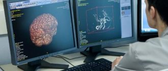
What does MRI and CT show?

As a result of diagnostics using a nuclear resonance machine, visual images of the area under study are obtained. Pictures are taken in color. Different tissues of the body are painted in different shades, the pathological area receives a different color in comparison with normal tissues. Thanks to this, diagnosticians determine the localization of the abnormal zone even at the earliest stages of its inception.
Not only the presence and location of lesions is determined, but also the presence of metastases and the degree to which they are equipped with blood vessels.
Both computed tomography and magnetic resonance imaging are capable of diagnosing the presence of oncology in organ bodies. When examining the affected areas, the following pathologies are often discovered:
- Primary neoplasms. According to statistics, this is the rarest type of gray matter abnormality, when the spread of aggressive cells is observed only within the brain body.
- Cancerous lesions of a secondary nature. If the disease is present in other parts of the body, cancerous “shoots” often spread to the head area.
- Malignant pathologies. With this anomaly, rapid, uncontrolled growth of “killer cells” occurs, which displace healthy tissues and grow into them without clear demarcation.
- Benign lesions. Unlike the previous type, these areas have clear boundaries and sizes, are separated by their own membrane from the gray matter and do not have penetrating processes.
The reasons for suspecting an anomaly are the following symptoms:
- CNS disorders.
- Gradual increase in intracranial pressure.
- The appearance of constant pain due to displacement of a healthy part of the organ.
Symptoms related to the appearance of a malignant neoplasm include:
- increasing, dull pain in the head;
- regular attacks of nausea and vomiting in the morning;
- lightheadedness, dizziness;
- loss of orientation in space;
- convulsive conditions;
- hearing loss or decrease;
- decreased level of vision, feeling of “interference” in front of the eyes.
Stages of brain tumor in adults
A typical stage for a cancerous lesion determines treatment options, prognosis, and typical signs and symptoms.
There are 4 stages in total - from the mildest to the extremely severe. The first stage is when one of the parts of the brain is affected by a small neoplasm, a nodule, there is no penetration into neighboring tissues, there is no pressure on the surrounding areas of the brain.
The second stage - the tumor grows slowly, but there is penetration into neighboring areas;
The third stage - the tumor changes its structure, cells divide faster, neighboring sections and tissues grow;
Stage four – the tumor is large, grows into neighboring brain structures, and distant metastases are possible.
What does a tumor look like on an MRI of the brain?
If you compare benign and malignant pathologies, they will look different on the tomograph images.
New growths of the first type are visible as darker areas compared to normal areas. Only a professional physician can distinguish them. This formation is distinguished by its slow growth rate, clear contours, and lack of “migration” beyond the affected area. The disease does not spread to adjacent cells and other organs.
The most common classes of benign formations:
- Glioma of the first stage of development. This name is due to the fact that the tumor develops from glial cells of the gray matter.
- Meningioma.
- Schwannoma. This disease affects only the auditory nerve, without spreading to adjacent tissues.
- Hemangioblastoma. It differs in that it affects only the vascular network.
How to identify a cancerous lesion in a tomograph image? Firstly, the contours of such formations are quite blurred. Secondly, the color of the problem area completely matches the color of healthy tissue, which is a characteristic feature in this situation. Thirdly, metastases are very clearly visible.
If cancer is suspected, specialists usually prescribe an examination with intravenous administration of a contrast agent. It is this that colors the affected areas, which become visible even at the earliest stage of appearance.
Diagnostics
The first examination is carried out by a neurologist.
The doctor evaluates general complaints, conducts an examination, examines muscle tone, reflexes, communication, emotions and cognitive functions. EEG and echo-encephalography are also performed. A visit to an ophthalmologist is indicated to assess the condition of the fundus, acuity, and visual fields. Suspicion of a tumor formation is an indication for a CT or MRI of the brain. Angiography of cerebral vessels, PET scan and other complementary examinations may be prescribed. In some cases, a biopsy can help determine the type of cancer, but it is not possible in all cases.
Modern methods of treatment
“The volume of treatment is selected depending on the course of the brain tumor,” says oncologist Alexander Seryakov.
– The most effective is surgical removal of the tumor. The use of surgical microscopy and navigation allows for radical removal of the tumor and minimizes injury to healthy tissue. If the tumor cannot be completely removed, after its partial removal, the patient is prescribed radiation, as well as chemotherapy, targeted (“targeted”) or immunotherapy. For small malignant tumors (up to 3 cm), stereotactic radiosurgery is possible (using Cyber-Knife linear accelerators or Gamma-Knife gamma machines). To remove tumor tissue, many beams of radiation are used at once, which are directed at one point or collected into one beam. Its direction will constantly change during the irradiation session, but the beam necessarily passes through the tumor tissue.
For large tumors, and especially in cases where it is impossible to surgically remove the tumor, classical external beam radiation therapy is used. In some cases, it is used after surgery to reduce the risk of relapse (regrowth).
Chemotherapy is carried out with cytostatic drugs, taking into account the type of tumor. The effectiveness of chemotherapy increases significantly if it is combined with radiation therapy.
Targeted therapy in combination with radiotherapy and chemotherapy improves survival of patients with aggressive brain tumors. Most often used in the treatment of glioblastomas.
Currently, methods of using immunotherapy for malignant tumors (antitumor vaccines, immunotherapy drugs) and its integration into existing standards of treatment are being considered.
Can an MRI show a brain tumor?
Based on the results of magnetic resonance imaging, doctors can identify changes in tissue structure. Brain tumors on MRI are determined by direct and indirect signs. Doctors suspect a pathological formation based on the type and uniformity of changes in the MR signal:
- hypo- (weaker than surrounding areas) or hyperintense (response stronger than from normal areas);
- heteromodified (different characteristics are mixed);
- isointense (the tumor looks like other tissues, but differs from the normal architecture of the brain).
There are also indirect signs. The latter give reason to suspect a brain tumor against the background of an isointense tissue response or confirm its presence with an altered MR signal.
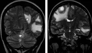
Multiple metastases in the brain substance on MRI without contrast
Secondary manifestations of pathology may be:
- lateral dislocation of the median structures;
- displacement of the vascular bundle;
- changes in the size and deformation of the ventricles;
- axial dislocation;
- occlusive hydrocephalus due to blockage of the cerebrospinal fluid pathways;
- displacement or deformation of the basal cisterns;
- cerebral edema in the tumor area and around it (local, generalized, total, periventricular).
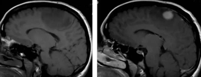
Pre- and post-contrast images of a malignant brain tumor
Using MRI for brain cancer, the localization of the tumor, the degree of its invasion into neighboring structures, features of the blood supply, stage of the disease and the presence of metastases are revealed. To obtain additional information, scanning is carried out in various sequences (T1, T2, FLAIR, diffusion-weighted, etc.), in native mode and after contrast.
Prevention of brain tumors in adults at home
Prevention of brain tumors, according to oncologist Alexander Seryakov, generally includes:
- healthy lifestyle;
- optimal physical activity (preferably in the fresh air);
- complete rest;
- giving up bad habits (for smokers and alcohol abusers, the likelihood of developing brain tumors increases by almost 30%);
- a diet rich in fruits and vegetables;
- limiting stressful situations (or changing your attitude towards negative circumstances).
Symptoms of brain cancer
Malignant tumors account for almost half of the identified intracranial neoplasms. Common manifestations of pathological changes in the brain can be:
- headaches, often in the morning, with nausea and vomiting;
- vertigo (dizziness of central origin);
- impaired coordination of movements;
- deterioration of vision, hearing;
- speech disorders;
- irritability;
- drowsiness;
- convulsions;
- behavioral disorders;
- weakness in the muscles of the face or limbs;
- numbness, paresthesia in certain parts of the body;
- mental disorders, etc.
The clinical picture of tumors differs depending on the location, size, and type of tumor. Pathology is differentiated based on data obtained from instrumental and laboratory examinations and histological analysis of tissues.
Based on how a brain tumor looks on an MRI, doctors make assumptions about its nature. There can be no unambiguous statements, since the verification of formations is carried out using histological analysis. Characteristic signs of malignant brain tumors on MRI images are:
- their shape is incorrect;
- unclear contours of neoplasms;
- pronounced perifocal edema;
- mass effect (displacement of nearby structures);
- hydrocephalus;
- heterogeneous accumulation of contrast (sometimes they do not accumulate at all);
- “variegation” of the tumor (hemorrhages, necrosis and/or cysts in the structure), etc.
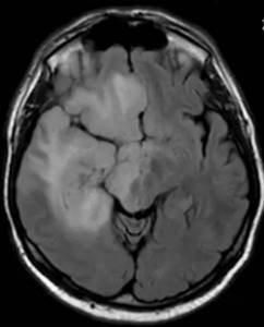
Glioblastoma on brain MRI without contrast