Adults
Children
Doctors Cost
Price list Doctors clinic
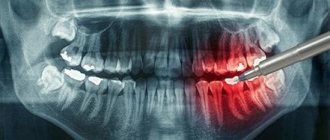
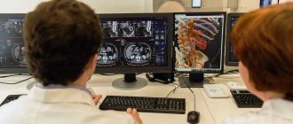
Cone beam computed tomography (CBCT) is one of the most accurate radiation diagnostic methods. Allows you to obtain a three-dimensional image of teeth or any part of the maxillofacial area. Using the data obtained, the specialist will make an accurate diagnosis and develop the most appropriate treatment regimen.
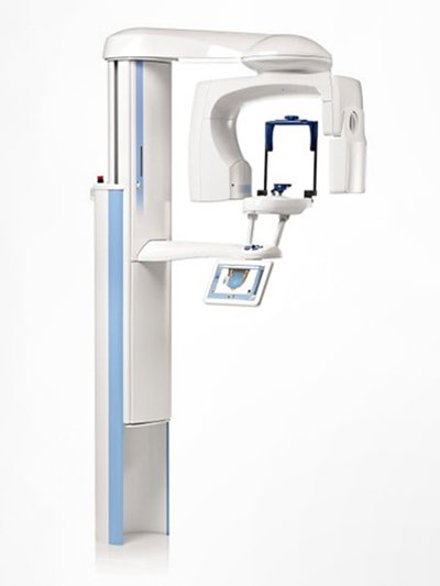
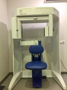
In the building of the Family Doctor clinic on Usacheva, a modern X-ray cone-beam computed tomograph ORTOPANTOMOGRAPH OP300 Maxio from Kavo is installed. It is designed for use in dentistry and otolaryngology.
Indications for CBCT
In dentistry, there are many indications for cone beam tomography. The main ones include the following:
- planning orthopedic or orthodontic treatment, dental implantation;
- planning endodontic treatment (filling root canals), as well as assessing treatment results;
- making decisions on osteoplastic operations;
- control of the position of implanted implants;
- making a decision before complex removal of impacted teeth;
- diseases of the temporomandibular joint;
- assessment of the consequences of injuries suffered, etc.
This method can also be used in the complex diagnosis of diseases of soft tissues and jaws.
Computed tomography of teeth
CBCT (cone beam computed tomography) allows you to create very informative 3D images that differ from conventional X-rays in a number of ways:
- High image quality.
- Possibility of creating a three-dimensional image.
- High research speed.
- No overlap of anatomical structures.
- Significant reduction in radiation exposure due to the speed of examination.
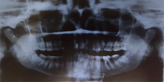
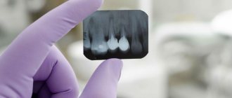
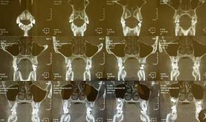
Panoramic photograph on film
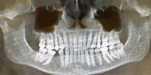
Panoramic image from CBCT
The doctor who owns such an instrument sees the features of the patient’s anatomy: the number of teeth, the location of the teeth in the bone, the angles of inclination of the roots of the teeth, the volume and quality of the bone tissue of the jaws, the size of the jaws and their position in the skull, the condition of the bone structures of the temporomandibular joints, size respiratory tract. This is important when planning orthodontic treatment.
This study makes it possible to make linear measurements of the length of tooth roots, the thickness and density of bones, the location of the maxillary sinuses and bone canals in which blood vessels and nerves pass. The latter is very important if the patient is undergoing implantation or treatment of tooth roots. CBCT allows you to identify pathological processes in teeth and jaws (inflammation, cysts, neoplasms). Visualization of the nasal septum, maxillary and frontal sinuses, paranasal sinuses and nasal passages allows us to identify pathology and refer the patient to an ENT doctor.
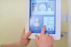
No special preparation is required to conduct a CBCT examination. The patient can be referred for diagnostics directly on the day of the initial examination. The study is carried out in a sitting position for 20-30 seconds in comfortable conditions. A sensor rotates around the face, capable of detecting rays from an ionizing source. Sensors transmit information to a computer. The signal is processed by the program. The results are recorded on a CD. It is possible to send pictures by email.
Contraindications to the study:
- Pregnancy (the X-ray dose in the machines is very small, but it can be potentially dangerous for the development of the fetus).
- Pronounced pain syndrome.
- Hyperkinesis is a neurological disease when the patient is unable to remain in the required position for at least a minute or two.
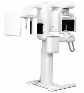
CT scanners used in our clinics
The ORTHODONT CENTER clinics use three types of devices:
- On Tula - KaVo 3D eXam / i-CAT.
- On Tushinskaya - VATECH PaX-i3D.
- On Novoslobodskaya - Planmeca ProMax 3D.
Planmeca ProMax 3D
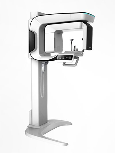
VATECH PaX-i3D
KaVo 3D eXam / i-CAT
Advantages of the method
The diagnostic capabilities of CBCT are wide: the method allows you to accurately assess the anatomical features of the root canals, alveolar processes, bone tissue, joints, etc.
Main advantages of the method:
- Minimum radiation exposure. A lower dose of radiation, compared to a standard x-ray, allows you to minimize harm to health and repeat the diagnosis more often if necessary.
- Speed of the procedure - the examination takes no more than 90 seconds.
- Lack of special training.
- Visualization of the study area in volume.
- Possibility of additional processing of the obtained results.
Based on the information obtained as a result of the examination, the doctor can choose the most appropriate treatment method and predict the results of therapy.
Preparation and execution
One of the advantages of the method under consideration is the absence of the need for preliminary preparation - if prescribed, the patient can immediately undergo the procedure without leaving the clinic. The only condition is the removal of jewelry, as well as the removal of removable metal prosthetic structures. In situations where the patient wears fixed metal-ceramic prostheses, the doctor should be notified in advance, who will change the basic settings of the equipment.
Based on the location of the area being examined, diagnosis is carried out in a sitting or standing position. In any case, a mandatory condition is to wear an apron with a lead layer on the chest, which protects the patient’s body from radiation.
To obtain a three-dimensional image, an X-ray tube is used, rotating around a given area and directing the rays - with subsequent retransmission to the screen of a stationary PC. The shooting speed is about 30 pictures per second, the total duration of the diagnosis is no more than two minutes, during which the patient must remain motionless.
Advantages and disadvantages
Positive aspects noted by specialists with experience in using CBCT include:
- Speed of obtaining diagnostic results;
- Minimum radiation dose compared to conventional x-rays;
- No need for additional preparation;
- Accuracy of readings obtained in three-dimensional projection.
Among the disadvantages, it is worth noting the costs associated with the purchase of equipment for computed tomography, as well as training of doctors to obtain access to the protocol. This fact determines the insufficient prevalence of equipment in both public and private clinics.
Model range of dental tomographs. Review
At the moment, there are dozens of models of dental computed tomographs on the CBCT market. In Russia, devices made in Japan (Morita), Germany (Kavo), Finland (Planmeca) and South Korea (Vatech, Genoray) are popular. Units vary in price, patient positioning, image resolution, functionality, software capabilities, dimensions, and warranty terms.
Despite the fact that the first dental CBCT devices were developed in Japan, currently this technology is most actively developed by manufacturers from Europe (Germany, Finland) and South Korea. Dental tomographs from these manufacturers differ from their analogues in more understandable software (maximally adapted to the needs of the dentist), ease of operation, relatively compact size and affordable cost.
Table 1. Comparison of characteristics of dental tomographs KAVO, Planmeca, VATECH, Genoray
| Characteristic | KAVO (Germany) | Planmeca (Finland) | VATECH (South Korea) | Genoray (South Korea) |
| Scan time | 8-20 sec | 18-26 sec | 10-24 sec | from 7.7 sec – fastest scanning |
| Functions: CBCT, orthopantomography, TRG (cephalostat) | + | + | + | + |
| Positioning | standing and sitting | standing and sitting | standing and sitting | standing and sitting |
| Cephalostat scanning areas | 22*26 | 27*30 | 23*27 | 23*24 |
| Voxel size | 80-400 microns | 75-400 microns | voxel size (mm) 0.2/0.3 mm | 70-400 microns – highest resolution |
| Image detector | CMOS | CMOS | CMOS | CMOS |
| Current strength RT | 2-16 mA | 1-16 mA | 4-10 mA | 4-12 mA |
| RT voltage | 60-95 kV | 54-90 kV | 50-90kV | 60-90 kV |
| Focal spot | 0.5 mm | 0.5 mm | 0.5 mm | 0.5 mm |
| Shooting at low radiation dose | + | + | + | + |
South Korean Papaya 3D units produced by Genoray are in high demand among dentists. GENORAY Corporation is one of the three largest manufacturers of diagnostic X-ray equipment in Korea. The company is a full-cycle enterprise, that is, it specializes not only in the development of equipment and the production of components, but also in the assembly of medical equipment.
Distinctive features of the Papaya 3D Genoray CBCT scanner
- The maximum resolution in its class
e is 70-120 µm: for accurate diagnostics in endodontics and examination of the upper and lower dentition. - There is no need to purchase additional licenses for the software:
while with other manufacturers this needs to be done when the number of users increases. - Extended warranty of 5 years:
while the warranty for tomographs from other manufacturers does not exceed 12-24 months. - Auto positioning for all types of studies:
the function facilitates and speeds up the procedure. - SMARF algorithm:
reduces artifacts caused by foreign bodies made of metals so that they do not spoil the quality of images. - Modular architecture:
the basic device can be easily retrofitted with a cephalostat, and the design also allows for expansion of the scanning area - without replacing the device. - Maximum comfort for patients:
during the procedure, the patient is face to face with the doctor, which facilitates interaction with the patient and speeds up the examination. You can also set a voice notification about the beginning and end of the procedure. - Ability to examine patients with disabilities,
including people in wheelchairs.
Recommendations for purchasing CBCT dental tomographs
The choice of a CBCT device is made based on the needs of the clinic, software capabilities and equipment costs.
Thus, before purchasing a device, ask yourself the following questions:
- How much money are you willing to spend on a dental tomograph?
- Which specialists will operate the CBCT machine and what procedures will be performed?
- What are the dimensions of your office and are you limited in space?
- Why do I need this, and what benefits will I get from purchasing a CBCT?
Benefits of purchasing a dental tomograph
Increased income for the clinic
A CBCT device is a profitable investment for a clinic. The purchase of the device allows the clinic to expand its range of services with more popular and expensive diagnostic procedures. So, if an orthopantomogram in Moscow costs from 1,000 to 3,000 rubles, then 3D tomography costs from 2,000 to 10,000 rubles. At the same time, a CBCT device pays for itself as quickly as an orthopantomograph - due to the high price of the procedures and the large number of indications for the procedure (see above).
If the device performs 5 procedures per day (at an average price of 4,500 rubles for examining two jaws), you will pay for a mid-price dental tomograph costing 6-8 million rubles. in just 1 year. The second year of operation will bring you 8,212,500 rubles. (4500*5*365) – with the same frequency and cost of procedures and excluding expenses.
Own diagnostic center at your disposal
If you purchase a dental tomograph with a cephalostat and an orthopantomography function, then you have a full-fledged diagnostic machine at your disposal. You close the cycle of treating and diagnosing patients within your clinic. Thus, you not only attract new clients, but also eliminate the possibility of losing a patient by sending him to another dental center for diagnostic procedures.
More patient trust
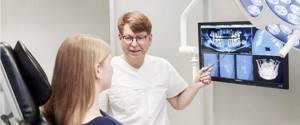
CBCT images display pathological areas and anatomical features of the dental-facial system so clearly that they become understandable even to non-specialists. The patient should no longer take the doctor's word for it. For example, a doctor can clearly demonstrate to a patient the number of canals in a tooth that require filling, and thereby justify the cost of the procedure.
After canal treatment, implant installation or surgical treatment, the doctor can take an image showing the positive results of his work, which increases the patient’s confidence and motivation to continue treatment or rehabilitation procedures in your clinic.
Increasing the prestige of the clinic and increasing the professionalism of specialists
By installing the device, you will dramatically increase the status of the clinic and the level of your staff: when such equipment appears in the clinic, dentists, learning how to use it, improve their professional level.
Protection from lawsuits
During forensic medical proceedings, visual CBCT images can play an important role, namely, confirming the absence of a medical error. Moreover, all information obtained during examinations is stored in the database of the CBCT computer system: the doctor can access it at any time.
Types of modern CBCT machines
Modern dental tomographs differ in scan volume (field of view or FOV), the presence of a cephalostat and patient positioning.
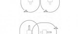
Field of View (FOV)
CBCT machines are classified by field of view or scanning height as follows:
- For a localized area (dentoalveolar complex): about 5 cm
- For one jaw, upper or lower: 5-10 cm
- For both jaws: 7-10 cm
- For the maxillofacial area: 10-15 cm
- For the craniofacial region: more than 15 cm
Field of view (FOV) is one of the most important parameters of a CBCT scan and depends on the size and shape of the detector, as well as the features of the collimator (a device that blocks X-rays that do not pass through the area of interest).
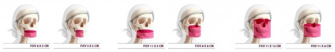
The larger the maximum field of view, the higher the installation cost will be. For this reason, before purchasing a device, you need to decide what shooting modes you will need so as not to overpay for equipment. For example, for implantology, an FOV of 8×8 (in some cases – 10×10) is sufficient; for otolaryngology and maxillofacial surgery – from 10×15. For devices with modular architecture, the choice of FOV can be expanded in the future.
Equipment with cephalostat
The cephalostat is a separate “arm” of the dental tomograph. It is used by orthodontists to create teleroentgenograms—images of frontal and lateral projections of the skull. The photographs allow you to study the position of the jaws relative to the skull and each other, as well as the inclination of the teeth and are mandatory when planning the installation of braces.
There are the following models on the market: with cephalostat in the basic configuration, with the possibility of retrofitting, and without the possibility of retrofitting.
Clinics where an orthodontist works require a device with a cephalostat. If orthodontic services are not provided, but are planned in the future, the medical center requires a dental tomograph with the ability to retrofit with a cephalostat.
Patient position
There are three different types of CBCT platforms, depending on the patient's position:
- Sitting position
- Standing position
- Patient supine position
Devices that scan the patient while standing are more suitable for wheelchairs and take up no more space than orthopantomographs. Sitting units are considered more comfortable, but they take up more space and make the examination of patients with disabilities more difficult. CBCT devices with the patient in the supine position occupy the largest area and volume and are not adapted for examining all patients with physical disabilities.