directions
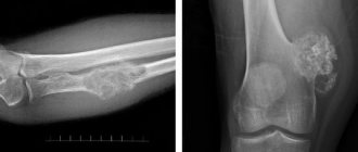
X-ray of the lower extremities is a non-invasive, informative and very simple method of examining the leg, which allows diagnosing injuries of varying severity, damage, congenital and acquired diseases.
The very concept of radiography of the lower extremities is broad, because this can mean the examination of various lower extremities: foot, toes, knee joint, etc. Depending on the problem that has arisen in a particular part of the leg, the doctor will order an x-ray of a certain part of the lower extremities and identify the pathology.
Currently, work is underway on the website to change the price list; for current information, please call: 640-55-25 or leave a request, and an operator will contact you.
Prices for services
- X-ray of the hip joint in direct projection 900a
- X-ray of the ankle joint in direct posterior projection 900a
- X-ray of the knee joint in direct projection 900a
- X-ray of the patella 900a
- X-ray of the calcaneus in direct projection 900a
- X-ray of the toe in direct projection 900a
- X-ray of the hip joint in lateral projection 900a
- X-ray of the femur in direct posterior projection 900a
- X-ray of the femur in lateral projection 900a
- X-ray of the calcaneus in axial projection 900a
- X-ray of the knee joint in lateral projection 900a
- X-ray of the calcaneus in lateral projection 900a
- X-ray of the tibia-fibula in the lateral projection 900a
- X-ray of the ankle joint in lateral projection 900a
- X-ray of the lower leg in direct posterior projection 900a
- X-ray of the lower leg in lateral projection 900a
- X-ray of the foot in lateral projection 900a
- X-ray of the foot in frontal projection 900a
- X-ray of the phalanges of the toes in a direct projection 900a
- X-ray of the pelvis laying according to Launstein 1350a
- X-ray of the foot in oblique projections 1350a
- X-ray of the pelvis in direct posterior projection 1350a
The information and prices presented on the website are for reference only and do not constitute a public offer.
Preparation for the procedure
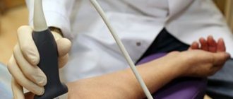
Angiography of the aorta and vessels of the lower extremities requires compliance with certain preparation rules:
- In 2-3 days, the patient undergoes a series of laboratory tests: general blood test, biochemistry, coagulogram, test for HIV, hepatitis, syphilis, Rh factor.
- One week before the procedure, it is prohibited to use blood thinning medications.
- Two weeks before the procedure you need to give up alcoholic beverages.
- A few hours before the test you need to refuse food and water. If the procedure is scheduled for the morning, then the last meal before it should take place the night before, no later than 18:00.
- Also the night before, the patient is asked to take an antihistamine (to minimize possible allergic reactions to the contrast) and a sedative (to relieve anxiety).
Our clinics in St. Petersburg
Structural subdivision of Polikarpov Alley Polikarpov 6k2 Primorsky district
- Pionerskaya
- Specific
- Commandant's
Structural subdivision of Zhukov Marshal Zhukov Ave. 28k2 Kirovsky district
- Avtovo
- Avenue of Veterans
- Leninsky Prospekt
Structural subdivision Devyatkino Okhtinskaya alley 18 Vsevolozhsk district
- Devyatkino
- Civil Prospect
- Academic
For detailed information and to make an appointment, you can call +7 (812) 640-55-25
Make an appointment
We draw your attention to the schedule of technological breaks in the CT and X-ray rooms.
Today, an X-ray examination of the lower extremities can be done in almost all clinics, hospitals and private medical institutions. But most often you will need to make an appointment with the specialist who prescribes the examination. Or you will have to wait in line at the city emergency room. The condition of the equipment used for X-ray examination is also important. The equipment must be modern, safe and clearly show the identified pathologies, lesions and injuries in the image.
You can take an x-ray of your legs in the emergency room of a multidisciplinary medical center, where you will be given first aid by experienced and competent traumatologists, orthopedists and radiologists, they will quickly and efficiently take x-rays of your legs using a safe high-tech X-ray machine Clinomat (made in Italy), they will identify and diagnose a disease or injury and prescribe an effective treatment.
Pain when walking, swelling of the leg, burning, limited movement, pain and crunching when bending - all these unpleasant symptoms do not arise without reason. If there was no direct damage to the leg, i.e. injuries, it is imperative to find out the cause and begin treatment in a timely manner. But even a careful examination by a competent specialist does not always help determine the source of the disease. As a rule, with such signs, the doctor will most likely order an x-ray.
How are x-rays taken of the bones and joints of the arms and legs?
An X-ray of the bones of the legs and arms takes on average no more than five to ten minutes and does not require the use of painkillers, since it is an absolutely painless and safe procedure.
Tubular bones are most often examined. To obtain high-quality images, two perpendicular projections are used. The main condition for the patient is to remain motionless during the shooting. The procedure includes the following:
- The limb being examined must be freed from clothing and any metal objects;
- The limb is placed in the center of the X-ray cassette and the rays are directed at it at the appropriate angle;
- The radiologist takes pictures in such a way that adjacent joints are also clearly visible. This approach allows for high-quality visualization of the entire bone and ensures that adjacent joints and bones are not damaged.
If it is necessary to conduct a functional study, the patient is asked to flex/extend the joint of the area being examined or provide a load on the limb being diagnosed. There are often cases when, due to illness or injury, the patient cannot achieve the required position of the arm or leg. Then CELT radiologists use special projections, not forgetting to maintain the perpendicular position of the cassette relative to the X-ray emitter.
Reasons for contacting a radiologist and traumatologist in the area of the lower extremities
- sudden lameness;
- joint pain;
- pain when walking;
- characteristic clicks or crunches when bending or straightening the leg;
- weakness in the limbs;
- injuries and mechanical damage, etc.
In all of the above cases, an x-ray is required - a picture of the leg, which will help identify bone disorders, assess the condition of the joints, diagnose tumor processes, inflammatory processes of soft tissues, dislocations, subluxations, fractures, tendon ruptures and other injuries, developmental pathologies in children, etc.
The causes of ailments affecting the lower extremities can be factors such as a sedentary lifestyle, excessive stress on the legs, poor nutrition, hypothermia, metabolic disorders, excess weight, various injuries, including birth, infectious diseases, etc.
Types and features of radiography of joints and bones of the arms and legs
The procedure can be carried out in the usual way, or maybe using a contrast agent. If the patient has indications, the diagnostician can conduct functional studies of the joints and determine their ability to flex/extend. When examining the bones and joints of the lower extremities, a static load is used, which allows one to identify a number of important diagnostic data.
The use of techniques using a contrast agent allows for targeted examination of not only bones, but also joints, along with blood vessels and lymph nodes.
| Methodology | Indications for use | Peculiarities |
| Pneumoradiography | Examination of the soft tissues of the hands and feet. | Involves the use of air or gas as a radiopaque agent. |
| Angiography | Assessment of the condition of the blood vessels of the upper and lower extremities. | Contrast study of vascular functions, which allows you to determine the state of blood flow and identify any existing pathological processes: narrowing, aneurysms, obstructions. |
| Arthrography | Determination of the condition of articular joints. | A contrast agent is injected into the joint cavity, the patient is asked to make movements involving it, and his condition is studied. Most often used to diagnose large joints of the upper and lower extremities. |
| Fistulography | Study of pathological fistula tracts. | Such x-rays of the joints of the arms and legs, as well as their bones, make it possible to detect infections and determine the consequences of injuries. The process uses various contrast agents: iodine-based, water-soluble, oily or aqueous suspensions of barium sulfate. |
| Lymphography | Assessment of the condition and identification of pathologies of the lymph nodes of the arms and legs. | It is aimed at determining the state of the lymphatic system of the arms and legs and involves the introduction of contrast directly into the lymphatic vessels, followed by tracking its movement using X-rays. |
X-ray of the lower leg
Posted at 16:20h in Services by doctor
As a result of the transition to upright walking, the legs took on the main load. The lower limbs (legs) perform the function of support and movement of a person. Each of the two human legs consists of 26 bones and, of course, muscles. The structure of the lower extremities is conventionally divided into three main parts:
- femur (femur and patella, which protects the knee joint);
- shin (tibia and fibula);
- feet (consist of many small bones)
Due to the complexity of the function performed and constant loads, the lower extremities are often susceptible to injuries of both a traumatic nature and various kinds of diseases. A very effective way to confirm a preliminary diagnosis based on clinical signs and symptoms is x-ray diagnostics.
How unsafe is an X-ray of the legs and how can you be treated with an X-ray?
Everyone knows about the dangers of ionizing radiation. However, everything depends on the degree of radiation and the total radiation exposure, because such radiation tends to accumulate in the body. That is why, after each X-ray diagnostic procedure, a note is made on the corresponding accounting sheet about the amount of radiation dose received. An average person who goes to the dentist several times a year and takes an X-ray and undergoes a routine fluorography procedure will receive, in addition to natural background radiation, exposure equal to approximately 15 mSv. And a dose of less than 10 μSv may not be taken into account at all.
Restrictions on the procedure may be associated with:
- severe condition of the patient, especially if bleeding occurs;
- pregnancy;
- under 15 years of age;
- significant total radiation dose received previously
However, given the insignificant radiation exposure of X-ray diagnostics of the legs, the information content of the method should prevail over the desire to avoid radiation.
Recently, thanks to the development of low-dose X-ray equipment, such a common disease as a bone growth on the surface of the heel bone in the form of a spike (wedge), also known as a “heel spur,” is effectively treated with the help of X-rays. Modern devices make it possible, firstly, to reduce radiation exposure, and secondly, to act purposefully, that is, without affecting the areas of other organs. This method makes it possible to very quickly remove pain and stop pathological processes. The dose of radiation received during the procedure does not exceed 10 μSv (0.01 mSv).
Possible complications of angiography
After angiography
A number of complications may arise, regardless of the doctor’s qualifications and what equipment was used (although it is better to trust your health to experienced doctors and modern technology). Among the main complications:
- Allergic reaction. It is usually associated with the patient’s individual intolerance to the components of the contrast agent. To avoid or minimize this complication, it is recommended that an allergy test be performed before the x-ray.
- Bleeding localized at the puncture site. This complication can be avoided by correctly applying a tight bandage immediately after removing the catheter.
- Swelling or hematoma at the puncture site.
- The development of renal or heart failure is extremely rare, although it cannot be excluded.

