Sign up
Price
Our doctors
Ultrasound of joints in adults and children shows specialists the condition of all their constituent structures - ligaments and tendons, cartilage and muscle tissue, synovial bursae. In a few minutes, the procedure allows you to create a reliable picture of any pathology affecting these areas of the human body. The action of an ultrasound device is based on the reflection of a special type of waves from the biological media of the body; the signal that is obtained when affecting healthy tissue will be different from the signal from pathological areas. The procedure, which is painless and easy for the patient, helps to identify changes and deformations in bone tissue and accurately assess the processes occurring in articular cartilage. At the same time, early diagnosis of diseases allows you to promptly begin therapy and obtain the most positive treatment results.
ULTRASOUND OF JOINTS AVAILABLE AT BRANCHES:

Ultrasound of joints in the Primorsky region
Address: St. Petersburg , Primorsky district, st. Repisheva, 13

Ultrasound of joints in the Petrograd region
Address: St. Petersburg , Petrogradsky district, st. Lenina, 5

Ultrasound of joints in Vsevolozhsk
Address: Vsevolozhsk , Oktyabrsky Prospekt, 96 A
Unlike other types of medical diagnostics, which provide a static image, this type of examination allows you to monitor the functioning of the joints in dynamics, which is critically important for identifying pathology.
How is ultrasound performed?
Ultrasound of the knee and other joints in the Art Medic clinic is carried out according to standard international protocols. Typically, the study is carried out in a resting position, then functional diagnostics are shown in dynamics (flexion - extension). For example, during a spinal ultrasound, the patient is asked to tilt their head forward and back to examine the cervical vertebrae.

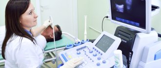
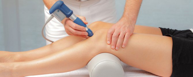
A special conductive gel is applied to the skin of the body, which provides an airless environment. This is necessary so that the ultrasonic waves do not attenuate. The doctor moves a special sensor over the patient’s body in the place where the joint is projected onto the surface. The image is displayed on the screen of the ultrasound machine.
Ultrasound is often performed not for diagnosis itself, but as a treatment control. For example, when administering drugs intra-articularly, the doctor focuses on the image on the monitor of the ultrasound machine, so as not to damage the surrounding tissue with the syringe needle.
When is it indicated to do an ultrasound of the joints?
The procedure is carried out to identify defects in the structure and functionality of the shoulder and elbow joints, hand and wrist, knee and hip joints, and ankle. Situations in which this type of wave diagnosis is indicated include:
- joint area injuries;
- subjective painful sensations;
- restrictions in mobility (contracture);
- swelling of soft tissues and changes in their color;
- autoimmune pathologies;
- disturbances in the functioning of the endocrine system;
- compaction of soft tissue elements;
- rheumatic ligamentous defects;
- arthrosis and arthritis, as well as osteoarthritis;
- hygroma;
- bursitis.
The procedure is also indicated for suspected fractures and dislocations; in the presence of bruises and sprains of elements of the musculoskeletal system.
In newborns and infants, the procedure is done to assess the development, condition and topography of the hip joint to exclude dysplasia or congenital hip dislocations.
Indications for the procedure
The main types of injuries, as well as pathological processes for which this examination is prescribed:
- mechanical injuries,
- diagnosis after suffering from Lyme disease,
- arthrosis,
- arthritis,
- overweight,
- incessant pain
- endocrine disorders,
- inflammatory processes,
- immobility.
In addition, ultrasound examination can also be prescribed for preventive purposes after 45 years.
Using this method you can identify:
- Synovitis, ruptures of the synovial membrane, hemarthrosis;
- Various inflammations and damage to ligaments (chronic tendonitis, rupture of tendons or ligaments);
- Damage to the meniscus (cysts, ruptures of its horns);
- The presence of loose bodies in the joints;
- Various bone pathologies;
- Tumors.
Types of ultrasound of bones and joints
Ultrasound of the elbow joint. It is used as a method of recording the dynamics of the process where lesions have already been identified (for example, with inflammation of cartilage tissue, with the appearance of fluid in the joint capsule, in the presence of a Baker's cyst). There are categories of people who are at risk for developing pathologies in this particular anatomical area - tennis players (there is even the concept of “tennis elbow”) and musicians. During work, all of them produce multiple flexion-extension movements, creating increased loads on the musculoskeletal system. People at risk are recommended to undergo preventive diagnostics 1-2 times a year for early detection and treatment of ailments.
Ultrasound of the hip joints. Visualization of articular cartilage, ligaments, and soft tissues is used to detect a wide range of pathologies associated with destructive or degenerative changes. Problems in the pelvis and hips also appear after blows, falls, injuries or prolonged compression. It is possible to carry out the procedure if a characteristic crunch appears in the joints during movement, or with spasms of the striated muscles of the thighs and buttocks. For newborns, the main purpose of wave diagnostics of this type is to recognize congenital femoral dislocation; the pathology is common and is detected in 15 out of every 1000 young children.
Ultrasound of the knee joint. This type of functional-organic diagnostics is prescribed for sprains, connective tissue defects, and arthritis. The study visualizes the ligaments and their contours (straight or deformed), shows the condition of the bone cortical layer, the amount of synovial fluid and pathological inclusions, if any, and demonstrates the condition of the meniscus. As a method of occupational examination, the procedure is used for people at risk - these are wrestlers, skiers, gymnasts, speed skaters and snowboarders. For these people, the load on the knee joints is very high.
Ultrasound of the ankle joint. Hardware diagnostics make it possible to differentiate sprains and ruptures of ligaments and tendons, identify deformation of the structural elements of the joint, changes in shape, soft tissue disintegration, the condition of the heel bursa and bone structures adjacent to the ankle.
Ultrasound of the shoulder joint. The wave diagnostic technique will help determine how damaged the cartilage, bone or soft tissues are after injuries, bruises, falls, or prolonged compression of the shoulder girdle area. The procedure is prescribed when early signs of inflammatory lesions appear and when there is destruction (swelling, redness, hardening) of the soft tissue elements of this anatomical area.
What does the diagnosis show?
An ultrasound of the joints shows what is happening to them at the moment. With the help of an examination, injuries, pathologies, cysts, degenerative and inflammatory processes can be identified. Let's look at each point in more detail.
Pathologies in the joints
Ultrasound of joints is also done to identify pathologies. These can be transformations after operations, inflammatory processes, dystrophic changes.
Based on the results of the examination, heterogeneity of the structure, hyperechoic formations, edema, displacement of ligaments and tissues can be revealed. These changes may indicate degenerative processes.
Detection of a cyst
A cyst is a hollow tumor, usually filled with fluid. Using ultrasound, you can identify a cyst in the joints and evaluate its development. Most often, cyst removal is only possible through surgery. If left untreated, it can grow and/or become malignant.
Injuries
Joint injuries can occur for various reasons: a fall, high load, awkward movement. During ultrasound diagnostics, it is possible to identify the consequences of these injuries: rupture of ligaments, tendons, hematomas.
Degenerative processes
Factors in the development of degenerative diseases are osteoporosis (decreased bone density) and wear and tear of the joint with subsequent destruction of cartilage. Mostly elderly people are susceptible to these processes. Subsequently, various types of arthrosis arise. Ultrasound clearly shows these processes and the condition of the joints (for example, narrowing of the gap between the joints, irregularities).
Joint inflammation
Inflammation is a normal reaction of the body to the ingress of pathogenic bacteria. For any manifestations of inflammatory processes in the joints (pain, swelling, redness, limited mobility, crunching, fever), you must immediately consult a doctor to undergo an examination and receive the necessary treatment. Neglected inflammatory processes lead to chronic diseases (gout, various arthritis) and even disability. During an ultrasound of joints and limbs, inflammation is indicated by free fluid in them.
Cost of ultrasound of joints:
| Services list | Price in rubles | |
| Saint Petersburg | Vsevolozhsk | |
| Ultrasound of the knee joints | 1300 | 1200 |
| Ultrasound of the elbow joints | 1200 | 1000 |
| Ultrasound of hand and wrist joints | 1200 | 1000 |
| Ultrasound of the shoulder joints | 1200 | 1000 |
| Ultrasound of foot joints, ankle joints | 1300 | 1200 |
| Ultrasound of the hip joints | 1300 | 1200 |
The prices indicated on the website are not a public offer. Check the cost with the administrators.
Where in Khabarovsk can you get an ultrasound of the knee or shoulder joint?
Medical offers its patients all types of ultrasound diagnostics at reasonable prices. We can perform an ultrasound of the ankle, shoulder, knee or spine directly on the day of your visit. You don't even need a referral from your doctor to do this!
To make an appointment, call 8 (4212) 50-30-038;. You can also order a call back absolutely free by leaving a request on our website. The clinic administrator will contact you as soon as possible and arrange an appointment at a convenient time for you.
Medical is located at: Khabarovsk, st. Panfilovtsev, 38. Detailed directions are presented on our website.
Features of diagnostics
The study is carried out without preliminary preparation. The method can be used both in the presence of complaints of pain, limited mobility and discomfort, and without clinical manifestations (for preventive purposes). When examining the lateral and anterior parts of the knee joints, the patient is lying on his back with his legs straightened. To examine the menisci, you need to flex your limbs. Inspection of the posterior sections is carried out in the prone position.
During the examination, the diagnostician examines both joints at once, even if only one hurts. It uses several accesses:
- front;
- medial;
- lateral;
- rear.
The anterior approach allows you to display the condition of the patellar ligaments, patellar bursa, fatty tissue, and subpatellar bursa.
Using the medial approach, the specialist examines the inside of the capsule and the meniscus and lateral ligaments. In this case, the patient is in a supine position with his legs extended. This access allows you to assess not only the condition of soft tissue formations, but also large bones, hyaline cartilage, determine the volume of effusion and the amount of natural lubrication.
Inspection from behind helps to assess the condition of the neurovascular formations, the posterior parts of the menisci, the head of the gastrocnemius muscle, the cruciate ligament, and tendon tissue.
Antero-longitudinal projection
In the anterior longitudinal projection, the sensor is placed above the patella. The specialist evaluates the shape and structure of the suprapatellar bursa, which looks like a triangle, determines the presence of pathological fluid, and examines the contours of the tibia and patellar ligaments.
Anterotransverse projection
Using the anterior transverse projection, the doctor evaluates the contours of the femur and suprapatellar bursa. The sensor is located above the patella. The synovial membrane is not visible. By lowering the transducer downwards, the anterior condyles of the tibia can be examined.
Lateral medial and posterior projections
During an ultrasound of the lateral medial projection of the knee, you can examine the joint spaces, medial meniscus, tibial and femoral condyles, and ligaments. When examining in posterior projections, the doctor has the opportunity to examine the bottom of the popliteal fossa, the lateral meniscus, hyaline cartilage, tendons, and the condyles of the tibia and femur. There is high-quality visualization of the neurovascular bundle located in the popliteal fossa.
Advantages of this diagnostic method
Diagnostics using ultrasound has a great advantage compared to other types of studies, therefore it is prescribed most often. Among the advantages it is worth highlighting:
- The procedure is completely painless.
- Non-invasive - for diagnosis there is no need to make punctures, incisions or injections.
- No exposure. If we compare X-rays and ultrasound, the latter technique has zero radiation exposure. This advantage also allows for repeated testing during treatment.
- The ability to examine not only bones, but also soft tissues. If it is possible to prescribe an ultrasound of the knee, then in terms of information content the doctor will prefer it to an x-ray.
- High diagnostic accuracy, which is improving as modern ultrasound diagnostic stations are created.
- Low price. This indicator compares ultrasound of joints favorably with MRI or tomography.
The combination of positive qualities allows the use of sonography, including for diagnostics in children, and the procedure can be repeated as many times as desired.
Indications
Depending on the indications, the patient is prescribed an ultrasound of the joints of the upper and lower extremities, as well as the lower jaw joint of the bones. The reason for the study is the patient’s complaints, indicating the presence of pathologies associated with degenerative changes and disturbances in the functioning of bone joints:
- joint pain of unknown origin;
- various injuries in the joint area;
- swelling, deformation, crunching when moving;
- decreased or difficult physical activity;
- age-related changes;
- diseases of the endocrine glands, often accompanied by articular pathologies;
- suspicions of inflammatory processes and tumor formations.
This study is often prescribed for puncture of joint tissues and fluids. There are no contraindications for ultrasound examination of joints; the only limitation may be the complete lack of mobility of bone joints, which makes it difficult to assess the tissues surrounding the joint.
What diagnoses can be made based on the results of the study?
Depending on what the ultrasound shows, the doctor can diagnose diseases such as:
- Osteoarthritis.
- Knee arthrosis.
- Arthritis, synovitis, tendinitis.
- Disease associated with the meniscus.
- Various ligament injuries, such as sprains.
- Fracture, both open and closed.
- Congenital disorders of the knee joint. Various neoplasms.
The advantage of ultrasound examination is that the patient can receive the examination results five minutes after the procedure. Based on the results of the ultrasound, the doctor who gave the referral for this procedure can confirm or refute the preliminary diagnosis.