The bronchi and lungs are part of the life-supporting respiratory system of the body. They are located in the chest cavity. The bronchi make up the so-called bronchial tree, through which air enters the lungs.
From the main wide bronchus, narrower ones extend to the right and left lungs, which, branching, end in small-caliber bronchi called bronchioles.
The lungs are a paired organ. On the outside, each lung is covered with a special serous membrane (pleura). The pleura also lines the walls of the chest cavity. Between these two layers of pleura is the pleural cavity. Each lung is divided into lobes: in the right lung there are 3 (upper, middle and lower), in the left there are two. Each share is divided into segments.
Lung tissue is not dense; it contains a large amount of air. It is in the alveoli of the lung that gas exchange occurs between the air entering from the outside during inhalation and the blood in the capillaries.
What does MRI of the bronchi and lungs show?
Many clinicians do not consider MRI of the bronchi and lungs an alternative diagnostic method, given their airiness. However, MRI is an informative and safe diagnostic method, which justifies its advantages over other classical (x-ray) methods accompanied by radiation exposure.
It is the absence of ionizing radiation that makes MRI an indispensable method for studying the lungs and bronchi when it is necessary to examine children and people with a significant decrease in the number of leukocytes in the blood; for diseases requiring repeated monitoring.
MRI of the bronchi and lungs can also be prescribed in cases where the information obtained by x-ray examination is not enough for an accurate diagnosis.
MRI is based on recording nuclear magnetic resonance: the response signal of hydrogen protons contained in the tissues and fluids of the body in an electromagnetic field. By changing the mode of the radio waves sent, the doctor receives information that is processed by the computer. The advantage of the method is the ability to obtain slices in 3 projections.
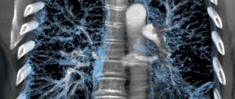
A variation of the MRI method for examining the lungs is MR angiography - a study of blood vessels without the use of contrast.
MRI of the bronchi and lungs allows:
• identify foci of inflammation and their location; • detect a tumor at an early stage, assess its nature and stage, metastasis to lymph nodes and germination into blood vessels; • detect an abscess (abscess) in the lung or a cavity (cavern); • identify accumulation of exudate, blood or pus in the pleural cavity; • diagnose tuberculosis at an early stage.
MRI is also used when planning lung surgery and to monitor the results of surgery.
Advantages of Sofia Cancer Center
The Sofia Cancer Center has created comfortable conditions for receiving and conducting diagnostics. For the convenience of the patient, individual support throughout the clinic is provided,
Advantages of the Sofia Cancer Center:
- the best equipment;
- everything in one place: clinic, diagnostic department, hospital, pharmacy, etc.;
- the possibility of obtaining consultation after MRI diagnostics;
- 30 years of unique experience;
- modern interior, like in a five-star hotel;
- experienced doctors, leaders of world medicine;
- full compliance with Russian and international standards (including JCI).
Indications and contraindications
MRI is prescribed for the following indications:
• pleurisy (inflammation of the pleura without effusion of exudate or with effusion); • neoplasm in the mediastinum; • lung or bronchus cancer; • pleural tumor; • effusion in the pleural cavity of unknown origin; • cystic fibrosis (hereditary disease with severe respiratory dysfunction); • enlargement of intrathoracic lymph nodes; • atelectasis (collapsed lung or part of it); • tuberculosis or suspicion of it; • acute and chronic respiratory failure; • sarcoidosis (systemic inflammatory lung disease); • sequestration of the lung (a developmental anomaly in which a section of the lung is formed separately from the bronchus); • abnormalities of the pulmonary vessels; • pulmonary artery aneurysm (expanded lumen of the vessel in a limited area); • pulmonary embolism (blockage of a vessel with a blood clot, air or piece of tissue); • pneumofibrosis (proliferation of connective tissue against the background of an inflammatory chronic process); • solitary pulmonary nodule less than 3 cm (single round formation on the periphery of the lung tissue);
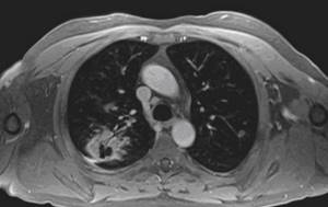
MRI is more informative than CT in assessing respiratory function, in diagnosing circulatory disorders in the lungs, and in differential diagnosis of the causes of atelectasis.
The capabilities of MRI for visualizing bronchioles and alveoli and their diffuse changes are limited. Therefore, emphysema and obstructive pulmonary diseases, bronchiectasis are best diagnosed using CT. The central bronchi are clearly visible on MRI images: you can see both their expansion and thickening of the wall.
Contraindications:
• presence in the body of an implant made of ferromagnetic metals; • cardiac pacemaker and other electronic systems in the body of the subject; • shot, bullets, fragments in muscles (under the influence of a magnet they can move and damage tissues and blood vessels); • pregnancy (there is no sufficient data on the absence of exposure to the magnetic field on the fetus); • lactation (after applying contrast, feeding with mother's milk can be resumed after 2 days); • claustrophobia (fear that occurs in a closed space); • renal failure (the use of contrast is contraindicated).
How does a CT scan differ from an X-ray of the lungs?
Computed tomography is a modern method of radiological diagnosis of various diseases, which is based on radiography. . The method was developed and proposed by Nobel Prize-winning scientists G. Hounsfield and A. Cormack in 1972. Classical radiography was invented in 1896; it was most often used in dentistry and for the study of the lungs, since at the turn of the 19th-20th centuries. mortality from pneumonia, tuberculosis and asthma was extremely high.
The key difference between digital x-ray and computed tomography of the lungs is the trajectory of the x-rays and the imaging technique. In conventional radiography, X-rays pass perpendicularly through the area of interest only once, so the radiograph is a two-dimensional, single-layer image. Lung X-ray is the most affordable examination, which is often prescribed first if the patient has signs of pneumonia, tuberculosis, obstructive pulmonary disease, or tumors. The problem with this type of diagnosis is that, for example, with pneumonia, only stage III and IV lung damage can be reliably determined on an X-ray, and shadows from large organs can obscure other tissues.
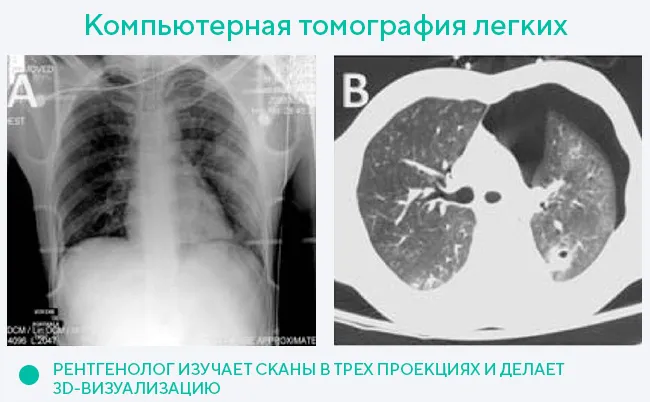
CT scans are distinguished by higher image clarity and information content. During computed tomography, the X-ray tube, together with sensitive sensors, makes several revolutions along a spiral path, scanning the area under study. The CT machine takes many scans up to 1 mm thick, on the basis of which a three-dimensional model of the lungs, blood vessels, organs and bones of the chest is recreated in high resolution. Thus, after computer processing of images, tissues and organs can be examined in three projections; the effect of overlapping shadows from organs in the case of computed tomography is absent.
The high image clarity of computed tomography is associated with the diagnostic technique and the physical properties of the radiation. X-ray has a 20% attenuation coefficient, while tomography has a 0.5% coefficient, and therefore a higher resolution.
Both x-rays and computed tomography can be done with contrast. X-ray or CT scan of the lungs with contrast will help visualize blood vessels and tumors. However, primary differentiation of neoplasms into benign and oncogenic is possible only within the framework of CT, which is also related to the quality of the images.
Since a chest X-ray is essentially 1 picture, and there are many CT scans, the radiation from a CT scan of the lungs is higher due to multiple exposures. On average, a patient receives 0.1 mSv of radiation during one lung X-ray procedure, and 2.5 mSv during a lung CT scan. However, this dose of ionizing radiation is safe for the patient. It is permissible to do a CT scan of 5 zones per year. When referring for one or another radiographic examination method, doctors are always guided by the criterion of feasibility and patient safety.
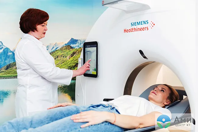
In a specialized facility, the procedure is performed on a new generation Siemens Somatom go.Now device with reduced radiation exposure.
Preparation
The study does not require special preparation. It is better to carry out the procedure on an empty stomach or you can have a light breakfast 2 hours before. You should not use cosmetics containing metal components.
Before the procedure begins, it is necessary to remove hairpins, jewelry, any type of jewelry, piercings, and watches. Before the procedure, the patient is put on a disposable gown. If you want to stay in your clothes, you should make sure that there are no metal fittings in them, and empty your pockets of glasses, coins, lighters, and credit cards.
How is diagnosis carried out?
If you carry out the procedure following all the rules, all symptoms and tests will be determined correctly. At the same time, preparing for the procedure does not require much effort:
- The procedure does not require abstinence from food and drinks. You can eat and drink and exercise. Some experts recommend spending time before the procedure in peace and quiet.
- You should come to the procedure in comfortable clothes, without jewelry or jewelry. In some cases, the patient may be offered a disposable gown.
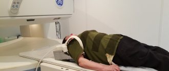
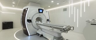
- The device itself consists of glass and a scanner. Before starting the procedure, the patient must take the most comfortable position, since it will not be possible to move or move during magnetic resonance imaging.
- The doctor must secure the patient's body using special belts. This is necessary in order to get the clearest, most detailed and high-quality images.
- After this, the table with the patient moves into the apparatus tunnel.
- Scanners analyze tissue, after which the data is sent to a specialist’s computer.
- Some studies may require contrast. This is carried out according to indications. If necessary, the patient is injected with a special substance intravenously (in the hand or forearm).
- After this, the radiologist draws conclusions and interprets the data.
It is worth noting that during the procedure the doctor monitors the patient from the next room. The procedure itself lasts approximately 30-60 minutes on average. In some circumstances, the study may take longer.
Progress of the procedure
The patient signs written consent to conduct the study. The subject is placed on a special table, which is rolled into the chamber of the apparatus. During the entire examination, which lasts about half an hour, the patient must lie still so as not to distort the quality of the images.
During operation, the tomograph makes quite loud sounds (if desired, you can take earplugs in your ears before starting the study). During the procedure, two-way communication between the patient and the doctor or operator is possible. There is an alarm button to stop the study if the patient’s well-being worsens.
In some cases, it is necessary to conduct an MRI with contrast of the bronchi and lungs for a more accurate diagnosis. Contrast (usually gadolinium) is injected into a vein. When using it, the procedure is extended by approximately 20 minutes.
Make an appointment
The clinic is located in the Central Administrative District (CAO) of Moscow, next to several metro stations: Novoslobodskaya, Tverskaya, Chekhovskaya, Belorusskaya and Mayakovskaya.
You can make an appointment by calling +7 (495) 775-73-60 or using the online form.
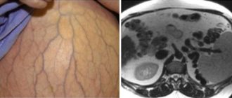
What can be seen on the results of a vascular MRI
As a result of scanning the vascular network, the doctor can examine the smallest sections of the vascular wall, assess their length, structure, and mark areas of damage. Due to the fact that the resulting image is three-dimensional, the images will clearly visualize blood clots, possible wall dissections, expanded or narrowed zones, as well as a tendency to sclerosis.
Thanks to the high technology of the method, the device makes it possible to study not only venous and arterial blood flow, but also to assess the condition of the capillary network. Despite the wide possibilities, studies of large-caliber vessels, such as those of the renal system, brain and legs, are most in demand.
Make an appointment now!
How to prepare for the examination
Preparation for vascular MRI does not involve following a diet or changing your usual daily routine. The patient must follow the instructions and recommendations of medical personnel:
- It is necessary to remove metal objects - accessories, jewelry, including piercings;
- The patient must undress to his underwear; some clinics suggest changing to disposable clothing;
- If administration of contrast is indicated, it is necessary to refrain from eating 2-3 hours before the examination.
Indications for vascular MRI
In general, magnetic resonance scanning is indicated for the following complaints:
- Recurrent severe headaches;
- Unreasonable dizziness;
- Decreased concentration and memory impairment;
- Convulsive syndrome.
The list of diseases and conditions that are indications for magnetic resonance imaging is quite wide. This includes head injuries, the presence of pathologies of the circulatory system, as well as:
- Stroke and heart attack;
- Suspicion of atherosclerosis;
- Examination for the presence of neoplasms;
- Inflammatory processes localized in blood vessels;
- Aneurysms, stenoses;
- Thrombosis;
- Vasculitis;
- Meningitis.