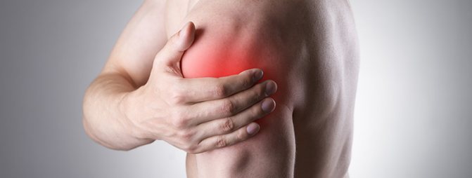
MRI of the shoulder joint is a method for diagnosing diseases of large joints, which has many advantages. It does not require the painful procedures required for endoscopic examination or analysis of intra-articular fluid. MRI is a very safe procedure due to the lack of radiation and can be used even during pregnancy and in children. Magnetic resonance imaging is based on the use of magnetic fields and radio waves, the interaction of which allows you to obtain detailed images of the area under study. Using them, doctors can easily determine what disease is bothering the patient.
The structure and anatomy of the shoulder joint
The shoulder joint is the most mobile joint in humans, formed by the head of the humerus and the scapula. His lack of powerful ligaments is compensated by the muscles of the shoulder girdle and upper limbs surrounding the shoulder like a corset. On the one hand, this provides greater mobility, on the other hand, it explains why the shoulder joint is easy to dislocate.
Along the periphery, the joint is surrounded by short ligaments that form the articular capsule. It unites the joint into one whole and holds the articular surfaces of the head of the humerus and scapula in optimal position. The inner surface of the joint capsule is covered with a synovial membrane that produces fluid into the joint cavity, acting as a lubricant and nourishing the cartilage.
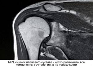
An MRI of the shoulder joints clearly shows all the anatomical structures that form the joint. The method allows you to assess the condition of the soft tissues and cartilage of the shoulder, ligaments, joint capsule, joint cavity, tendons and muscles.
Pros and cons of tomography
Let's start, of course, with the advantages.
MRI of the shoulder joint is an absolutely safe procedure. The tomograph generates harmless magnetic waves rather than aggressive X-rays. During the scanning, the integrity of the skin is not compromised, and after its completion, the person can immediately return to their usual activities without any restrictions.
The study is very informative. It allows us to examine in great detail the structure of a particular formation and identify changes in it even at the early stages of its development. The distance between the sections that are displayed on the screen during the procedure does not exceed a few millimeters, which reduces the chance of missing pathology to a minimum. The duration of the tomography is limited to 30 minutes, during which no additional effort is required from you. You just need to remain still and wait for the examination to complete.
As for the disadvantages of MRI, they can be counted on the fingers of one hand. First of all, this is the cost of the procedure. It cannot be called low, but the result obtained always justifies the money spent. Secondly, there are a number of contraindications for this type of diagnosis. These include claustrophobia, the patient’s heavy weight, as well as the presence of metal devices in his body (prostheses, cochlear implant, pins, staples, etc.).
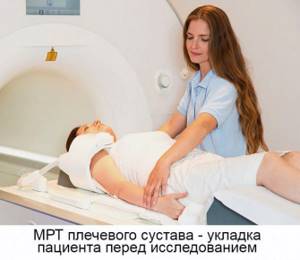
Carrying out an MRI procedure of the shoulder joint
What are the types of shoulder diseases?
All diseases of the shoulder joint can be divided into several groups:
- injuries (dislocations, fractures, ligament and tendon ruptures) and their consequences (instability of the shoulder joint, habitual shoulder dislocation);
- degenerative-dystrophic pathologies (osteoarthrosis);
- inflammatory diseases (arthritis, bursitis, tendinitis, synovitis);
- neoplasms.
MRI of the shoulder joint is used to diagnose all possible diseases arising in this area.
Shoulder injuries
Shoulder injuries are one of the most common reasons for visiting a traumatologist. As mentioned above, the characteristics of the shoulder joint contribute to frequent dislocations, ruptures of ligaments and tendons, which causes conditions such as habitual dislocation and instability of the shoulder joint. With direct physical impact, intra-articular fractures of the head of the humerus and the glenoid cavity of the scapula can be diagnosed.
Degenerative-dystrophic pathologies of the shoulder joint
Osteoarthritis or deforming osteoarthritis is a pathology that occurs when the nutrition of articular cartilage is disrupted. The direct cause of the disease may be trauma, inflammation, metabolic disorders, congenital predisposition, aseptic necrosis of the head of the humerus. As a result, the natural process of renewal of cartilage tissue slows down, which gradually becomes thinner and is replaced by rough connective and bone tissue. The person experiences pain, limited joint mobility, and impaired function of the upper limbs.
Inflammatory diseases of the shoulder joint
Inflammation of the joint capsule (arthritis) and the soft tissues surrounding the shoulder joint (bursitis, synovitis, tendinitis) can occur as a consequence of the underlying disease (rheumatism, rheumatoid arthritis), or as a result of infection, chronic or acute injury. In 15% of cases, it is impossible to establish the cause of this group of diseases.
Neoplasms
The shoulder joint can be affected by both primary tumors of soft tissues and bones, and by metastasis from distant foci. Benign neoplasms are also not uncommon.
Interpretation of information obtained after MRI
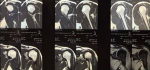
After the MRI is completed, decoding of the received information begins - this is done by an expert in the field of radiation testing. Often, the waiting time for results is no more than 120 minutes, but if there is a high level of workload, the patient will have to return for answers the next day. In the photographs after the MRI, the doctor will be able to see:
- Inflammatory processes occurring in the studied area, causing degenerative and destructive deformations of hard structures and cartilage (intensively highlighted in the image).
- Oncology appears as areas with a dense texture.
- In case of ligament rupture, displacement and changes in areas are visible.
- When a complete or partial fusion of the gap forms, there is a possibility of developing joint immobility.
- In situations where dead zones form, there is a danger of the spread of an infectious disease.
- The accumulation of fluid indicates bursitis.
Don't know where you can get an MRI? Visit the official website mrt-v-spb.ru! It is here that you will receive a lot of useful information: the number of vacancies, the cost of the procedure, addresses and contact numbers of clinics. In addition, our service has a special section that contains real reviews from patients of many city medical institutions. With the help of this data, you can quickly decide on the choice of organization for conducting MRI and other methods of checking the body. Also, our company has a hotline where specialists help clients with complex questions and applying for an appointment with a doctor.
Symptoms that warrant an MRI of the shoulder
Magnetic resonance imaging of the shoulder joints is indicated if the patient has the following complaints:
- pain in the shoulder joint, often worsening with movement of the shoulder;
- limited mobility, impaired function of the upper limbs on the affected side;
- swelling, redness of the skin in the shoulder joint;
- crunching, clicking, discomfort that occurs when moving the shoulder and/or in a certain position of the arm;
- neoplasms, nodes and bumps that are visible in the shoulder area with the naked eye or by palpation.
Procedure
The tomography procedure of the shoulder joint takes on average 20-40 minutes. The duration of the scan depends on the tomograph model. In the diagnostic room, the patient is placed on a tomography table. Then a special coil is placed over the examination area. The table is moved into the scanning part of the device and the screening procedure is started.
The main task of the patient during the examination is to remain completely still. This is largely a guarantee of quality research. The fact is that the tomograph is very sensitive to any movement. If the patient moves the shoulder during the scan, motion artifacts appear on the tomograms. They reduce the effectiveness of the study and the overall quality of diagnosis.
Questions about diagnostics
Dress code
You can enter the MRI room in any clothing that does not contain metal. When going to the clinic, it is best to wear loose, non-restrictive clothing without metal elements (zippers, rivets, hooks), in which you can lie comfortably. For women, we recommend bringing a T-shirt or not wearing a bra with metal wires or hooks.
Preparation
This tomography does not require any preparatory steps from the patient.
Is MRI harmful to health?
MRI is a completely harmless diagnostic method for the human body. This method of examination can be carried out at any age and for any disease an unlimited number of times, unless you have contraindications.
Contraindications
Some pacemakers and foreign objects in the body may pose serious limitations to tomography. In particular, cochlear implants, vascular clips, stents, heart valves and insulin pumps, pacemakers, neurostimulators, steel screws, staples, pins, plates, joint endoprostheses may be a contraindication to diagnosis. The patient must notify the radiologist about all implanted objects in the body. The diagnostician will be able, based on information about the composition and model of the implant, to assess the possibility of conducting diagnostics.
If you are having an MRI with contrast, be sure to report any allergies to medications or kidney problems. It is also necessary to inform the radiologist about a possible pregnancy.
Is it possible to do an MRI with braces and dental implants?
Dental implants and crowns are not a contraindication to magnetic resonance imaging. The magnetic field does not have any negative effect on them. Fixed brace systems can produce artifacts on tomograms during MRI of the head. If the light effect is too strong, the doctor will stop the study and offer the patient alternative diagnostic methods.
Is the device noisy?
Any MRI machine in working condition makes noises reminiscent of tapping. The open tomograph is one of the quietest installations. The noise from its operation is significantly lower compared to closed tomographs. If the sounds of the operating unit cause you anxiety, you will definitely be offered special noise-canceling headphones.
What should I do if I have claustrophobia?
An open tomograph is the optimal solution for patients suffering from panic attacks in a closed space. It is open on the sides on three sides and does not create a claustrophobic feeling.
Can I take sedatives before an MRI?
If you are a little nervous, before the tomography you can take mild sedatives, for example, valerian, motherwort infusion or afobazole. Taking sedatives does not have a negative impact on the quality of MRI.
Why is it important not to move during the test?
Any movement during the examination reduces the quality of the resulting images. Multiple motion artifacts may appear on the images, and the MRI results will be uninformative.
Can I do the research with an accompanying person?
Absolutely yes. You can invite any accompanying person from among your family and friends to the MRI room. It is important that your companion does not have metal implants or artificial pacemakers in his body.
In what cases is MRI prohibited?
MRI is a safe diagnostic method, but this is true if precautions are followed and there are no contraindications. Otherwise, the study may be harmful to health. MRI is prohibited if:
- the patient has a pacemaker, defibrillator, cochlear implant or any other medical device implanted in the body;
- the patient’s weight exceeds 130 kg, body circumference is more than 150 cm (a person of this size cannot physically fit into the device);
- the patient is a child under 5 years of age (children younger cannot remain still during the entire study, which is why MRI must be performed in a specialized children's hospital under anesthesia);
- the patient is a pregnant woman in the first trimester of pregnancy (the study is not recommended “just in case,” despite the fact that over many years of using MRI it has never become a source of health problems for the woman or fetus);
- there are metal foreign bodies in the patient's body - small metal shavings in the eyes, unrecovered bullets, shrapnel (this is especially important for patients who have undergone surgery on the brain and blood vessels - in such cases, doctors often use metal clips that are installed on the arteries to stop bleeding ).
When to carry out comprehensive research
Our body is a single system in which everything is interconnected. And if a pathological process occurs in one part of the body, there is no guarantee that over time it will not spread to neighboring formations. That is why it is sometimes impossible to limit oneself to one area of research. An example is rapidly growing tumors and their metastases, which can spread through the blood or lymph to any part of the human body.
And in cases of difficulties with differential diagnosis, it would not be amiss to examine several areas to make a single correct diagnosis. Remember that the sooner doctors detect pathology, the sooner treatment will begin!
MRI of the shoulder joint - preparation
MRI of the shoulder joints is performed at any time convenient for the patient; the examination does not require prior preparation. There is no need for a diet; you are allowed to eat and drink, but it is more comfortable to undergo the examination after a light rather than a heavy meal.
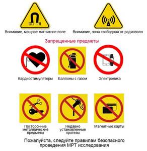
Since the equipment generates powerful magnetic fields during the examination, patients are prohibited from having any foreign metal objects, as well as electronic devices, for their own safety. When approaching a tomography machine, metal can be forcefully attracted to the magnet of the machine and cause injury or cause a burn, and a cell phone or other gadget may malfunction or even catch fire.
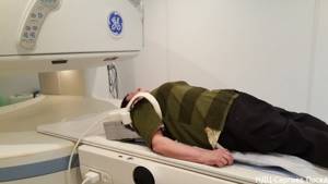
How is an MRI of the shoulder performed?
The study is carried out in a separate room where a tomograph is installed - an MRI device that looks like a large cylinder with a cylindrical hole in the center. This hole or channel of the tomograph is equipped with a movable table. At the beginning of the procedure, the patient is placed on it in a supine position. Additional coils are installed on the shoulder area - they help make the image clearer. Headphones are put on the patient's ears - during operation the device makes a lot of noise, and this can frighten a sensitive person. You can also communicate with the operator and listen to music through headphones - some of our patients take their favorite songs with them on a flash drive to relax during the procedure. After installation is completed, the table moves into the apparatus tunnel to begin the examination.
All that is required of the patient is to remain still and follow all the operator’s instructions. After completing the study, the table moves out, after which you need to wait 15-30 minutes until the decoding of the images is completed.
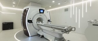
Expert equipment
The Clinical Hospital on Yauza uses the latest generation 1.5 T Philips Ingenia fully digital magnetic resonance imaging scanner, which provides:
- Reduced scanning time (less time to lie in the magnet)
- The maximum possible diameter of the tunnel (73 cm) - the patient’s waist circumference (circumference) is up to 225 cm
- Highest quality images
- Full range of studies due to maximum equipment
- Additional information through advanced software
- Environmental control system (music from the patient’s playlist, choice of lighting, personal climate control)
- Automated quality control system for research and descriptions
- IT platform with triple control of research results - with the support of professors, leading specialists from Russia, Europe and Israel (“second opinion”)
What does an MRI of the shoulder joint show?
Patients are always interested in what an MRI of the shoulder joint shows. Knowing this, we decided that simply making a conclusion and handing over the images is not enough - every visitor to our center receives a personal consultation with an MRI diagnostic doctor after the examination. We will definitely tell you in detail if an MRI of the shoulder joint shows signs of the disease. You will be able to look at the pictures with explanations from a specialist doctor, find out what disease the detected changes correspond to, and which doctor you should contact for further treatment.
Carrying out MRI with contrast
The doctor may determine the need for a study with a contrast agent if a tumor is suspected or its presence is definitely established. If the tumor is operable, it is important to determine its exact boundaries and the distribution of pathological cells. The procedure is called indirect MRI arthrography.
Gadolinium is used as a contrast agent. Its amount is calculated by a specialist based on the body weight of the subject. This type of diagnosis takes a long time, because for correct results it is necessary to create a load on the joint. To do this, after administering the drug, the patient must move the shoulder joint for 20 minutes.
Next, magnetic tomography is performed and pictures are taken in which the blood supply network is clearly visible and the location of the tumor is clearly visible.
MRI or CT scan of the shoulder joint, which is better?
Computed tomography or CT is another diagnostic method that is similar in appearance but completely different from MRI. Unlike MRI, CT uses x-rays, making it better suited for diagnosing diseases of the bones that make up the shoulder joint. Cartilage, muscles, and ligaments are practically invisible on CT images. We can conclude that MRI is ideal for diagnosing diseases of the joint itself and its soft tissues, while CT is prescribed mainly for fractures of the head of the humerus and scapula.
Indications for magnetic resonance imaging of the shoulder

The patient may be referred to undergo this type of study for various indications. Thus, MRI is performed when there are signs of the development of a pathological process in order to either detect the presence of the disease or completely exclude it. In addition, the procedure is carried out to determine the severity of the injuries received and the exact location of the damage.
A doctor can write a referral to a patient if the following disorders are suspected:
- in order to identify the extent of shoulder injuries;
- in case of damage to soft shoulder tissues;
- in the presence of a tumor in the shoulder (tomography makes it possible to identify clear boundaries of the tumor);
- when joint mobility decreases due to injury;
- for arthritis, arthrosis, osteoarthritis;
- when joint swelling occurs;
- in order to monitor the effectiveness of the prescribed treatment in the postoperative period.
In most cases, the study is prescribed once, although if there are serious indications, this method can be carried out an unlimited number of times.
Patients do not have to worry that frequent scanning will harm their health, since tomography is absolutely harmless. Thus, for progressive chronic lesions of the shoulder, when systematic monitoring of the dynamics of the treatment is required, MRI of the shoulder can be prescribed very often.
The MRI method detects even minor changes in hard and soft tissues, which makes it possible to monitor the healing process after surgery, and in patients with chronic diseases, the dynamics of changes.