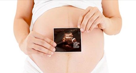
Of course, for any expectant mother, pregnancy is a very specific and unusual period in life, during which too many changes occur in a woman’s body to simply not notice. Unfortunately, neither the negative influence of the environment, nor the impact of chronic diseases, nor the possibility of accidentally “catching” the virus can be avoided. During pregnancy, it is highly undesirable to take medications or undergo many medical procedures, as this may affect the development of the fetus. But there are also exceptions.
Why do the eyes bleed and the baby that matured in... the ovary
There are many examples where magnetic resonance imaging (MRI) made it possible to identify hidden pathologies or, on the contrary, to refute a previously made diagnosis and avoid unnecessary surgery.
By the way, the latter is also very relevant, especially against the backdrop of increasing cases of surgical aggression towards patients. You can find many sites on the Internet that talk about MRI as an advanced and very effective diagnostic method. But we decided to start with examples that actually took place, which the gynecologist at the Medservice clinic told us about.
Pregnancy control after cancer
In any case, if a woman previously suffered from cancer, was cured, waited the appropriate period and became pregnant, she needs to strictly monitor her own condition for all nine months. In no case should you neglect examinations, which should be regular.
By seeing oncology specialists, you can be sure that your health is under complete control. Doctors prescribe the necessary diagnostic measures and monitor the general condition of each patient. We are located at: Moscow, 2nd Tverskoy-Yamskaya lane, 10. You can make an appointment using the feedback form on the website or by phone.
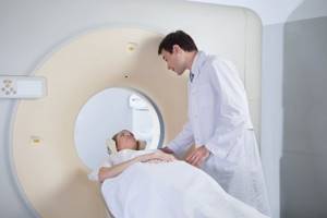
MRI and pregnancy
Despite the fact that MRI is partially contraindicated during pregnancy, there are cases when it saves the lives of both mother and child. In one of the regions of Udmurtia, a woman who was 32-33 weeks pregnant came to the maternity hospital with complaints of abdominal pain. She was hospitalized due to risks of premature birth.
The results of the examination and ultrasound of the pelvic organs did not allow us to determine treatment tactics and make a decision - to allow childbirth or to prolong treatment. The lady was sent for an MRI, where it was revealed that the pregnancy was ectopic - ovarian. The lady was sent for surgery. Only a miracle and an MRI saved both mother and child.
Why do pregnant women still undergo MRI?
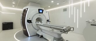
Many types of diagnostics are also contraindicated during pregnancy, but, for example, ultrasound diagnostics (ultrasound) and MRI are carried out quite widely. Why? These diagnostic methods are based on the physical properties of sound waves in media of varying densities (ultrasound) and certain patterns of behavior of hydrogen atoms in a high-power magnetic field. Neither the first nor the second cause significant harm to the human body, which is why they are calmly prescribed by doctors to expectant mothers.
It is advisable to make MRI a habit
A lady consulted a gynecologist with complaints about the appearance of bloody, pink discharge from the genital tract. Her cheerfulness and youthful appearance beyond her years did not foretell any unpleasant surprises.
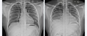
An ultrasound of the pelvic organs revealed a polyp in the uterine cavity. In order to clarify the diagnosis, the gynecologist referred the patient for an MRI, where complete destruction of the muscle tissue of the uterus was visualized.
Apparently, the process lasted two to three years; in fact, we are talking about the second or third stages of cancer. The patient was referred to an oncologist.
Neither ultrasound nor external examination made it possible to timely determine the presence of malignant tumors. MRI allows you to detect malignant and benign formations at the earliest stages, when their size does not exceed 0.5 mm. With ultrasound, the lower limit of accuracy is about 1 cm, but this is subject to the highly qualified doctor and the resolution of the equipment. It must be of an expert class. That is why it is very important to conduct MRI not only as prescribed by the attending physician, but also for preventive purposes. For example, during medical examinations.
Advantages of MRI in gynecology
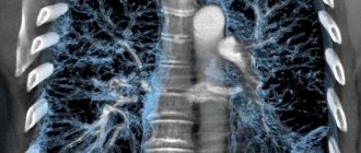
MRI allows you to obtain complete images of human organs and tissues at any level and in any given plane with an assessment of their functional integrity. If necessary, you can obtain layer-by-layer series of images with slice thicknesses from 4-5 mm to 0.8 mm. MRI with contrast provides a more accurate verification of the tissue identity of tumors, identifying small metastases at the earliest stages of development. In addition, tomography allows you to assess the structure and condition of bones, soft tissues, blood vessels, ligaments, and cartilage.
Is it possible to do a CT scan in the early stages?
Sometimes it happens that the procedure is performed on a woman who does not yet suspect she is pregnant, since the period is 4 weeks or even less. But to be on the safe side, it is best to visit a gynecologist before a CT scan, so that after the procedure you do not have to think about the consequences of a CT scan in the early stages of pregnancy. It is also worth noting that the embryo at such stages is not yet as sensitive to radiation as at a period of 9 to 15 weeks, when all vital organs are formed. Therefore, there is every chance that in this case the consequences of CT scanning during early pregnancy will be either minimal or absent altogether. An ultrasound can be performed to identify abnormalities in fetal development.
, and based on its results, think about further actions.
When should an MRI be performed?
MRI should be performed if the presence of benign or oncological formations is suspected. Tomography of the pelvic organs with contrast is applicable for cancer patients to determine the area of spread of metastases and damage to neighboring organs.
MRI of the pelvis is indicated when identifying the causes of infertility in men and women (obstruction of the fallopian tubes), in case of injury, the presence of anomalies and structural pathologies, and inflammatory processes in the organs of this part of the body.
Tomography of the pelvic organs in women for use in the diagnosis of endometriosis. The endometrium is the hormone-sensitive tissue lining the uterine cavity that can implant in a variety of tissues, including the ovaries, fallopian tubes, bladder, peritoneum, rectum, and other distant organs. These fragments, in accordance with the phases of the menstrual cycle, undergo the same cyclic cycles as the endometrium in the uterus, which is accompanied by pain, an increase in the affected organ in volume, and bleeding. Endometriosis can lead to infertility. Diagnosing endometriosis and localizing distant metastases is quite difficult, but MRI provides such an opportunity.
When planning untimeliness, MRI allows you to assess the condition of the uterine scar in mothers who gave birth by cesarean section. Images in the axial and frontal directions make it possible to identify problem areas and assess the risks of scar failure, which is essentially an assessment of the risk of maternal death.
CT and X-ray of the jaw during pregnancy
The condition of the oral cavity and teeth is the most important factor affecting health and communication with other people. If there are problems (rotten teeth, inflammation or various types of infections), a person often experiences bad breath, which interferes with normal communication with friends and colleagues. In addition, the infection can quickly move down the esophagus, which already poses a danger to the internal organs. Therefore, you need to monitor the condition of your oral cavity every day and undergo appropriate treatment if necessary.
But any therapy or dental prosthetics requires preliminary diagnosis using X-rays or CT. And if for most people this procedure does not pose a danger, then for a pregnant woman this question remains open. There are many points of view on the topic - is it possible to take dental x-rays during pregnancy and how harmful is this procedure? Some experts say that this is an absolute contraindication to X-ray examination, while others are not so categorical in assessing the degree of risk. Let's look at this situation in more detail.

Content
- Is it possible for a woman to have a dental x-ray during pregnancy?
- In what cases is a dental x-ray required for pregnant women?
- Photos of pregnant women's teeth at different stages
- Is it possible to do a CT scan during pregnancy?
- Planning pregnancy after CT scan
- Basic preventive measures
- X-ray examination in St. Petersburg
Is it possible for a woman to have a dental x-ray during pregnancy?
Pregnancy is associated with a high consumption of nutrients and minerals, including calcium and magnesium. All of them are spent on the formation of the fetus. If a woman’s body does not receive them from the food she eats, then all useful substances are taken from tissues, including teeth. Even a healthy woman begins to have problems, and if the patient had dental problems before, then during pregnancy they become significantly worse. Enamel demineralization and other consequences occur.
For the treatment to be correct, the specialist needs a clear x-ray, but how dangerous is this during pregnancy? Ideally, any exposure that could be harmful to the health of the fetus should be avoided by treating the child at the planning stage. But situations are different, so the question of whether pregnant women take dental photographs remains relevant.
Here you need to take into account the type of X-ray machine with which the study will be carried out. Conventionally, they can be divided into 2 types:
- Old Soviet X-ray machines. Doha radiation exposure is about 7 μSv (for the lower jaw) and 15 μSv (for the upper jaw). In older equipment, these parameters are even higher - 20 and 30 μSv, respectively. This is a very large dose that can harm the child's health.
- Modern visiographs. These devices emit significantly less radiation. The average radiation dose is about 2-3 μSv.
Modern devices generate a narrower and more even beam, avoiding exposure to surrounding tissue. The impact on it is quite comparable to radiation from a computer or the sun, so it is absolutely safe.
In what cases is a dental x-ray required for pregnant women?
Dental treatment “blindly” without detailed photographs – this approach can hardly be called professional. An image of the area being examined allows the doctor to see the localization of the inflammatory process, nerve bundles, dental canals and other important nuances.
A dental photograph during pregnancy is taken in the following cases:
- Incorrect growth of "eights".
- When you need to determine the length and shape of root canals (for pulpitis).
- There are suspicions of periodontitis.
- Injuries and fractures of the tooth root.
- During the formation of granulomas, cysts and other neoplasms.
Based on this, we can say that a dental x-ray during pregnancy is a completely common procedure that is performed when indicated. But this needs to be done only on modern equipment.
Here are a few rules that pregnant women need to know:
- Dental X-rays during pregnancy cannot be done using film X-ray machines. The optimal choice is a computer visiograph. Its radiation dose is approximately 10 times less than that of older devices.
- The stomach and chest must be covered with a lead apron.
- Before the study, you must inform yourself about your pregnancy and give the exact date.
Photos of pregnant women's teeth at different stages
The first 12 weeks are a vital period in the formation of the fetus, when all the internal organs are formed. It is at this time that there is the greatest likelihood of pathologies developing. Therefore, a dental x-ray during pregnancy in the 1st trimester is undesirable, but if there is a real threat to the mother’s health, the procedure is performed. The decision must be made by the attending physician, who evaluates all possible risks.
Experts recommend taking dental x-rays during pregnancy in the 2nd trimester. The fetus is protected by the placental barrier, the formation of organs is almost complete, and the mother’s well-being becomes more stable.
The 3rd trimester is a period when the sensitivity of the uterus significantly increases to any external influence. In the later stages, an X-ray examination is not advisable, but if the patient suffers from acute pain or there are other emergency situations, then the procedure is possible.
Is it possible to do a CT scan during pregnancy?
Computed tomography is a highly informative diagnostic method based on X-ray radiation. The diagnostic value of this study is higher than that of a standard x-ray, but many are interested in the issue of harm from CT scanning during pregnancy.
If we talk about absolute contraindications, then the first trimester is an undesirable time for carrying out any procedures that, to one degree or another, may affect the condition of the fetus. There are some cases when a woman’s condition is in critical condition and urgent diagnostics are necessary, then a positive decision is made to carry out the procedure (if it is not possible to use alternative diagnostic methods).
Computed tomography during pregnancy is never performed when examining the pelvic organs, hip joints and abdominal cavity. This is explained by the fact that when examining these areas, the body receives the highest dose of radiation. Plus, the fetus is in close proximity to targeted X-rays.

Planning pregnancy after CT scan
Many women are interested in the question: how long after it is possible to plan a pregnancy if a CT scan has been performed? Experts say that if the woman was not pregnant during the procedure, then there are no special restrictions. X-rays do not have a negative effect on the condition of the egg or sperm in men (if the radiation dose is not higher than the permissible norm).
X-rays do not tend to accumulate in the body. In other words, their influence ends exactly at the moment when the tomograph or X-ray machine is turned off.
How long after a CT scan can you get pregnant? Despite the above, experts recommend waiting about 2-3 months before conceiving a child. These are normal precautions that are especially relevant if the area was examined in the immediate area of the reproductive organs.
Basic preventive measures
To avoid exposure to x-ray radiation during pregnancy, it is necessary to promptly identify diseases even before the planning stage of conception, as well as take preventive measures. These include the following recommendations:
- Dental care should be carried out not only with standard brushing, but also with additional means (rinses, dental floss, etc.).
- Pregnancy planning must include oral sanitation.
- Visit to the dental office (at least 2 times a year).
- Adding foods that contain large amounts of vitamins and minerals to your diet.
X-ray examination in St. Petersburg
We talked about whether it is possible for pregnant women to have dental x-rays and at what time it is most dangerous for the child and the mother herself. In general, opinions about the dangers of this research are based on the practice of past years, when old X-ray machines were used, the radiation dose of which was much higher. Now
X-ray - we invite everyone to undergo examination using CT or X-ray using modern equipment. Minimal radiation exposure eliminates the negative impact on the health of the woman and her fetus (subject to precautions). Before the examination, be sure to tell about the exact timing of pregnancy.
Sign up for a study by phone
+7 (812) 332-52-54
Contraindications
Despite the fact that MRI is considered one of the safest diagnostic methods, there are some contraindications to its use. We are mainly talking about the presence in the patient’s body of foreign bodies that contain metal particles, including implants, dentures, shunts, dental crowns, insulin pumps, pacemakers, and vascular clips. It is impossible to carry out the procedure if the patient’s weight exceeds 130 kilograms, since the tomograph chambers are quite narrow. MRI is not recommended in the first weeks of pregnancy. But for serious indications, the study is possible.
As for claustrophobia, a good solution is to study with open-type tomographs, one of which is installed at the Medservice clinic.
In what cases should an examination not be carried out?
When prescribing an MRI of the uterus and ovaries, the following features are taken into account:
- pregnancy due to harm to the fetus from the contrast agent;
- breastfeeding a child due to the penetration of a contrast agent into breast milk;
- chronic renal failure due to increased load on the kidneys;
- individual intolerance to gadolinium.
An absolute contraindication for MRI of the uterus and ovaries is:
- the presence of foreign metal objects in the patient’s body;
- the presence of implants, for example, a pacemaker.