Today, the magnetic resonance imaging method is recognized not only as the most harmless, but also the most effective. It is capable of providing high-quality images of the deepest structures of the examined organ, allowing it to detect pathology at the stage of its development. However, this method requires complete immobility of the subject for 30–50 minutes. Moreover, not all segments of the population are able to remain motionless for such a long time. Then doctors do an MRI under general anesthesia.
Who needs an MRI under general anesthesia?
MRI under anesthesia is performed only in case of urgent need. Only to achieve absolute stillness at the moment of exploration. Because otherwise it will not be possible to obtain the accurate results necessary for examining the patient. In addition, there is an increased possibility of receiving an erroneous transcript, which will lead to incorrectly prescribed treatment.
Of course, most people can take into account the condition necessary for tomography, but there are segments of the population who find it difficult to cope with this task. These include:
- children under 4 years old;
- persons suffering from mental disorders;
- people with severe claustrophobia;
- patients with epilepsy;
- persons who have severe pain syndrome that does not allow them to assume a calm body position;
- patients with paresis who suffer from involuntary movements of the limbs.
Magnetic resonance imaging
At Ilyinskaya Hospital, the decision to conduct an MRI scan on a patient is made jointly by the attending physician and the radiologist. We always select the most informative method of radiation diagnostics and do not conduct studies “self-prescribed” by the patient.
- Our specialists
Advanced high-tech equipment is essential for diagnosing diseases. But even more important is the expert radiologist who interprets the images obtained from this equipment and answers the questions posed to him by the clinician. The specialists of the Radiation Diagnostics Department of the Ilyinskaya Hospital are involved in the international scientific and educational process and have the most current, constantly updated knowledge in their field of medicine. Our radiologists work closely with clinicians to provide accurate image interpretation and, in complex cases, participate in consultations with surgeons, internists and other specialists.
- Reduced MRI machine noise
Our MRI machine has a low-noise function (Silent Scan technology), for example, brain examination can be carried out completely silently, and more complex examinations (abdominal cavity, pelvis) can be carried out with minimal noise.
- Overweight patients
The MRI machine has an increased tunnel width of 70 centimeters and a body weight limit of 150 kg. This allows overweight people to undergo MRI examinations comfortably.
- Fear of MRI examination
The wide tunnel of the device and reduced noise make the MRI examination procedure easier for patients with fear of closed spaces and panic attacks.
- MRI under anesthesia
At the Ilyinskaya Hospital, it is possible to conduct an MRI examination under anesthesia (intravenous, mask). However, such a study is carried out not at the request of the patient, but strictly according to indications. The decision is made by the radiologist together with the anesthesiologist. The anesthesiologist-resuscitator remains with the patient during the examination and for the required time after awakening.
- Implants, endoprostheses, stents
Modern metal implants and endoprostheses installed in any part of the human body are not an absolute contraindication for MRI examination, since they are made of an alloy of non-magnetic materials. The patient's history of coronary artery bypass grafting and vascular stenting is also not a contraindication to MRI examination.
- Absolute contraindications
There are a minimum number of absolute contraindications to MRI examinations. These include: installed pacemaker (except non-magnetic ones); the presence of metal fragments in the heart, brain, near large vessels; presence of metal shavings in the eyes; the presence of a cochlear (hearing) implant and an implanted insulin pump.
- MRI examination in children
For children aged 0 to 5 years, MRI is always performed under general anesthesia. Before the study, consultation with an anesthesiologist is necessary. An anesthesiologist-resuscitator remains with the child during the entire study and for the required time after awakening. Children over 5 years old require an individual approach, and sedation is also used if necessary.
- MRI during pregnancy
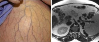
MRI during pregnancy is performed according to the mother’s vital indications. It is most difficult to conduct research in the second trimester, since at this time the fetus actively moves. This causes strong artifacts and the resulting image is difficult to interpret. In the third trimester, MRI examinations are most often performed to evaluate fetal malformations.
- MRI for intensive care patients
The radiology department of the Ilyinskaya Hospital has a full range of non-magnetic resuscitation equipment. There are no restrictions in MRI studies of intensive care patients. If the patient is on mechanical ventilation, he can be connected to a special non-magnetic ventilator and a study can be performed.
- Storage and transmission of diagnostic images
All received images are stored electronically and are always available. They are transmitted and viewed by specialists in an internationally accepted format (PACS). Clinicians at the Ilyinskaya Hospital have the ability to view images remotely through any device connected to the network. In an emergency, this allows surgeons and other doctors to quickly evaluate the images and make decisions.
- Second opinion
The PACS image storage system allows you to transfer research data to foreign medical institutions to obtain an independent expert opinion. Patients located in other regions of Russia have the opportunity to send their images via a secure Internet channel and receive an opinion from specialists at the Ilyinskaya Hospital.
- History of the invention of MRI
In 2003, scientists Paul Lauterbur and Peter Mansfield received the Nobel Prize for their invention of magnetic resonance imaging. in detail about the history of this outstanding discovery in this article.
Methods of administering anesthesia
The main task when performing an MRI using anesthesia is to temporarily turn off the consciousness of the subject. After the anesthesiologist examines the patient’s medical history and gives an adequate assessment of his well-being, he selects the most suitable option, which is used for medicated sleep. The methods of administering anesthesia for MRI include the inhalation technique, in the form of putting on a mask, and parenteral, administered intravenously.
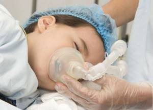
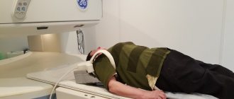
Today, children are often given anesthesia using a mask method for examinations.
Most often, a laryngeal mask airway is used during magnetic resonance imaging. Moreover, the anesthesiologist will remain close to the patient all the time, monitoring his cardiac and respiratory functions, and oxygen saturation of the body. If there is a need, then the person’s condition is corrected. When a study is carried out on patients with epilepsy, inhalation anesthesia is contraindicated for them. In this situation, it is necessary to administer medications only intravenously.
Important! At the end of the procedure, the subject remains under the supervision of a specialist until he fully returns to consciousness.
What types of anesthesia are used for MRI?
Depending on the specific case, the following types of anesthesia are recommended and used:
- sedation is the use of modern medications, with the help of which complete calm of the patient is achieved (medicines, for example, Relium, alprazolam);
- inhalation or intravenous, sometimes with artificial ventilation.
To determine the choice of a specific type of anesthesia, you should pay attention to the individual characteristics of the patient being examined. A qualified anesthesiologist will help you make the right choice, and the study will go well.
Advantages and disadvantages of using anesthesia
In order for MRI to give the most accurate results, it is necessary to remain motionless throughout the examination, and when using contrast agents, this can increase to an hour. Since not all people can stay in one position for a long time, they are diagnosed under general anesthesia. This is especially necessary for examining children, since their psyche cannot always withstand fear or the strong sounds of a working apparatus.
Of course, no person is immune from the negative effects of medical sleep. However, hypersensitivity to its administration, which can manifest itself in the form of an allergic reaction, up to anaphylactic shock, is only an individual reaction of the body. Exceptional reactions to them are possible, in the form of depression of respiratory function.
To perform an MRI, the doctor uses drugs that have a short-term effect with such advantages as:
- rapid removal from the human body;
- minimal effect on the central nervous system;
- rapid achievement of the desired result.
Patients suffering from epilepsy are given intravenous anesthesia only.
As a result of their short action, anesthesia drugs can even be used for diagnosis in children of the first year of life, whose nervous system is not fully formed.
How is anesthesia performed during MRI?
Most patients have the question: “How is anesthesia performed?”, “Is anesthesia scary or painful?”
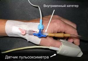
During sedation, the drug is administered intravenously using a special dispenser, and oxygen is inhaled through a light face mask.
There is a special dispenser that helps administer the drug intravenously to a patient for MRI under anesthesia.
Sevoran is a new type of inhalational anesthetic that is most often used for MRI under anesthesia. It is considered better than intravenous anesthesia due to the following qualities:
- non-toxic;
- does not cause allergies;
- quickly introduced into the body and excreted at the same pace.
A person’s condition largely depends on the characteristics of anesthesia.
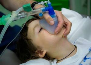
With Sevoran inhalation anesthesia, the patient immediately switches off. This important component allows for minimal influence on the child’s psyche, especially considering how violently children react to injections of various types.
How is it carried out and what types of anesthesia are recommended for adults? Sevoran is not enough for such patients. It serves only as a supporting component, and an intravenous anesthetic is required for complete anesthesia. A person regains consciousness literally a few minutes after completing the inhalation of Sevoran. Given the characteristics of anesthesia, complete recovery occurs on average within an hour.
A laryngeal mask is a special airway that is used during anesthesia. It eliminates tracheal intubation and other risks related to it. A laryngeal mask connected to the device reliably provides a person with a breathing mixture. And if the need arises, it allows for mechanical ventilation.
For some patients, the provision of MRI under sedation has been a lifesaver.
Contraindications for administering anesthesia
However, there are cases when the administration of anesthesia is contraindicated. Prohibitions include:
- severe developmental delay;
- absence of a swallowing reflex in the subject;
- severe lack of weight;
- impaired metabolic processes;
- presence of viral diseases at the time of the study;
- redness of the body of unknown nature;
- exacerbation of bronchial asthma;
- psychiatric diseases in the acute stage;
- severe pathologies of cardiac activity.
What complications can there be?
Over the past decades, evidence has emerged that the function of certain parts of the brain is impaired as a result of the administration of anesthesia in children under 4 years of age.
MRI for children
Usually this situation is accompanied by various disorders of attention and memory. However, these complications most often occur as a result of extensive surgery. In addition, the possibility of such a development of events due to surgical trauma cannot be excluded.
To induce medicated sleep during an MRI, specialized equipment is required. Conventional equipment used for inhalational administration of anesthesia does not work, since it is equipped with heart rate monitors and heart monitors, the effect of which is eliminated by the magnetic field of the tomograph.
In order to avoid negative consequences, it is recommended to conduct the examination in a medical clinic equipped with all the necessary equipment. If there are none, then the number of complications associated with the activity of the patient’s internal organs increases several times. This must be taken into account when deciding where to conduct this research.
A proper consultation with a doctor will help avoid complications from anesthesia.
It often happens that an MRI is necessary, but anesthesia is undesirable, often when diagnosing children. In this case, it is important to try to psychologically prepare the little patient. It is possible for the child to conduct the study next to his parents, who will lie on the couch next to the tomograph and, if necessary, can hold the baby to ensure complete immobility.
In addition, today there is high-field equipment with motor correction. During the examination, all interference caused by involuntary movement is eliminated using subsequent computer processing. Of course, we are talking only about minor movements. If the subject actively moves his limbs, it will not be possible to obtain clear images.
MRI under anesthesia: features
MRI examinations are performed in a strong magnetic field, which is why standard anesthesia equipment and monitors do not work. The MRI department of the Institute of Health clinic is equipped with special anesthesia-respiratory and monitoring equipment that allows you to administer anesthesia and monitor the patient’s condition. The study is carried out under the constant supervision of an anesthesiologist-resuscitator. To conduct the study, we use the safest anesthetic for inhalation (mask) anesthesia - Sevoran (ABBOT Laboratories USA).
Preparation for the procedure
Before performing an MRI under anesthesia, you must first visit the clinic for consultation. You should definitely come to it, as the doctor will be able to collect the information he needs regarding the patient’s health. Thanks to which he will be able to choose the right anesthesia and its dosage, thereby minimizing possible complications.
Important! During a conversation with a doctor, you need to inform him about the presence of allergic reactions to medications and food.
Before receiving anesthesia, if the examination is scheduled for the morning, you should take your last meal no later than 7 pm, and water no later than 20 pm. If the diagnosis is carried out during the daytime, then food is taken 6 hours before the test, water - 4 hours. This will help avoid nausea and vomiting. In addition, doctors recommend going to bed two hours later the night before, and getting up in the morning an hour earlier than your usual schedule.
Recording an MRI with contrast
To sign up for a study, choose any method:
- call the clinic +7 (495) 103-99-55,
- order a call back,
- leave a request for an appointment using a convenient form on the website:
SIGN UP
At the Kuntsevo Treatment and Rehabilitation Center, we have created all the conditions so that you can make a diagnosis from 8 am to 10 pm on weekdays and from 9 am to 9 pm on weekends.
Our center is within walking distance from the Molodezhnaya metro station.
If you are driving, a nice addition will be free parking for our visitors.
Recommendations
To make it easier to endure anesthesia during MRI, you must follow the following recommendations:
- You should undergo all the necessary tests prescribed by your doctor.
- You should inform your doctor in advance about all medications you take.
- You should not come to your procedure with makeup on.
- It is important to remove all dentures before the MRI.
- If the patient wears contact lenses, they must be removed and placed in a container.
- After anesthesia, you are prohibited from drinking water for half an hour.
- Within 24 hours after an MRI under anesthesia, it is not recommended to drive a car or perform any work that requires increased attention.

It is important to properly prepare for diagnostics in order to get the correct result without the need for repeated examinations.
If all the doctor’s recommendations are followed and the medical staff act competently, it is completely safe to conduct a tomographic examination under anesthesia, which will help to make a timely diagnosis and undergo effective treatment.
What happens during diagnosis
In order to conduct an MRI with sedation, certain preparation will be required:
- testing and therapeutic examination, if necessary;
- Before diagnosis, you should not eat for 8-9 hours;
- You can drink tea, juice or water two hours before the test, but not later (the volume of liquid you drink should be no more than 200 ml);
- Before an MRI session with sedation, you should wash off all cosmetics that have been applied to your face and remove all jewelry, especially metal ones.
If lenses are used to improve vision, the doctor should know about it.