- Recovery after the procedure
- Diagnosis sequence
- What pathologies can be identified
- Advantages of the International Center for Health Protection
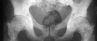
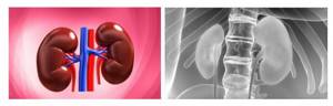
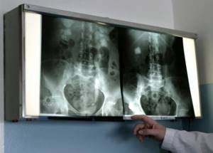
To study the condition of the kidneys, urography is performed. This is an X-ray examination of the genitourinary system, allowing you to see the location of organs relative to each other and bone structures. An x-ray of the kidneys records their size, shape and developmental abnormalities. It allows you to identify tumors, stones, calcifications, and foci of inflammation. This instrumental technique has enormous diagnostic value. With its help, you can make an accurate diagnosis in a timely manner and prescribe adequate therapy.
The essence of this type of diagnosis comes down to the introduction of a contrast agent into the circulatory system. After the procedure, a series of urograms is performed, which allows you to see on the screen and printed images all the existing abnormalities in the functioning of the kidneys. Despite the X-ray radiation, this method is safe, since the dosage of rays is minimal. It is applicable to patients of any age, only in newborns it is replaced by ultrasound.
Types of urography
- Survey urography. Using this type of x-ray, parasitic diseases, neoplasms, foreign bodies and kidney stones are detected. This is usually the first examination prescribed to a person if he suspects problems with the functioning of the renal structures. This method allows you to additionally examine the bones of the skeleton, assess the general condition and activity of the remaining genitourinary organs: ureters, bladder.
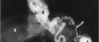
- Excretory urography. Allows you to set the filling time and intensity of the pelvis and bladder with liquid. The procedure is also called intravenous urography. A patient with an empty bladder is given a contrast agent and pictures are taken while the kidneys draw from the blood and store the contrast. Thanks to its use, this method is characterized by higher information content compared to survey urography, which consists of conventional radiographs.
What is an X-ray of the urinary organs?
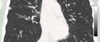
The examination method is based on the filtration capacity of the kidneys, the functions of releasing metabolites and excreting processed substances. Classic X-rays cannot display some features of the condition and structure of the organs of the urinary system, so the patient is injected with a special contrast agent to be able to study them. These drugs are filtered by the kidneys and appear on the image as dark areas with clear contours. A number of requirements are put forward for their properties, without compliance with which the effectiveness of the research may be reduced. Contrast agents should not accumulate in tissues or be metabolized in the body.
They should have low nephrotoxicity and normal levels of X-ray contrast.
Contrast agents can be:
- ionic;
- nonionic.
The first include high-osmolar monomers and low-osmolar dimers: Urografin, Isopak, Yodamide, Trazograph, Telebrix, Hexabrix. Nonionic drugs are represented by low-osmolar monomers and isosmolar dimers. Among such drugs are Lopamiro, Vizipak, Omnipak, Ultravist.
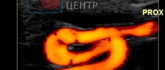
The introduction of contrast, among other things, allows you to obtain a clear image of hollow organs, unlike ultrasound or classic x-rays.
Indications for x-ray of the urinary system
The procedure is prescribed by the attending physician if the patient has the following disorders:
- pathologies of the genitourinary apparatus;
- inflammatory process occurring in the urinary tract;
- abnormal changes in bladder function;
- chronic kidney disease;
- urolithiasis - urolithiasis.
An examination is also recommended for abnormal prolapse of renal structures, benign and malignant tumors, and in case of failures and disturbances in the excretory function of organs.
If there is blood in the urine, the patient suffers from abdominal and lower back pain, there have been injuries to the urinary organs, he is also recommended to undergo urography in Moscow to clarify the clinical picture. Sometimes doctors use such diagnostics in combination with other methods, for example, to clarify the results of ultrasound of the urinary tract. To monitor the effectiveness of surgical treatment, urography is also prescribed, the price of which starts from 1000 rubles .
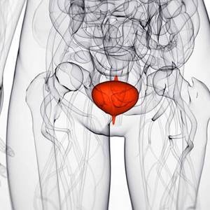
Features of bladder cystography
An X-ray of the bladder is highly informative and is prescribed by a urologist or surgeon. Cystography is carried out with the addition of a solution, which is administered in 2 ways: the first is ascending (using a catheter, 150-200 milliliters of the substance is administered through the urethra), the second is descending (intravenously). After intravenous administration, you should wait 30-45 minutes for the bladder to fill.
The following drugs can be used as contrast: urografin, iodamide, triomblast. X-ray with contrast helps to detect serious diseases: vesicoureteral reflux, fistulas, pathological narrowings, chronic cystitis (inflammation of the bladder), malignant and benign tumors, bladder diverticula, neoplasms, stones.
Content:
- Features of bladder cystography
- Indications and contraindications
- Alternative research methods
- X-raying of the bladder for children
- How is cystography performed on adult patients?
- Decoding images
Contrast x-rays are prescribed to assess the condition of the bladder and ureters. Using the procedure, you can identify the cause of urine incontinence and analyze the excretory function of the kidneys. 25-30 minutes after the administration of the contrast agent, with normal kidney function, only remnants of the contrast will be visible in the pelvis and calyces. If a large amount of the substance remains in the pelvis and calyces, this will indicate slow excretion of urine.
Contraindications and restrictions
X-ray examination is not performed if the patient:
- acute or chronic renal failure is diagnosed;
- allergy to iodine, which forms the basis of the contrast agent;
- severe pathologies of the urinary organs;
- diseases of the cardiovascular, respiratory system or liver in the stage of decompensation;
- blood clotting is impaired.
Kidney x-rays are not given to pregnant and breastfeeding women. But if the patient’s life and health are at risk, the doctor may decide to send the patient for examination, but this happens in exceptional cases.
For women there is another conditional limitation - the menstrual cycle. A planned study is best carried out at the end of menstruation, if there is no need for urgency.
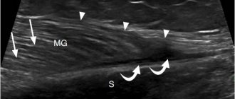
If it is impossible to conduct urography, the subject is prescribed diagnostic procedures that replace it: MRI, ultrasound, CT.
Preparation for the procedure
Preparatory measures for intravenous urography require special attention from the patient. Before the event, the intestines are cleansed with an enema or by taking medications that promote soft, comfortable bowel movements.
The day before the examination, you should adjust your diet. It is necessary to increase the volume of non-carbonated water consumed. It is advisable to exclude from the diet foods and drinks that increase the formation of gases in the intestines. These are sweets, bread, pastries, fruits with a high sugar content, carbonated drinks, cabbage, peas. On the day of diagnosis, a small portion of breakfast is allowed. 3 hours before the start of the event, completely refuse to eat. These actions are aimed at ensuring that the research is extremely effective and the results are reliable.
Many clinics do a test to determine if you are allergic to the contrast agent. To do this, the patient is injected into a vein with 1–3 ml of the substance; the exact dose depends on the drug used. Sometimes such a test can be replaced by a skin test, when iodine is applied to the skin.
Side effects when using contrast
Side effects occur after urography rarely. The following patient reviews have been recorded:
- iron taste in the mouth after radiography;
- transient mild rash;
- slight swelling of the lips.
Such symptoms can be eliminated by taking antihistamines, but usually the contrast itself is quickly eliminated from the body, which allows you to return to your normal rhythm of life. The procedure does not cause pain in patients and is rarely accompanied by adverse reactions. If you feel unwell or have complications, you should immediately inform your doctor.
Side effects and contraindications
Most patients tolerate the procedure painlessly, and side effects are extremely rare. However, sometimes the following phenomena are possible:
- iron taste in the mouth;
- slight rash and swelling of the skin (reaction to contrast agent);
- a sharp decrease in pressure, difficulty in inhaling and exhaling.
The following are contraindications:
- period of gestation and breastfeeding;
- renal failure;
- unconscious state of the patient.
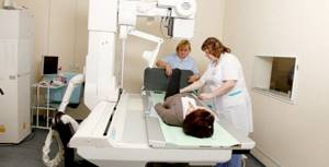
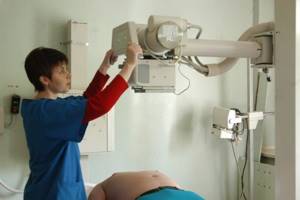
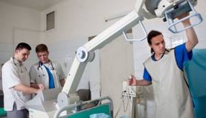
Diagnosis sequence
The event takes place in a room equipped for this purpose. The patient must sign a formal consent to perform the study.
Before you start you need:
- remove all metal objects and jewelry;
- take a sedative for severe anxiety;
- lie down on the table (sometimes the examination is performed in a standing position);
- follow the diagnostician's instructions.
Next, a survey of the kidneys is performed, after which an iodine-containing drug is slowly injected into a vein in the elbow. This takes 2–3 minutes. This technique minimizes the likelihood of discomfort for the patient.
The first image is taken 5–6 minutes later. If the functions of the renal structures decrease, the picture is taken after another 10–15 minutes. Next, images are taken over a period of 45–60 minutes. The doctor determines the required number based on the individual situation of the patient. Typically 3-5 images are obtained per session.
The injected contrast helps the medical staff monitor the period during which it will be excreted by the renal structures. It allows you to assess the condition of the urinary system, identify tumors and stones at an early stage.
After completion of the event, the diagnostician draws up a conclusion and gives the results to the patient. Making a correct diagnosis is possible only after a thorough study of a series of urograms obtained.

What pathologies can be identified
Subject to the conditions and preparation requirements, the event makes it possible to detect deviations:
- injuries of the genitourinary apparatus;
- congenital developmental anomalies;
- the presence of stones in the urinary system;
- benign or malignant formations;
- dyskinesia of the urinary tract.
After the study, you can clarify the diagnoses: hydronephrosis, pyelonephritis, urolithiasis, kidney tuberculosis, genitourinary tract infections, arterial hypertension, cysts, cancer.
Alternative research methods
If there are contraindications, the patient may undergo alternative diagnostics, for example, magnetic resonance imaging or pneumocystography. The peculiarities of pneumocystography are that gas is injected instead of a contrast solution. Sometimes lacunar cystography is prescribed - a combined procedure, during which a specialist injects 15-20 milliliters of contrast and 180-200 cubic centimeters of gas.
Another effective diagnostic method is voiding cystography, which is performed during urination. During the procedure, the work of the ureters is monitored, and areas of contrast leakage can be detected.
Cystography and other manipulations are needed to determine in detail the parameters and location of the bladder, identify pathologies and anomalies, stones and sand, ruptures, thickening of the walls of the internal organ, disruption of the urinary system and other dysfunctions.
An X-ray of the bladder is performed if ultrasound and cystoscopy fail to help make the diagnosis. The procedure is not recommended without indications, since it is painful, and the patient often feels discomfort during it.
Advantages of diagnostics in the multidisciplinary clinic “International Health Center”
If you need kidney urography, prices for this examination method start from 1,000 rubles. The list of procedures performed is reflected in the price list.
Our advantages:
- qualified doctors who have undergone training in Russia and abroad;
- high-precision equipment that allows you to obtain detailed images;
- respectful attitude of employees at all levels;
- convenient location of the clinic in the center of the capital.
We are increasing the availability of modern medicine; our clinic’s pricing is transparent. We are ready to explain what the cost of services consists of.