The body of the fair sex is a special mechanism in its structure, characterized by excessive fragility. That is why it requires increased attention and timely care. When situations occur that were previously not characteristic of the nature of a particular organism, the question arises of what to do.
In this case, you need to promptly seek help from a specialized ultrasound diagnostic center and make an appointment. Thanks to the research results, it is possible to completely cure ailments of the reproductive system in women.
Advantages of ultrasound diagnostics in gynecology
Using an ultrasound scanner, a specialist analyzes the condition of the pelvic organs, blood flow in the veins and arteries, identifies the presence of tumors, evaluates the relative position of organs and the presence of inflammation.
- Low cost of the procedure - unlike, for example, MRI and other research methods;
- Ultrasound is considered one of the most accessible methods, and a wide range of the population with different levels of income can be examined;
- High information content of the method;
- Applicable not only for studying the pelvic organs, but also for analyzing the development of the fetus inside the uterus.
- Has virtually no contraindications
- Portability - if necessary, the procedure is carried out not only in the office, but also in an ambulance or at home.
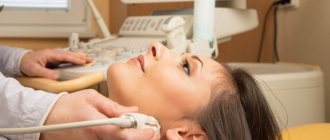
Ultrasound of the mammary glands - WHO recommends that women be examined twice a year!
The examination allows you to see not only the condition of the breast tissue, but also the regional lymph nodes. Thanks to this, it is possible to evaluate lymph flow, identify all kinds of neoplasms, even the smallest tumors and lumps less than 5 mm, and also examine all areas of the mammary glands.
A woman does not need to prepare in any special way for an ultrasound of the mammary glands, however, when prescribing the procedure, it is worth taking into account the characteristics of the menstrual cycle. So, to obtain the most accurate and informative results, the study should be carried out in the first half of the cycle, that is, on days 5–10.
Indications for undergoing gynecological ultrasound
Ultrasound diagnostics is distinguished by highly accurate results. Ultrasound is based on ultra-high frequency sound waves (ultrasound), with the help of which an idea of the current state of health of the patient’s internal organs is formed.
Using an ultrasound of the female genital organs, you can determine the state of pregnancy, the period of conception, and the health of the fetus. In a non-pregnant woman, the uterus, cervix, and ovaries are examined using ultrasound. Also examined are organs close to the reproductive system - the bladder and various parts of the intestines.
As a rule, gynecological ultrasound is prescribed to determine pathologies in the female body, as well as to monitor the course of pregnancy and monitor the effectiveness of the chosen treatment. A planned study, without additional indications, is carried out once a year.
Preparation for ultrasound in gynecology
There is no need to be afraid of this medical procedure. Ultrasound is quite painless and not harmful to the female body. Some patients experience discomfort that goes away immediately after a gynecological ultrasound.
Many girls are afraid that during the initial ultrasound examination they may be diagnosed with any abnormalities or pathologies, and then they will be sent for further, more serious examination. Remember, lack of treatment can be much worse than a serious examination. Take care of your health and undergo gynecological ultrasound regularly - 1-2 times a year.
In many cases, the effectiveness and correct determination of health status depends on preparation for gynecological ultrasound diagnostics.
Follow these rules before undergoing a pelvic ultrasound:
- Vaginal ultrasound (transvaginal) is performed on an “empty” bladder - you are allowed to drink 1-2 glasses of water a few hours before the examination, or empty your bladder by natural urination before the procedure.
- Ultrasound through the surface of the abdomen (transabdominal) is performed for girls who have never been sexually active, pregnant women in the 2nd and 3rd trimesters of pregnancy, and patients after transvaginal examination to clarify the data. This type of study is performed on a full bladder. To fully display the state of the reproductive system, the girl needs to drink 1.5-2 liters of pure still water an hour before the examination. In this case, you cannot urinate until the procedure is completed.
An additional condition for ultrasound examination is sometimes an empty bowel. This will significantly simplify the procedure and reduce the likelihood of erroneous results. For several days, you should exclude or limit as much as possible from your diet foods that cause increased gas formation. These are fruits, high-fiber vegetables, legumes, bakery and confectionery products, kefir and milk. It is also possible to use laxatives or use an enema (microenemas).
Remember, laxatives should be taken at least 12 hours before the ultrasound and only on the recommendation of a doctor.
All necessary consumables for an ultrasound examination (disposable diaper, condom for ultrasound, shoe covers, disposable napkins, etc.) are already included in the cost of the examination and are provided to the patient immediately before the procedure.
How does the procedure work?
Modern methods of ultrasound examination allow the procedure to be carried out as comfortably and without pain as possible. The algorithm for carrying out the manipulation depends on its type. Transabdominal examination is carried out as follows:
- the patient takes the required position and exposes the abdomen;
- the doctor lubricates the sensor with a special conductive gel and moves it over the abdomen.
Diagnostics can last from 5 to 20 minutes. A transvaginal examination involves inserting a probe into the vagina. Its diameter does not exceed 2 cm, so the patient does not feel pain during insertion.
An important aspect is the issue of hygiene. The doctor places a special thicker condom, designed for ultrasound, on the sensor. Only after this, a gel is applied to the sensor, which improves the conductivity of ultrasonic waves. For any type of study, data is displayed on a monitor that is synchronized with the sensor.
Duration of preparation and implementation
The timing of ultrasound in gynecology depends on the menstrual cycle, since various pathologies are easier to diagnose in the early stages of cyclic changes in the female body.
If the patient does not have any indications, chronic or congenital pathologies, infertility and the girl does not plan to become pregnant, she is recommended to undergo an ultrasound examination at the beginning of the menstrual cycle (5-7 days of the menstrual cycle). This allows you to fully examine the condition of the ovaries, endometrium, and cervix. At this time, a specialist can create a complete picture of the state of a woman’s intimate health and partially assess the reproductive function of the body.
If there are acute indications, ultrasound can be performed on any day of the menstrual cycle, including directly during menstruation.
If any pathology is detected in the patient during an ultrasound examination, she is recommended to consult an appropriate specialist (gynecologist, gynecologist-endocrinologist, urologist, surgeon, etc.). And also, undergo a re-examination after treatment. An additional ultrasound should always be performed before performing surgical interventions, be it curettage of the uterine cavity, removal of polyps, coagulation of cervical pathology, etc. Thus, the specialist can confirm or refute the diagnosis, diagnose possible contraindications or indications for the surgical procedure.
Recommendations for conducting an ultrasound examination of the pelvic organs
The main recommendation that should be followed when conducting an ultrasound examination of the pelvic organs, as well as when performing an ultrasound examination of other organs: it is better to carry out the examination by specialists with extensive practical experience. The more practice a doctor has in a particular field, the more changes he sees, which corresponds to the expression: “The doctor sees what he knows.”
The same applies to ultrasound during pregnancy. Many non-profiled clinics cannot identify various problems during pregnancy, pathologies of fetal development and placenta. Therefore, it is recommended that fetal ultrasound be performed only by qualified specialists.
It is recommended to keep a menstrual diary so that at your appointment you can confidently tell when your menstrual cycle began and ended.
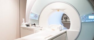
Ultrasound of the abdominal organs is the most important examination for middle-aged women
This procedure for women approaching the age of 40 is just as important as a pelvic ultrasound. Ultrasound of the abdominal cavity involves scanning the liver, spleen, kidneys, pancreas, gallbladder with bile ducts, genitourinary system, blood vessels, and retroperitoneal space.
The doctor evaluates parameters such as position, size, internal structure of organs, the presence of pathological inclusions, developmental anomalies, inflammatory changes typical of chronic and acute diseases, traumatic and parasitic injuries.
Ultrasound of the abdominal organs allows you to diagnose a number of pathologies:
- acute and chronic hepatitis;
- cirrhosis;
- fatty infiltration;
- cysts;
- benign and malignant neoplasms;
- abscesses;
- cholelithiasis;
- acute and chronic cholecystitis;
- disturbances in the outflow of bile;
- acute and chronic pancreatitis;
- developmental anomalies;
- cholestasis;
- infectious and inflammatory processes;
- signs of hypertension;
- the presence of plaques, stenoses, blood clots.
Which method of ultrasound to choose?
Ultrasound is performed in several ways: transabdominal and transvaginal sensors. As a rule, the doctor chooses a certain method depending on the woman’s condition and his qualifications.
The transabdominal method is indicated for girls who are not yet sexually active, as well as for women who have large abdominal masses, including the pelvis.
The main method of conducting gynecological ultrasound throughout the world is considered to be a transvaginal probe. This method allows you to avoid many inaccuracies and errors that are made when examining the pelvic organs through the abdominal region.
In many modern clinics, both methods are combined to obtain the most reliable results.
There are also other types of ultrasound examinations: Doppler ultrasound, 2D, 3D and 4D monitoring.
2D, 3D and 4D monitoring
Using these diagnostic methods, you can see not only the intrauterine development of the fetus, but also diagnose the slightest changes in the female body. For this study, slightly larger sensors are used that transmit signals to a special module. It summarizes the signals and displays a three-dimensional image on the screen.
In the case of 3D monitoring, the image can be viewed from all sides using different viewing degrees. In 4D visualization, the examination is recorded on a memory card or external media in video format. The patient and the attending physician can see the object in motion. This allows you to accurately determine the location of the pathology and its shape.
See your gynecologist regularly and don't forget to get an annual checkup. Take care of your health.
There are contraindications, consultation with a specialist is required.
List of articles
Additional tests and examinations for women from 30 to 40 years old
At this age, a woman needs to undergo the following examinations (in addition to the previous list) regularly every 1 - 1.5 years:
- blood lipid profile study;
- blood sugar;
- measurement of body mass indexes;
In middle-aged women, the risk of heart attacks and strokes increases due to the increased formation of atherosclerosis. Timely diagnosis and correction with medications and diet will prevent this threat.
At the age of forty, the likelihood of developing breast cancer increases significantly, so an ultrasound scan that can detect the slightest lump or tumor is extremely necessary. At this age, a set of examinations will help prevent chronic pancreatitis, peptic ulcers, cholelithiasis and type 2 diabetes mellitus.