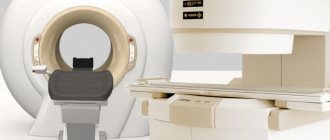
Magnetic resonance imaging (MRI) is a non-invasive diagnostic method that allows you to establish a diagnosis with maximum accuracy and develop an effective treatment regimen.
To visualize the brain and its detailed examination, exposure to powerful magnetic fields and high-frequency pulses is used. The computer program then processes the results obtained and produces information in the form of a picture that can be transferred to paper or electronic media.
The procedure is completely painless and safe for humans. During an MRI scan, the patient is not exposed to x-rays. The examination can be carried out repeatedly, even for children over 3 years of age and senior citizens. Using MRI, it is possible to detect a variety of brain diseases in the early stages of development, which is the key to their successful treatment in the future.
Indications
The resulting layer-by-layer images of brain structures allow specialists to study in detail the condition of tissues and blood vessels, identify the pathological focus, its location, shape, size, assess the degree of prevalence, etc.
No other diagnostic method (radiography, ultrasound or computed tomography) provides as much information and in such a volume as MRI.
You can undergo the examination yourself or as prescribed by a neurologist in the following conditions:
- constant (frequent) headaches, dizziness;
- suspected brain tumor (regardless of malignancy);
- examination before brain surgery;
- assessment of the condition of structures and meninges after surgical treatment;
- infectious diseases (encephalitis, meningitis);
- decreased vision and hearing not associated with eye or inner ear disease;
- acute cerebrovascular accidents (stroke);
- neurological disorders (frequent fainting);
- pathological changes in blood vessels (aneurysm, stenosis);
- diseases of the nerves (auditory, visual);
- deterioration of memory, concentration;
- congenital malformations of the brain;
- diffuse (extensive) changes (Alzheimer's disease, multiple sclerosis);
- epilepsy (including post-alcohol epilepsy);
- various injuries;
- monitoring (control) of the condition of the cancer site after treatment;
- inability to perform a computed tomography scan.
In addition to the above conditions, children are also prescribed MRI in the following cases:
- delay in psychomotor development;
- speech disorders (stuttering);
- inappropriate behavior in everyday life and society without justified reasons;
- convulsive syndrome;
- frequent fainting conditions.
Requirements for the placement and organization of CT work
1. The X-ray department (office) is not allowed to be located in residential buildings and children's institutions. (The X-ray computed tomography room also applies here) (clause 3.1.*)
2. It is not allowed to place X-ray rooms under rooms where water can leak through the ceiling (swimming pools, showers, restrooms, etc.). It is not allowed to place the X-ray treatment room adjacent to wards for pregnant women and children (clause 3.4.*)
3. The composition and area of the premises of the X-ray computed tomography (X-ray) room are specified by the organization that manufactures the computer tomograph in the form of a project proposal, which is taken into account when developing the cabinet design, but does not replace it. This clause also applies to the placement of other types of foreign-made X-ray machines, the documentation for which contains the company’s design proposals (clause 3.10.*)
IMPORTANT! Specialists will provide all technical requirements for the installation of equipment proposed for delivery, which will be specified in the technical specifications and plans for the preparation of premises made for specific premises in medical institutions, in the form of a design and technological proposal.
4. The width of the doorway in the RCT treatment room must be at least 1.2 m with a height of 2.0 m, the size of the remaining doorways is 0.9 x 1.8 m. (Dimensions are given in the final version) (clause 3.12.* )
5. The door from the treatment room and the control room to the corridor should, for reasons of fire safety, open “towards the exit” (during the evacuation), and from the control room to the treatment room - towards the treatment room (clause 3.26.*)
Composition and area of CT rooms for routine examinations**
The composition and area of the premises are specified by the design assignment and the requirements of the equipment manufacturer
| Room | Area, m2) |
| Procedural | 22 |
| Control room | 8 |
| Generator/computer room | 8 |
| To process research | 8 |
| Doctor's office | 9 |
| Cabinet for undressing | 4 |
| Viewing room | 6 |
Contraindications
The procedure is based on the effect of magnetic fields, so MRI is strictly prohibited for people with pacemakers, ferromagnetic endoprostheses, metal staples on brain vessels, electronic implants in the middle ear, and foreign objects (metal fragments) in the tissues of the eye. The presence of these objects can affect the examination result, and the magnetic field, in turn, can disrupt the operation of, for example, a pacemaker. It is also not possible to perform an MRI on people who are overweight (the diameter of the MRI capsule tunnel is 60 cm).
Relative contraindications (conditions in which MRI is considered possible):
- pregnancy (first trimester);
- claustrophobia (fear of closed spaces);
- wearing braces and dental crowns;
- using an insulin pump;
- tattoos with paint containing iron;
- metal foreign objects not localized in the head area.
Preparation
MRI is not time-sensitive and can be performed both in the morning and in the afternoon/evening hours. No special preparation is required. The patient only needs to have identification documents (passport), a referral from a doctor and the results of previous examinations, if any.
Eating and taking medications before the procedure is not prohibited. If claustrophobia is present, the patient is given light sedation (injection of a sedative) immediately before the examination.
MRI placement requirements
1. Magnetic resonance imaging departments are not allowed to be located in residential buildings and in built-in and attached premises (clause 2.9.*)
2. A magnetic resonance imaging (MRI) scanner can be placed as part of the radiology department (clause 10.14.2.*)
3. Diagnostic MRI rooms (departments) are not allowed to be placed adjacent (horizontally and vertically) to wards for pregnant women, children and cardiac patients
4. Shielding is carried out using a Faraday cage, taking into account the power of the tomograph. The design of the walls, ceiling, floor, doors, windows in the treatment room must ensure that the levels of the electromagnetic field in the adjacent rooms are reduced to acceptable values (Appendix 8*). Sound insulation of walls, ceiling, floor, doors, windows of the technical room and diagnostic room must be carried out in accordance with calculations of the acoustic influence of the equipment and ensure hygienic requirements for noise in adjacent rooms (Appendices 9 and 10*)
Composition and area of premises of the MRI room***
The composition and area of the premises are specified by the design assignment and the requirements of the equipment manufacturer
| Room | Area, m2) |
| Diagnostic/Procedural (RF cabin - Faraday cage) | 25 |
| Control room (operator room) | 10 |
| Preparatory room | 12 |
| For undressing patients | 2 |
| Technical room | 20 |
| For meals | 10 |
IMPORTANT! It is not allowed to locate significant masses of metal, transformers, electrical devices of significant power, elevators, high-voltage power lines and electrical cables near the MRI procedural rooms. Cars, trams and other objects moving in close proximity can also have an undesirable impact.
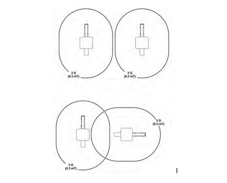
Mutual placement of two MRIs
When placing multiple MRIs, the placement should be as shown below
Sanitary and hygienic requirements for MRI and work organization
1. The magnetic resonance imaging unit must be located on the first floor of an isolated compartment of the existing medical and preventive building or in an extension to it, or in an isolated building constructed specifically for this unit. It is allowed to place MRI as part of the X-ray diagnostic department.****
2. To place the tomograph, a room must be selected whose load-bearing structures are capable, with an established safety margin, of withstanding the load created by the equipment included in the product, taking into account the load from service personnel, patients, etc.****
3. Tomograph power supply:****
— it is recommended to carry it out via a separate feeder (input), not electrically connected to the institution’s network and household networks; — to weaken external electromagnetic fields, the diagnostic room should be shielded using a Faraday cage. This shield must provide electromagnetic interference suppression of at least 80-100 dB in the frequency range 6.0-6.4 MHz. The screen is made of copper foil; - doors and windows of the diagnostic room must also be shielded; — the entry of equipment power supply lines, including lighting, as well as signal lines into the diagnostic room must be carried out through a filter panel; — all ducts or ducts for air conditioning and electrical panels in the diagnostic room must be made of non-magnetic materials; — for the normal functioning of the MRI, the presence of large metal masses and high-power power lines in the immediate vicinity of the magnetic system should be avoided.
4. The technical room (which is the main source of noise) should not be located in adjacent rooms to the control room, doctor’s office and patient rooms. When placing air or water cooling equipment, noise and vibration protection measures must be taken to ensure noise and vibration levels within the remote control room in all adjacent rooms.****
5. The control room must be located in a separate room with natural light. The use of the control room for receiving patients is not permitted.****
6. When performing angiography, a procedural area of 12 square meters is required. m, in which it is necessary to comply with the sanitary and epidemiological regime of aseptic premises.****
IMPORTANT! MRI should not be placed in a residential area; stairwells, elevators, and patient rooms for pregnant women and children should not be adjacent. People with pacemakers, insulin pumps and magnetic prostheses are not allowed to be in an area with a magnetic field strength of 0.5 Tesla or higher.
Brain MRI technique
The examination can be carried out both on an outpatient basis and in a hospital setting. The duration of the procedure is from 15 minutes to half an hour.
The patient is placed on the sliding table of the MRI unit. To ensure complete immobility, the patient’s body is fixed with bolsters and belts, and the head is fixed with special clamps.
If it is necessary to administer a contrast agent (for more effective visualization of the structures being examined), the nurse installs an intravenous catheter for the patient.
Important! The administered substance is absolutely safe and does not in any way affect the health and well-being of the patient.
After this, the table, together with the patient, slides inside the magnetic capsule, and the medical staff leaves the office. A two-way communication system involves communication between the doctor and the patient. At certain moments the doctor may ask you to hold your breath or inquire about the well-being of the person being examined. The patient, in turn, reports all his sensations and changes in health status by pressing the button to turn on the intercom inside the capsule.
At the end of the study, the patient is released from the fixing devices, the intravenous catheter is removed and asked to wait for a while outside the office doors while the doctor performs a preliminary analysis of the images. If poor quality images are obtained, the procedure may need to be repeated.
Regulatory documents SanPiN
* SanPiN 2.1.3.2630-10 “Sanitary and epidemiological requirements for organizations engaged in medical activities.” ** SanPiN 2.6.1.1192-03 “Hygienic requirements for the design and operation of X-ray rooms, devices and the conduct of X-ray examinations.” *** SanPiN 158.13330.2014 “Buildings and premises of medical organizations. Design Rules” (approved by order of the Ministry of Construction and Housing and Communal Services of the Russian Federation dated February 18, 2014 N 58/pr). **** Information and methodological letter of the Office of Rospotrebnadzor for Moscow dated 01.08.2007 N 9-05/122-486 “Sanitary and hygienic requirements for magnetic resonance imaging scanners and organization of work” 01.08.2007. Category: Moscow. Document as of August 2014.
Protection against X-ray radiation must comply with the requirements of legislation and regulatory documents in force in the Russian Federation (incomplete list):
— SanPiN 2.6.1.2523-09 “Radiation Safety Standards (NRB-99/2009)”;
— SP 2.6.1.2612-10 “Basic sanitary rules for ensuring radiation safety (OSPORB-99/2010).”
Possible complications during and after MRI
To date, no complications have been observed after MRI of the brain. During the procedure, the patient may experience psychological discomfort from being in a confined space. This condition is easily eliminated by pre-administration of sedatives.
Children are required to be provided with headphones or earplugs to drown out the noise of the operating scanner. Adult patients usually request them themselves. In some clinics, light music is played for the patient to create a favorable atmosphere inside the capsule.
At the moment the contrast agent is injected into the vein, the patient may feel a rush of heat or, conversely, a feeling of cold flowing through the veins. Some people experience an iron taste in their mouth. This is a normal reaction and there is no need to worry about it.
During insertion and removal of an intravenous catheter, the subject feels physical discomfort, the same as when an injection into a vein. But all this is survivable and does not require special treatment. Even a small hematoma at the puncture site will resolve on its own in a few days, leaving no mark on the skin.
It is possible that the temperature of the area of the body under study may increase; with MRI of the brain, the head. This is also only a temporary effect that will not cause harm to health. But if this causes great concern to the patient, he is simply obliged to inform the doctor about his condition.
There is also a small risk of developing an allergic reaction (hives or itchy eyes) or nausea after contrast injection. In this case, the medical staff of the MRI room will immediately provide the necessary medical assistance.
Alternative Methods
Magnetic resonance imaging has left radiography and ultrasound far behind in terms of information content and accuracy of results. Only computed tomography (CT) can compete with MRI. But there are significant differences between the two modern diagnostic methods.
- MRI is good at identifying the pathology of soft tissues and blood vessels of the brain, CT scans at the bone structures of the skull and cavities filled with cerebrospinal fluid (CSF);
- MRI does not carry radiation, CT uses radioactive radiation;
- The quality of MRI images is much better, since they are not subject to the shielding effect of the skull bones, as with CT;
- Gadolinium-based MRI contrast is much less likely to cause allergic reactions than iodine-based CT contrast.
Despite the abundance of advantages, MRI is not always indicated for everyone. Therefore, computed tomography, as well as other, slightly outdated, research methods are widely used as an alternative method in modern diagnostics.
MRI at the Elena Malysheva Diagnostic Center
Remember that timely and high-quality MRI is the key to successful treatment in the future. An inexperienced radiologist or outdated equipment can cause incorrect diagnostic results and, as a result, ineffective treatment.
Qualified specialists at the Elena Malysheva Medical Center with many years of experience devote exactly as much time to MRI examinations as is necessary for a thorough diagnosis of the body, and the conclusion will be ready within an hour! Take care of your health, and we will save your time!
Sign up for an MRI at the Elena Malysheva Diagnostic Center near the Baumanskaya metro station (see map) by phone: 8 or leave a request on the website.
Baumanskaya metro station (see map) 8
leave a request on the website