Computed tomography is a non-invasive form of examination that provides layer-by-layer diagnostics of the internal parts of the body of the object being studied. The technique involves the penetration of X-ray radiation through anatomical objects of different densities.
The absorption of rays occurs with different activity indices, which depends on the density of the scanned area. The procedure is one of the most effective technologies in the field of medical research, capable of detecting diseases at various stages of development. Multispiral CT scans for cancer show the structure of solid tumors. The examination allows us to study changes in the size of the lesion during treatment.
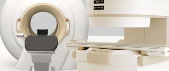
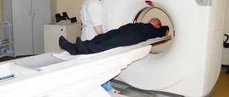
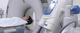
What does a CT machine look like?
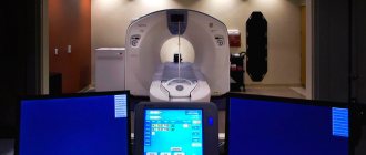
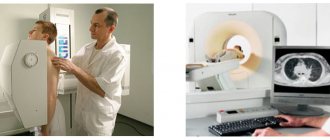
The computer device looks like a rectangular installation with a tunnel inside the central part. The patient is placed on a sliding table passing through the tunnel area. Scans throughout the examination are generated using a narrow rotating beam of radiation and a group of sensors located on the scanned ring (gantry). A set of devices responsible for image processing is installed in an adjacent room, where a specialist controls the operation of the scanner and monitors the examination process.
How does computed tomography work?
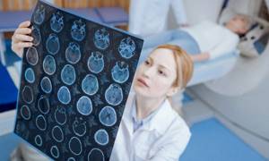
The principle of scanning is based on determining the difference in the reduction of ionizing radiation by various tissues, processing the generated information by a computer using mathematical formulas, images (slices) of the scanned areas of the patient’s body on the monitor with further interpretation by a radiologist.
During its infancy, the diagnosis made an explosion in medical diagnostics, as it became possible to visualize the layer-by-layer thickness of the human body without the use of surgical instruments or an optical probe penetrating inside.
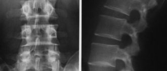
The pelvic CT method steadily occupies a leading position in the examination of various ailments - cancer, pathologies of the respiratory organs, abdominal cavity, and bones.
Characteristics available for display during computer research:
- Indicators of radiation captured by the sensor;
- Parameters recorded at the output of the tube used for radiography;
- Localization of scanning elements at any time.
Other data is generated by processing the original values. Most scan slices are perpendicular to the vertical axis of the body.
To obtain a cross-section, a full 360-degree rotation is made around the object under study, the layer thickness is pre-set. In a standard device, rotation occurs without stopping, the rays are distributed fan-shaped.
The electrovacuum device and the detector are connected, they work simultaneously: the beam beam is emitted and recorded by receiving devices located on the reverse side, almost synchronously. Fan distribution is carried out at an acute angle (up to 60 degrees), depending on the specific apparatus.
One picture is taken when the tube passes through a complete ring: the coefficients of reduction in emissivity power are recorded at a huge number of points (at least 1400).
What does the power of the tomograph affect?
The higher the amount of Tesla in the device, the clearer the image obtained during diagnostics will be. This is explained simply - the higher the magnetic contrast, the more clearly human tissue is visible.
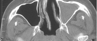
High-power tomographs make it possible to examine the smallest structures. The slice thickness for this study is 1 millimeter. This makes it possible to see the beginning of the development of the disease. The information obtained is valuable, since the disease is easier to eradicate at the initial stage. For oncological diseases, diagnosis using a high-power tomograph is important. It allows you to stop the disease at the beginning of its development and prescribe the correct treatment. Timely detection of abnormalities in the body can save the patient’s life. Low-field tomographs do not provide a clear image due to low spatial resolution.
The higher the power of the device, the higher the cost of the study. 1.5 Tesla MR tomographs are considered the most optimal in this regard. They allow high-quality diagnostics of the human body for a relatively low fee.
On low-field devices, the image quality is low and blurry. When reviewing the results, the doctor may doubt some points. A good specialist will insist on repeat research. This is necessary for further planning of treatment tactics.
To obtain a high-quality diagnostic result, it is more advisable to carry it out on a high-field tomograph. Thus, you can save money on repeated studies.
The time for diagnosing the body depends on the power of magnetic resonance imaging. This is one of the important parameters. There are patients who cannot remain still for a long time or who suffer from claustrophobia. For example, a study on a 1 Tesla magnetic resonance imaging scanner takes 15-20 minutes. And on a similar device with 1.5 Tesla, the time spent is 10-15 minutes.
What types of CT machines are there?
There are sequential and spiral tomographs. The first version of tomographic installations belongs to the initial generation devices. The devices provide the ability to construct only one cross-section at a time, obtained as a result of X-ray radiation. Today it is rarely used due to the long duration of the procedure and a decent amount of absorbed radiation exposure.
The type of device that involves the action of X-rays in a spiral, due to the mixing of the directions of the scanned tube, the table with the person, helps to obtain more information in a short period of time, the volume of irradiation exposure becomes less.
The devices are divided into single-slice devices, forming a single scanning layer in one full circle; multi-slice, allowing you to create multiple slices.
The main advantages of multislice CT devices: increased examination speed, the ratio of useful signal to noise level, reduced ionizing effect on the patient, increased anatomical coverage area, minimal examination duration, improved image quality.
Today's medicine more often uses devices with the number of layers from 16 to 64. 16-slice CT installations are endowed with speed indicators that are 24 times higher than single-slice ones and four times faster than 4-slice equipment. The scanning time is noticeably reduced (by 30 times), the dose of CT rays is reduced due to a decrease in exposure, and motion artifacts are barely visible. To create high-resolution images and reduce the time spent on diagnostics, scanners are being introduced with an increase in the number of slices to 64.
The latest scanners are of great importance for high-quality tomography of all parts of the body, especially those that are constantly in dynamics - the heart, bone joints. The efficiency of image formation has made it possible for 64-slice CT machines to become a replacement for classical methods of checking the functionality of organs - the introduction of cardiac catheters and angiography. It turns out to carry out an examination of the coronary vessels, abdomen, lower pelvis, and chest in a matter of seconds. Contrast-enhanced CT makes it possible to display the smallest elements of the vascular system of the brain, kidneys, tumor formation, damage inside joints, violations of the integrity of bones, complex injuries, and other acute pathologies.
There are 320-slice representatives, the diagnostic capabilities of which are even difficult to imagine.
What is more effective: 3T or 1.5T magnetic resonance imaging (MRI)?
Is it true that a 3 tesla device is twice as good as a 1.5 tesla device? If we take into account only the field strength - of course. In the world of sales and marketing, too. However, in terms of visualization, throughput in terms of earnings - absolutely not. Before you invest more money into opening a center with a 3 Tesla machine, you should think about what you are going to do with it, how it can be useful to you and how it will not.
Cost effective systems
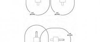
Without imposing a percentage, it is safe to say that a 1.5 Tesla MRI machine is suitable for most MR scans. The 1.5 T short bore machine remains the standard, most used magnetic resonance imaging scanner. This does not mean that 3 Tesla systems have not caught on, but return on investment, throughput, staffing, and other factors should be taken into account. Silence the noise or turn down the volume? During an MRI scan, there is always noise in the image. Much of this noise comes from the patient's body, as well as from the electronics of the MRI machine itself. It is important to get the "signal" that creates the image, not the "noise" that can affect the quality of the image. 1.5 and 3 tesla devices cope with this, but to varying degrees. Young children tend to be very noisy. If they get together, for example for a birthday, the excitement makes them even noisier. Games can keep them occupied for a while until the party is over. For the occasion, if you want to play musical chairs, you have two options to make everyone hear the music:
— Make the sound louder
- Calm children
The operation of a 3 Tesla MRI machine is in many ways similar to the operation of a stereo system playing music for children at maximum volume. Essentially, this way you get more signal - the higher the field strength, the more molecules resonate, drowning out the noise. The 1.5 Tesla system with a multi-channel coil works largely on the principle of “calming children”. Coil elements allow the examination to be carried out closer to the body, which reduces the amount of noise in the image.
Clarity, speed, need
Two parameters come to mind when thinking about 3 Tesla machines: clarity and scan time. Simply put, 3 Tesla systems, having a higher field strength, increase the signal (creating the image), and therefore the clarity of the image at a certain scanning speed. However, you can't get the best of everything at once, so MRI studies present a trade-off between scan time and image quality. Thus, depending on the technology, your bandwidth needs, and other factors, the advantage may be in one direction or another. The bottom line is that you will still get quality images on a 1.5T system using multi-coil technology - but the scan time will be longer than 3T. Conversely, you can reduce scan time on a 1.5 Tesla machine, but the image quality will be slightly worse. It all depends on the type of research.
Demand Offer
If you are doing research that requires the smallest details (complex brain work is one of the categories where a 3T machine is really needed), or you have a need to see a maximum number of patients in a day, you are inclined to purchase a 3 Tesla system, then you should plan everything in advance. Such devices are expensive - even on the secondary market you can pay twice as much for them as 1.5T, and yet they are difficult to find. Take the time to find a system and make sure your space is suitable for it. Remember: the strength of the electromagnets used to lift cars in junkyards is about the same as a 1.5 Tesla machine. And a 3 Tesla system has twice the magnetic field strength! Make sure to follow all safety precautions on site! If your research is less detailed, or the pace is less strenuous, a 1.5 Tesla system may give you everything you need. These systems are much more accessible, as are spare parts for them, as well as service engineers to maintain them. As with the 3 Tesla magnet, you must ensure that your facility is ready to accommodate the machine. Failure to take proper precautions can result in costly damage and serious injury.
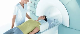
What is the difference between an open tomograph and a closed one?
According to their design features, tomography devices are produced in closed and open versions. The closed-type installation has a tunnel shape designed to place the object under study inside it.
Doctors often encounter unpleasant situations when examining patients suffering from claustrophobia; certain difficulties arise during computer diagnostics of children, the elderly, and overweight people.
Open tomography devices include equipment that emits a beam of X-rays in a spiral pattern. The advantage of the design is the absence of a fenced tunnel, which makes it possible to examine obese clients and facilitates the procedure for men and women who are afraid of limited space.
A spiral tomograph makes it possible to significantly increase the speed of the patient scanning process and increase the quality of the resulting image.
Disadvantages of a high field
It is useful to know that an increase in power does not increase the quality of diagnostics in the same proportion as the cost. Medium-class devices (1.5 Tesla) allow you to carry out all the necessary studies, including 2-3 mm sections quite quickly - 10-15 minutes for each part of the spine.
The injector, or automatic syringe, provided in expensive modifications, is not as necessary as in CT scanners - the volume of contrast agent in MRI does not exceed 10–20 ml, and it is easy to introduce it manually.
The higher the power of the device, the more it heats the implants, so high-field diagnostics are not indicated for patients with endoprostheses.
Thus, tomographs of different power differ in purpose, information content, contraindications, and cost. Therefore, it is advisable that the specialist, when referring for an MRI, indicates the class of equipment on which the procedure should be performed.
Which type of CT scanner is better?
Which CT is better? Many diagnostic centers use spiral and multi-slice units in their work. Typically, people are offered diagnostics using devices with a number of sections from 8 to 64. A number of highly specialized medical clinics use 128-slice equipment.
It is better to check the type of computed tomograph to perform the examination with the treating doctor. To analyze hard tissue injuries, it is reasonable to register for the study at a facility equipped with 16-32-slice units. If you need to study vascular canals, the heart, and various organs, the best option would be 64-slice MSCT tomographs.
Classification of tomographs
Based on the number of units of measurement of magnetic induction, different types of tomographs are distinguished:
- 0.4 T;
- 1 T;
- 1.5 T;
- 3 T;
- Devices with more than 3 Tesla.
The differences between these devices are in power and quality of work. In medical practice, MRI 1.5 Tesla is considered the most popular. They are able to conduct high-quality diagnostics of all human organs, tissues and blood vessels. It is the tomograph that is able to detect the presence of a tumor in the body, its size and area of spread.
3 Tesla devices are more powerful. They are able to diagnose all hard-to-reach structures of the human body. But such a need arises in isolated cases. The disadvantage of a 3 Tesla device is the cost of its research. The 1.5 Tesla MR tomograph does an excellent job of examining the body. Diagnostics with a 3 Tesla device is usually not required.
Note!
All tomographs with a field power of more than 3 Tesla are used only for scientific purposes. Thanks to them, scientists conduct research at the molecular level.
The disadvantage of high-field MRIs is that they are all closed-type. This is explained simply - to achieve the result, the area of the magnetic field must be maximum. For people suffering from claustrophobia, examination on such a device is a test.
Recently, MRI scans in the form of a tube have become increasingly common, in which the legs and head of the subject remain open, unless this contradicts the doctor’s prescription. Diagnosis carried out in this way is much easier for the human psyche.