MRI of the stomach and intestines: diagnostic advantages
A study using magnetic resonance imaging combines harmlessness for the patient (there is no negative impact of radiation, no violation of the integrity of the body is required) and high information content for the doctor (good image contrast, characterization of the functional characteristics of organs, the ability to construct a three-dimensional image).
Another advantage is the possibility of comprehensive diagnostics with determination of the thickness of the intestinal and gastric walls, the position of the gastrointestinal tract loops, as well as assessment of the condition of the lymph nodes, vessels, fatty tissue and organs of the retroperitoneal space located in the immediate vicinity.
The thin gastrointestinal tract is difficult to access for research; pathological processes occurring in it can rarely be detected at an early stage. Solving the problem is often only possible with the help of an MRI of the intestine. What the examination result shows - inflammation, an oncological or ulcerative process, and forms the basis of the diagnosis, predetermining successful treatment.
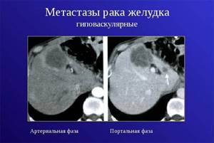
How are gastric cancer metastases diagnosed?
To search for metastases in stomach cancer, the following diagnostic methods are used:
- Computed tomography is good at detecting bone metastases, but can also show lesions in soft tissue.
- MRI is a safe examination using a magnetic field that helps detect metastases in soft tissues. In this regard, it is more accurate than CT.
- Positron emission tomography is a study during which a special mark is introduced into the body - safe radioactive sugar. Since tumor cells are actively multiplying and need a lot of energy, they accumulate this sugar, making them visible in pictures taken with a special apparatus. There are devices with which you can perform PET and CT simultaneously, this helps to get a more detailed picture.
- Chest X-ray is used to look for metastases in the lungs.
- Sometimes there is a need for diagnostic laparoscopy - a procedure during which the doctor makes a puncture in the wall of the abdominal cavity and inserts an instrument with a video camera ( laparoscope ) inside. This helps to assess the extent of tumor spread and detect secondary lesions in the peritoneum and internal organs.
A biopsy of the metastatic lesion can be performed. When examining the tissue under a microscope, tumor cells characteristic of stomach cancer are found in it. In order to select the optimal treatment, molecular genetic analysis is performed for certain marker substances:
- HER2 is a receptor on the surface of cells that stimulates their proliferation. In cancer, HER2 activity may be increased.
- PD-L1 is a protein that can interact with immune cells and suppress their activity. It belongs to a class of substances called checkpoints .
Reasons for prescribing magnetic resonance imaging of the stomach
MRI of the stomach and intestines is not a primary diagnostic method. The study is always preceded by a consultation with a doctor, who determines the feasibility and how often this procedure can be performed on a particular patient.
Diagnosis is prescribed by a gastroenterologist, oncologist, as well as a therapist or pediatrician. After laboratory tests and other methods (ultrasound, fibrogastroscopy), a preliminary diagnosis is made. Only after this they resort to a more accurate study, which makes it possible to visualize in detail the organ of interest and its condition in order to make a final diagnosis.
The immediate reasons for prescribing this method of examination may be:
- Suspicion of an oncological process.
- Gastrointestinal bleeding.
- Inflammatory, ulcerative processes.
- Metastases of tumors of other localization.
- Anomalies in the development of the digestive tract.
Prognosis for gastric cancer with metastases
The five-year survival rate for stage 4 gastric cancer is 5%. This means that only five out of a hundred patients diagnosed with the disease will still be alive after 5 years.
On average, after 3 months from the moment of diagnosis of gastric cancer with metastases, half of the patients remain alive. The prognosis is worse if the cancer has spread to the bones and liver: with such metastases, half of the patients die within 2 months.
| More information about the treatment of stomach cancer at Euroonco clinics: | |
| Treatment of stomach cancer | |
| Oncologist-gastroenterologist | 5100 rub. |
| Chemotherapy appointment | 6900 rub. |
| Emergency oncology care | from 11000 rub. |
| Radiologist consultation | 10500 rub. |
Book a consultation 24 hours a day
+7+7+78
Magnetic resonance method for diagnosing the gastrointestinal tract
The magnetic tomography method is one of the safest diagnostic methods and does not cause side or long-term reactions. The parameters of the magnetic field created during the operation of the tomograph are within acceptable values for humans.
The reasons why an intestinal MRI may not be suitable for a person are cost, expediency in making a diagnosis, as well as contraindications. It is unacceptable to perform tomography on people with foreign metal objects inside the body (fragments, bullets, knitting needles, pins, plates), stimulators and pacemakers (pacemakers, neurostimulators).
What does MRI of the stomach and intestines show?
If it is necessary to make a differential diagnosis in controversial cases, an MRI of the intestine is performed to assess the condition of the organs before surgery, monitor the treatment provided, as well as the ineffectiveness of other research options. Moscow has a wide network of clinics where you can undergo this examination.
What can be seen in tomography images:
- Features of the shape and size of the gastric organs.
- Signs of inflammation of the mucous membrane.
- Anomalies in the development of the esophagus, partial narrowing of the lumen, obstruction of patency.
- Foreign bodies, injuries, bleeding, acute appendicitis, proctitis, gastritis.
- Benign neoplasms (lipoma, hemangioma, fibroma, neurogenic mediastinal tumors, leiomyoma).
- Malignant neoplasms (lymphoma, carcinoid, gastrointestinal tumors).
- Localization and prevalence of metastases of cancer processes.
- Condition of large vessels, including the veins of the esophagus, disorders of the blood supply to the gastrointestinal tract (ischemia), lymph nodes.
- Crohn's disease, celiac disease, infectious lesions, abscesses, polyps, hematomas, diverticulitis.
What to choose: MRI of the intestine or colonoscopy
It is impossible to say for sure which is better: MRI of the intestines or colonoscopy. Each method has its own advantages in diagnosis depending on individual tasks.
Unlike tomography, colonoscopy is an invasive method associated with side effects: the possibility of organ damage, bleeding, and gastrointestinal infection. At the same time, colonoscopy is performed using a fiber colonoscope equipped with a video camera, with the ability to take material for a biopsy or perform minor operations (removal of polyps, cauterization of the walls).
Magnetic resonance diagnostics in this case acts solely as a visualization method, after which a colonoscopy can be prescribed, which combines the functions of accurate diagnosis and treatment.
The choice of examination option (MRI of the intestine or colonoscopy) is made by the attending physician, based on the clinical picture, contraindications and missing information in making a diagnosis.
Features of preparation for magnetic resonance diagnostics
The highest quality images are obtained from a properly prepared subject. Preparation begins a few days before the study.
You should adhere to a low-carbohydrate diet and consume a minimum amount of foods that promote gas formation - legumes, fruits, bread, cabbage, onions, juices, carbonated drinks. If a person is constipated, then care should be taken to cleanse the body by taking laxatives or an enema.
A light dinner is advisable the evening before the procedure, and on the day of the procedure it is better not to have breakfast if the procedure is performed in the morning. If later, a light breakfast is allowed.
The doctor may give an intravenous sedative if the patient is prone to seizures, agitated, or nervous, as this will make it difficult for the patient to remain still during an MRI of the bowel. What the monitor shows in case of improper preparation: unclear picture, distortion of images by the contents of the gastrointestinal tract. This makes assessment difficult and re-examination may be required.
Interpretation of MRI results of the abdominal organs
The radiologist begins receiving images on the computer even before the end of the study, while the patient is undergoing the procedure. At the DiMagnit center, the doctor begins to interpret the results without waiting for the end of the procedure. The images received on the computer are immediately examined for the presence of pathologies. This allows you to significantly reduce the waiting time for a conclusion after the study. . This helps you save a lot. To draw up a final conclusion about the area being examined, the doctor will need about 30-40 more minutes after completing the study.
MRI of the abdominal organs is a safe procedure that allows you to accurately identify abnormalities in the functioning of internal organs. There is no need to be afraid of the procedure - it is absolutely painless. The results of the study can be obtained and transferred to the attending physician on the same day.
| MRI of the pancreas in Rostov-on-Don |
| MRI of the abdomen with contrast |
| MRI of ureters |
| What does an MRI show? |
| MRI of the kidneys with contrast in Rostov-on-Don |
| MRI of the liver, gallbladder and pancreas |
Progress of the gastrointestinal tract examination
After a conversation with the doctor, the patient begins to prepare for the procedure: he takes off all jewelry and metal objects, leaves his mobile phone and other devices, and changes into light changeable clothes.
The person sits on the tomograph table and takes a comfortable position in which he will need to remain motionless throughout the entire examination. While moving the table inside the tomograph body, noise may appear - this is normal.
The patient is alone in the office; if discomfort occurs, he has access to an intercom to communicate with the staff.
If the doctor has prescribed an MRI of the intestine with a contrast agent, then it is administered on the table immediately after the first scan. After repeated examination, the procedure is considered completed.
To which organs does stomach cancer metastasize?
In 2021, a group of scientists from Germany, Sweden and Finland conducted a study, the results of which identified the most common locations of gastric cancer metastases:
- Liver - 48%.
- Peritoneum - 32%.
- Lungs - 15%.
- Bones - 12%.
The location of metastases depends on the type of tumor. Thus, with cancer of the cardia (the junction of the esophagus with the stomach) in men, tumor cells more often spread to the lungs, nervous system and bones. Tumors in other parts of the organ tend to metastasize to the peritoneum. Signet ring cell carcinoma most often metastasizes to the peritoneum, bones and ovaries, and less often to the lungs and liver. Single metastases are usually found in the liver and peritoneum, while metastases in the lungs are often combined with metastases in the liver.
Tomography of the gastrointestinal tract with contrast
Despite the fact that tomography occupies a leading position among other diagnostic methods in its ability to clearly visualize the internal structures of the body, its capabilities can be expanded.
When performing an MRI of the intestine with contrast, you can examine small malignant tumors at the initial stage of development, assess the rate of metabolic processes in tissues, and examine blood vessels in detail.
The contrast agent enhances the signal received by the sensors from the body. For these purposes, gadolinium compounds are used for intravenous administration.
It should be remembered that MRI of the intestine with a contrast agent is not performed on pregnant women, people with allergic reactions to the components of the drug, or people with chronic renal failure.
Together with intravenous contrast, in some cases they resort to the infusion of 1-2 liters of liquid, which is poorly absorbed in the gastrointestinal tract. By filling the lumen and straightening the intestinal loops, the liquid provides a more detailed view of its walls. Additionally, medications are prescribed that slow down peristalsis (movement) in the gastrointestinal tract.
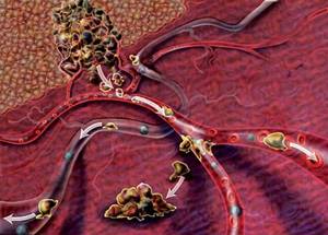
Why does stomach cancer metastasize?
Many molecular mechanisms are involved in the process of metastasis; there are still many gaps in current knowledge about them. It is known that malignant stomach tumors develop from special stem cells . Certain subtypes of stem cells make cancer more likely to metastasize.
In general terms, the process of metastasis occurs as follows:
- The primary tumor in the stomach gradually grows and invades surrounding tissue.
- Some cancer cells break away from the primary site and enter the blood or lymphatic vessels.
- With the flow of blood or lymph, tumor cells migrate to other organs, settle in small blood vessels, and penetrate through their walls into the surrounding tissues.
- Such a “journey” is dangerous for cancer cells, many of them die. But, if conditions are favorable, the cancer cell attaches itself to a new location and forms a microscopic secondary focus.
- Metastasized cells can remain inactive for a long time, for years. At a certain point in time, they can begin to actively multiply and secrete substances that stimulate the formation of new blood vessels necessary for the growth of tumor tissue.
Even after the patient has undergone treatment and is in remission, some microscopic metastases may remain in the body. Over time, they can cause a relapse.
Results of magnetic resonance imaging of the stomach
After scanning is completed, the signal received by the sensor is converted and processed by computer. The output is images of a given number of slices of a certain thickness.
After analyzing the data, the radiologist draws up a conclusion based on the MRI of the stomach and intestines. The conclusion shows:
- Size and shape of organs, blood vessels.
- The presence of pathological formations, changes in tissues indicating the development of the disease, the degree and volume of the lesion.
With this conclusion, the patient is sent to the attending physician for an accurate diagnosis.
All images and descriptions are available digitally. They are recorded on a disk or USB flash drive and can be sent to the patient’s email.
Symptoms
Often, the first symptoms of stomach cancer appear in the later stages, when metastasis has already occurred. Symptoms depend on which organ the cancer cells have spread to:
- In the peritoneum : abdominal pain, abdominal enlargement due to the accumulation of fluid inside ( ascites ), loss of appetite, causeless severe weight loss.
- In the liver: loss of appetite and weight loss, dark urine, enlarged abdomen, jaundice, pain in the upper abdomen on the right (under the right rib), nausea, vomiting, increased sweating.
- In the lungs: chest pain, persistent chronic cough, blood in the sputum, wheezing, shortness of breath, weight loss.
- In the bones: pain, pathological (from slight effort) fractures.
- In the brain: headaches, nausea, vomiting, weakness, numbness in the arms and legs, impaired coordination of movements, personality and behavior disorders, speech, swallowing, urinary and stool incontinence.
All of these symptoms can be caused by other diseases.
How often can you undergo a magnetic resonance examination of the gastrointestinal tract?
It is not difficult to understand how often this study can be carried out, based on the distinctive feature of the phenomenon of nuclear magnetic resonance that underlies tomography. The magnetic field interacts with hydrogen atoms, which are part of the human body. The resulting energy is released into space, recorded by sensors, without causing any harmful effects on the body.
During scanning, a person is not exposed to ionizing radiation, which does not create a limit on the number of examinations. If it is necessary to monitor the dynamics of the development of the disease, track the stages of recovery after surgery or treatment, the doctor has no restrictions on the number of intestinal MRI procedures prescribed.