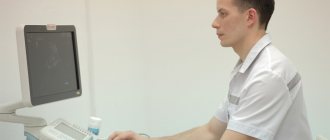
Ultrasound examination of the venous system involves obtaining an image of the lumen and wall of the vein, as well as the nature and direction of blood flow through it, using the analysis of reflected sound waves. Vein ultrasound is commonly used to look for blood clots, especially in the veins of the legs, varicose veins, venous valves, and abnormal backflow of blood (reflux).
For ultrasound of vessels and veins of the lower extremities, ionizing radiation is not used, so this method has no harmful effects. The technique for diagnosing the venous system is called ultrasound duplex (double) scanning, since a double amount of information is obtained. In the scanning mode, the two-dimensional structure of the venous vessel is studied, and in the Doppler mode, the speed and direction of blood flow through it is studied.
Ultrasound of leg vessels: the essence of the method
The idea of using ultrasound to study blood vessels was introduced after the substantiation of the Doppler effect, named after the Austrian physicist. It means a change in the frequency of waves perceived by the observer. Ultrasound of the vessels of the lower extremities is an informative, accessible and safe method. Therefore, it is used to identify numerous problems.
Types of research
Ultrasound is a non-invasive diagnostic method that can visualize small details and see structural changes. It has virtually no contraindications and is painless. To determine vascular diseases, three types of studies are carried out: two-dimensional Doppler sonography, duplex angioscanning, triplex scanning.
Two-dimensional Doppler ultrasound is an outdated technique that allows you to assess the condition of superficial and deep veins. It is used to identify primary signs of vascular diseases. The study is prescribed for people who experience fatigue when walking and increased sensitivity to cold. A diagnosis is necessary if swelling occurs in the legs and varicose veins develop.
Two-dimensional Doppler sonography is used to determine the patency of veins and assess the condition of the valves. During the study, the doctor does not receive direct information. He can judge the presence of diseases approximately, based on the nature and speed of blood flow, blood pressure indicators, frequency of the pulse sinusoid, the presence of tortuosity and kinks. The advantage of standard Doppler ultrasound is that the studies can be performed at the patient's bedside. The equipment is compact in size and easy to transport.
With the advent of informative and more accurate imaging methods, two-dimensional Doppler sonography has faded into the background. In modern medical centers, duplex angioscanning is performed. This is an effective technique, thanks to which phlebologists obtain reliable results. Using this method, doctors carefully examine the patency of the superficial and deep veins, assess the speed of blood flow and identify pathological areas.
Duplex angioscanning is a study that combines the advantages of classical ultrasound and energy mapping. When using color Doppler, doctors distinguish blood clots that are not visible under normal conditions. The research takes a long time. The specialist studies the structure of blood vessels in detail and draws conclusions. A color image appears on the monitor, allowing you to determine the speed and direction of blood flow, the condition of the venous valves, and the elasticity of the vessel walls.
During the study, functional tests are performed. This is an additional measure due to which abnormalities and vascular changes are detected. Duplex angioscanning is indicated for varicose veins, impaired blood outflow, and decreased lumen of blood vessels. This technique does not pose a danger to humans. During the examination, the specialist makes an accurate diagnosis and prescribes treatment.
Triplex scanning is an innovative method used to determine the condition of the vessels of the lower extremities. It combines the advantages of the previous two methods. Vascular examination using advanced equipment has significant advantages:
- high information content - the phlebologist identifies all kinds of defects: pathological tortuosity of blood vessels, blood clots, narrowing of the walls, aneurysms. The doctor determines the speed of blood flow and evaluates the structure of blood vessels
- efficiency - scanning devices operate in three modes. Therefore, experts determine the important anatomical features of the vascular system of the lower extremities: degree of tortuosity, patency, structure of the walls
- effectiveness - using triplex scanning, vascular pathologies are diagnosed in the early stages and an adequate treatment method is determined. It is often performed before surgery to help create a surgical plan. This way you can avoid complications and guarantee the best outcome.
Equipment in our clinic
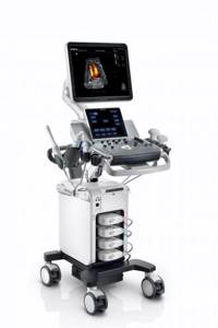
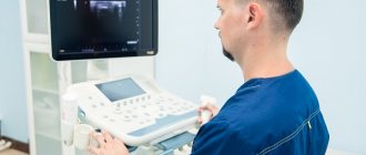
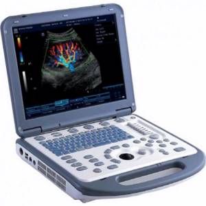
In our network of clinics there are more than 20 ultrasound diagnostic devices of a high and expert level, one of them is TOSHIBA VIAMO - a portable Japanese device of expert quality, which allows for a wide range of studies of the vascular system and internal organs. Excellent resolution for diagnosing venous thrombi. Work in Doppler mode and triplex scanning mode.
Mindrey DS 70 is a stationary ultrasound scanner of expert level. Allows you to conduct diagnostic studies of any complexity. Excellent assessment of blood flow in the vessels of the abdominal cavity, arms and legs, neck and brain. All types of sensors are used. Diagnosis of any pathology at an expert level.
Ultrasound scanning of veins is an effective diagnostic method with high clinical value. Correct ultrasound diagnosis of blood vessels is a key factor for correct treatment. For a long time, the diagnosis and treatment of venous pathology was carried out by eye, which led to unsatisfactory treatment results. Venous diseases require the most thorough ultrasound assessment by a specialist with significant clinical experience and working with high-quality diagnostic equipment.
Preparing for an ultrasound
Ultrasound examination does not require special preparation. Before the procedure, you do not need to limit yourself in food. Canceling medications that a person is taking also does not make sense. Medicines do not have a negative effect on ultrasound results.
Step by step procedure
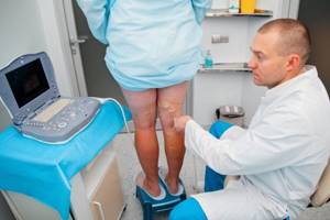
The study of the vessels of the lower extremities takes place in several stages:
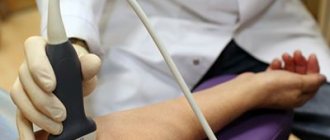
- the patient is freed from clothing below the waist (pants, shorts, tights, skirt)
- The doctor applies a small amount of gel to the legs, which increases the throughput of ultrasound.
- the person lies down on the couch with his stomach down and then turns over. The specialist examines the surface of the thighs and lower legs. At the final stage, the doctor examines the vessels in a vertical position. In this way, the blood flow of the vascular system is assessed
An ultrasound examination lasts more than half an hour. The procedure is absolutely painless. It does not cause any discomfort. During the diagnosis, the phlebologist examines the condition of the deep veins, arteries, venous valves, and junctions of large vessels. During the examination, information is displayed on the monitor. The specialist selects the frequency of ultrasonic waves, depending on the depth of the vessels. The doctor examines the clinical picture and makes the necessary conclusions.
Ultrasound of the veins of the lower extremities
Ultrasound detection of deep vein thrombosis
What does triplex ultrasound of the arteries and veins of the lower extremities show?
Treplex scanning of leg vessels allows the doctor to:
- quantify blood flow in arteries and veins;
- identify vascular disorders, including those caused by blood clots and atherosclerotic plaques;
- determine the presence of stenosis (narrowing) of the arteries, as well as their significance;
- determine the presence of varicose veins, its cause, severity, and the presence of valvular insufficiency of the veins;
- determine the presence of a blood clot, as well as measure its size, evaluate its structure and flotation (for example, with deep vein thrombosis);
- assess blood flow to organs and tissues;
- diagnose the presence of aneurysms
Modern vascular surgery is unthinkable without ultrasound of the vessels of the legs, since it allows us to identify predisposing factors for the development of circulatory disorders.
Indications for the study
Many factors negatively affect the functioning of the vessels of the lower extremities. These include heredity, a sedentary lifestyle, prolonged sitting and heavy physical activity. An important indicator that causes varicose veins is excess weight. Therefore, if you are overweight, the risk of developing diseases of the circulatory system increases.
Ultrasound of the vessels of the lower extremities is performed in the presence of the following problems:
- fatigue in the legs even after a short walk
- a feeling of heaviness that does not go away for a long time
- tingling in the lower extremities
- signs of varicose veins: formed nodules, mesh
- swelling of the legs (especially in the afternoon)
- numbness of fingers
- high blood sugar
- night pain in the lower extremities
Pregnancy is a special condition in which a woman rapidly gains weight. This negatively affects her health. Swelling of the legs, fatigue, networks of dilated vessels are the main symptoms indicating the presence of varicose veins. During pregnancy, the pressure of body weight on the legs constantly increases. Therefore, a woman should wear compression tights. These are products widely used in medicine. They prevent deep vein thrombosis, reduce swelling and provide a compressive effect.
Pregnant women should undergo ultrasound examination if vascular disease is present or suspected. The procedure does not have a negative effect on the fetus. After the diagnosis, the doctor identifies inflamed areas and assesses the condition of the blood vessels. If thrombophlebitis is diagnosed, medications that thin the blood are prescribed.
Ultrasound of the vessels of the lower extremities has no strict contraindications. Even children are allowed to do it. Timely examination helps to identify impaired blood supply, atherosclerosis, varicose veins and other diseases.
After diagnosis
Having completed the study, the doctor gives the patient the opportunity to wipe off the gel, while he himself fills out the study protocol. Ultrasound of arteries is a highly informative method. In the absence of significant pathology, or during a follow-up examination, such a diagnosis is final and the patient is released. If an ultrasound scan reveals surgical pathology of the arteries of the lower extremities, the patient is referred for additional research methods (MSCT of the arteries with contrast or MRI in angio mode). In case of critical or acute ischemia after ultrasound, the patient can be sent immediately to hospitalization and additional examinations are carried out in the hospital.
The ultrasound angioscanning protocol must be preserved, since the doctor may need to observe changes in the ultrasound picture over time.
Ultrasound equipment
To conduct research, devices are used that are designed for visual diagnostics of the human body. Ultrasound rooms are equipped with multifunctional equipment designed to meet stringent requirements. Modern models of devices have ultra-precise focusing and compact dimensions. They provide high quality images.
To improve performance, the devices are equipped with various modes. For example, two-dimensional visualization, as well as color and power Doppler. Many models are equipped with glare-free monitors that rotate freely around an axis and tilt in the desired direction. It is possible to connect several sensors simultaneously. The received information is recorded on the hard drive and stored in a database.
When developing ultrasound machines, new technologies are being introduced: grain suppression, image detailing, automatic image optimization, wireless information transfer. The scanners are easy to operate. If necessary, a specialist can adjust the settings. The image is instantly transferred to the monitor. The doctor examines the details and assesses the situation.
There are two types of equipment on sale: stationary ultrasound and portable ultrasound. Stationary devices are installed in rooms designed for ultrasound examinations of leg vessels. They are impressive in size and provide reliable diagnostics. Portable devices are portable and small in size. Such devices are designed to conduct research in any place: ward, ambulance, emergency department. They are indispensable in cases where it is necessary to urgently determine a person’s condition and take the right actions.
Deep veins
The main deep veins of the lower extremities follow the course of the corresponding arteries. The deep venous system of the leg includes the anterior tibial (AT), posterior tibial (PT) and peroneal veins (MF). In golin, these deep veins are present in pairs on either side of the artery. The posterior tibial vein receives blood from the medial and lateral plantar veins and drains the posterior leg and plantar surface of the foot. This vein lies behind the leg and joins the popliteal vein at the popliteal fossa. The anterior tibial vein is an ascending continuation of the dorsal vein of the foot. It runs along the anterior surface of the leg just above the interosseous membrane between the tibia and fibula and joins the posterior tibial vein to form the tibioperoneal trunk and popliteal vein. The peroneal veins run along the posteromedial surface of the fibula and join the posterior tibial vein.
The popliteal vein is formed by the connection of the anterior and posterior tibial veins in the lower part of the popliteal fossa. It rises along the back of the knee and the distal aspect of the anteromedial thigh. The popliteal vein is located medial to the artery, below the popliteal fossa, superficial to the artery and on the lateral side above the knee.
After the popliteal vein enters the adductor hiatus, it is called the femoral vein. The term superficial femoral vein is no longer recommended because this vein is not superficial, but deep. In the lower part it lies on the side of the artery; in the middle part, behind the artery; and in the upper part, medial to the artery. The deep femoral vein from the inner thigh, running along the deep femoral artery, joins the femoral vein and forms the common femoral vein, which is located medial to the common femoral artery. The inguinal ligament is a landmark that separates the common femoral vein from the external iliac vein (Figure 3).

Figure 3:
Schematic representation of the deep veins of the lower extremities. Although the veins of the lower extremities form a continuous structure, they are named individually according to their closest anatomical landmark.
The inguinal ligament (upper dotted line) is an anatomical landmark between the external iliac and common femoral veins, and the adductor interception (lower dotted line) is an anatomical landmark between the femoral and popliteal veins. IVC – external iliac vein; COF – common femoral vein; FV – femoral vein; DFE – deep femoral vein; PV – popliteal vein; ATV – anterior tibial vein; PTV – posterior tibial vein; FIV - peroneal veins. It is convenient to begin the examination of the patient in the supine position or the Fowler position. A reverse Trendelenburg position is also recommended if possible, as it facilitates venous filling of the lower extremities and dilates the veins. External rotation of the hip and slight flexion of the knee help relieve muscle tension and are good for both identifying the deep veins in the mid-thigh, popliteal fossa, and calf, as well as for the compression maneuver (Figure 4A).

Figure 4:
Demonstration of the uncompressed and uncompressed ultrasound image and corresponding sound window in a CT scan based on probe placement. A. _ The patient position and a schematic representation of the sensor location are shown in Figures B, C, D, E, and F. B . The common femoral vein is visible at the level of the inguinal ligament on the medial side of the common femoral artery, which is round and pulsatile. The vein is not visualized when compressed (arrows). As it travels down the common femoral vein, it bifurcates into the deep femoral vein (open arrows) and the femoral vein (arrows).
At the level of the inguinal ligament, in a transverse view, the common femoral vein on the medial side of the artery is accessible to the physician (Figure 4B). Information about the vein's blood flow patterns can be assessed longitudinally using Doppler ultrasound. From the common femoral vein, it bifurcates into the deep femoral vein and the femoral vein (Fig. 4C). At the more distal aspect of the medial thigh, only the femoral vein is visible (Fig. 4D).
As the probe approaches the popliteal fossa, the popliteal vein is visualized, which is superficial to the popliteal artery in the popliteal fossa (Figure 4E). Manual vein compression is recommended every 3-4 cm.
When examining the popliteal vein downwards, in the back of the leg, two posterior branching veins are found from a posteromedial approach. The vein along the tibia is the posterior tibial vein, and the vein along the posteromedial surface of the fibula is the peroneal vein.
After straightening the patient's leg, the anterior tibial vein can be visualized from the anterolateral approach (Fig. 5). The anterior tibial artery and vein can be found just above the echogenic interosseous membrane between the tibia and fibula. If the posterior tibial vein cannot be traced from proximal to distal, it may be traced upward from the posterior to the medial malleolus, where the vein is more superficially located.
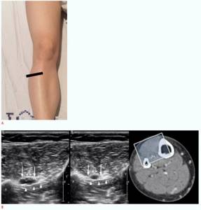
Figure 5:
Ultrasound examination of the anterior tibial vein. A. _ Straightening the patient's leg allows the anterior tibial vein to be approached from the anterior side. Above the interosseous membrane (arrowheads) between the tibia (T) and fibula (F), the anterior tibial vein (arrows) and artery (open arrows) are visible. The sound window is demonstrated using computed tomography.
Decoding and results
Ultrasound is an affordable imaging method that does not use harmful radiation. During the diagnosis, the doctor determines the thickness of the vascular membranes, pulsation index, and vascular resistance. The minimum and maximum speed of blood movement is also determined.
After conducting the study, the specialist receives accurate data that indicates the condition of the vascular system. It defines a number of important parameters and reflects them in the protocol. Based on such information, the doctor gives an opinion, according to which the vascular surgeon develops treatment tactics.
Using ultrasound examination of the vessels of the lower extremities, various diseases are detected:
- stenosis is a term that refers to the narrowing of blood vessels in the circulatory system. The development of such a pathology leads to undesirable consequences: muscle atrophy, ulcerative formations, pain. Stenosis appears in people who lead a sedentary lifestyle and are overweight. Diabetes mellitus also leads to changes in the structure of blood vessels
- Varicose veins are a serious disease accompanied by impaired blood flow. The trigger for the development of pathology is considered to be disruption of the venous valves with the subsequent occurrence of blood reflux. The main symptom of the disease is dilated saphenous veins. First, a person is bothered by a feeling of heaviness in the legs and a burning sensation, and then swelling appears. Pregnant women and people who spend a lot of time in one position (office workers, salespeople, drivers, bank tellers) are predisposed to varicose veins.
- Phlebitis is a disease in which the vein wall becomes inflamed. Its main symptoms include soreness, a slight increase in temperature, and redness of the skin. Phlebitis can be chronic or acute. The appearance of the disease is provoked by varicose veins, infection, and mechanical damage to the vessel. When diagnosing phlebitis, the doctor prescribes a set of therapeutic agents
- thrombosis - the formation of blood clots inside blood vessels. This is a pathological process because the free flow of liquid connective tissue through the system is limited. The key signs of the disease are severe swelling, pain and induration. Thrombosis may be asymptomatic. In such cases, the disease is detected by ultrasound. This problem cannot be ignored, because it leads to serious consequences.
Ultrasound of the vessels of the lower extremities is a study with which you can detect the disease in time and prescribe effective treatment. To carry it out, equipment with wide functionality is used. This is a guarantee that the results will be reliable.
General information for vein examination
Unlike arteries, veins have a weaker muscle layer with less elastic walls and are therefore difficult to visualize when the vein is compressed by the transducer (Figure 2A). The compressibility of the veins and the pulsation of the arteries may be a way to differentiate them. In addition, veins have valves that play an important role in preventing reflux of venous blood flow (Figure 2B). Normal flow is directed from distal to proximal and from superficial to deep. Typically, the vascular flow is not observed in gray tones, and the venous lumen is presented as anechoic. However, venous blood flow is sometimes available as an echo, which may reflect red blood cell aggregation and should not be confused with thrombosis. This phenomenon is especially common in conditions of slow venous blood flow. Continuous observation without probe movement or determination of luminal compressibility may help differentiate this condition from true thrombosis.

Figure 2:
Ultrasound examination of normal veins. A. _ Unlike arteries (open arrows), veins (arrows) have a weaker muscle layer with less elastic walls and therefore are completely compressed when compressed by the transducer. B. _
Veins contain valves that play an important role in preventing venous reflux. The choice of transducer for assessing leg veins is a trade-off between resolution and beam penetration. Generally, a frequency of 5 MHz or higher is recommended, but sometimes a lower frequency transducer is required for in-depth viewing in patients with obesity, edema, or prominent muscles.