For a person far from medicine, all ultrasound machines look the same. In fact, there are dozens of modifications of ultrasound devices and sensors that help doctors study any organs and tissues of the human body. Therefore, when making an appointment for an ultrasound, do not forget to ask which device will be used to examine you.
How an ultrasound machine works: the basics
The content of the article
Ultrasound diagnostics (sonography) is a method of examining the patient’s internal organs using ultrasound without the use of needles and other surgical instruments. It is the ultrasound examination that is accepted as the gold standard for primary examination throughout the world.
The ultrasound machine operates based on the piezoelectric effect. Inside the sensor, which is moved over the surface of the body, there are microcrystals of quartz, titanium or barium. When an electric current is applied, mechanical vibrations occur inside the crystals, which create ultrasonic waves with a frequency of up to 29 MHz. A special acoustic lens helps select a wave of a certain length. The higher the frequency of the ultrasonic wave, the more capabilities the device has.
Each organ or its department has an acoustic resistance unique to it. If the tissues to which the ultrasound wave is directed have different acoustic resistance (this is typical for lumps, cysts, neoplasms), one part of the wave is absorbed and the other is reflected.
The greater the differences in tissue, the greater the signal intensity. On the screen, areas that differ from neighboring tissues in density and other characteristics are displayed lighter and brighter. This effect is called echogenicity.
Sound wave
Sound waves come in 2 main types:
- Longitudinal - individual particles of the medium oscillate along the direction of the wave, which is typical for soft tissues, liquids, and gases.
- Transverse - individual elements are located in a perpendicular plane with respect to ultrasound waves, which is typical for solid structures, for example, bone tissue.
When individual parts pass along a longitudinal wave, different pressures are formed, which are associated with two main parameters:
- density;
- distance of elements from each other.
Ultrasound is capable of creating zones of high and low pressure, called alternating pressure.
What does an ultrasound machine consist of?
Despite some features and design differences, all ultrasound machines have the same components.
The “heart” of the device is an ultrasonic transducer, inside of which piezoelements such as quartz or barium crystals are placed. Under the influence of electricity that comes from the central processor, the crystals begin to vibrate and distribute ultrasonic signals around them.
The central processor makes all the calculations, and with the help of a pulse control sensor you can change the characteristics of the emitted ultrasonic pulses. An acoustic lens helps focus on a specific wave, and a sound-absorbing layer filters the displayed waves.
Thanks to the display, you can see a picture of the organ being examined and its surrounding tissues and structures. For better image quality, the ultrasound machine has a radio frequency amplifier, video and sound amplifier.
Using a cursor and keyboard, the specialist enters certain parameters or processes the received data. The reflected ultrasonic waves return to the transducer and are transmitted to the central processor. It calculates the speed of signal return and the distance from the sensor to the tissue.
The control sensor changes different scanning modes:
- mode A shows the amplitude of the reflected echo signal;
- M mode visualizes the organ in motion;
- Mode B displays a two-dimensional image that shows any changes in echogenicity. 20 pictures change per minute, which creates the illusion of movement;
- mode D is based on the Doppler effect, therefore it is used to study the patient’s blood flow.
All information is stored on a hard drive or CD or DVD. If desired, the client is given a printout or copy of the video recording (for example, the movements of the fetus - the unborn baby).
Characteristics of sound waves
Main characteristics of sound waves:
- amplitude (A);
- frequency (v);
- wavelength (λ);
- propagation speed (s).
The main parameter of any wave is its length, which is equal to the length of the path covered by the wave over a certain period of time, during which a full cycle of one oscillation occurs. For longitudinal waves, for example, this is a certain constant value of the distance through which compression and rarefaction zones alternate. For transverse waves, these are areas of up-down shear.
Types of ultrasound machines: not good and bad, but powerful and super powerful
If we consider the differences in the parameters and features of the image obtained on the monitor screen, then all ultrasound machines are conditionally divided into 3 categories:
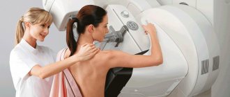
- 2D. This is a standard device that allows you to display an organ on the screen in two parameters - length and width. The picture turns out to be black and white, and it is difficult for a non-specialist to understand and see the pathology on the screen. However, there is enough information for an ultrasound specialist. He will notice various defects (cysts, fibroids, endometrial growth in gynecology, heart anomalies in cardiology, disturbances in the development of the brain in the fetus, its height and weight, the amount of amniotic fluid, etc.), so a two-dimensional ultrasound is required during pregnancy. For the pelvic and abdominal organs, a device with a frequency of 2.5 - 3.5 MHz is used. The procedure is completely safe for mother and child, but it helps to identify problems in the initial stages. It lasts no more than 15 minutes.
- 3D. It differs from a two-dimensional image in that another parameter is added - depth. A three-dimensional image appears on the monitor screen. If an expectant mother comes for the study, she will be able to see her baby’s face and also examine the structure of his body. The sex of the unborn child is determined with 100% accuracy using a three-dimensional apparatus. The duration of the 3D ultrasound procedure is about 50 minutes.
- 4D. This is a real hologram that makes it possible to see the baby in motion. If desired, parents can order a video recording of the examination. These are high-end ultrasound machines. The difference between them and 3D is that a three-dimensional image gives a picture of certain moments of the body position of the unborn child, and 4D shows a clear second-by-second video. In addition to pregnancy research, 4D devices are used in other areas of medicine. In urology it confirms an abscess of the prostate gland, in gynecology - even the smallest cystic formations, in ophthalmology - damage to the retina or eyeball, in oncology it will see the position of the vascular bundle relative to the neoplasm.
Ultrasound machines also differ in other characteristics.
By image quality:
- Conventional sonographs (have 16 transmission and reception channels).
- Medium technical class devices (over 32 channels).
- Ultrasound machines with increased capabilities (over 48).
- High-end devices (over 64).
- Expert class devices (several hundred channels).
The main technical parameter that distinguishes devices of different levels is the number of receiving and transmitting channels. The more there are, the higher the sensitivity and, accordingly, the resolution.
By specific application:
Ultrasound scanners. They work in 2D mode and give a two-dimensional picture. It has two operating modes: two-dimensional image (mode B) and one-dimensional echogram (mode M).
Highly specialized:
- Echoophthalmometer. Visualizes the structure of the eye in two- and one-dimensional images. In addition to modes B and M, it has mode D - spectral analysis of blood flow velocities using pulsed Doppler (PW) and continuous Doppler (CW).
- Fetal monitor. Measures the fetal heart rate. Detects pathologies of heart development in the early stages of pregnancy.
Ultrasound with Doppler
- with spectral Doppler (duplex devices). Display the work of blood flow in modes B, M and D;
- with color Doppler mapping. In addition to the same functions as a device with spectral Doppler, blood flow is displayed on a gray scale image of tissue. This is a rare device for specialized research.
Encephaloscope. This ultrasound machine is designed for neurosurgical research. Through the temple area, various brain structures are examined. The device operates on the basis of the transcranial method, which examines the characteristics of blood flow and identifies its disturbances. The encephaloscope records ultrasound signals reflected from various blood elements moving in the same direction. Then the received information is processed and displayed on the screen.
The brain absorbs much more blood than any other organ. In addition, it is very sensitive to hypoxia - lack of oxygen. Encephalography allows you to see the condition of the vessels and arteries that supply the brain, as well as identify pathologies such as abscesses, hemorrhages, cysts, hematomas, pertificates (deposition of calcium salts on the walls of blood vessels), gummas (scars), etc.
Sinuscope. This is a special ultrasound machine that examines the frontal and maxillary sinuses. It analyzes the ultrasound reflected from the walls of the nose. If the sinuses are full, a picture is displayed on the monitor screen in graphic form. The sinuscope helps to detect sinusitis, sinusitis, pharyngitis, and inflammation of the sinuses in the early stages.
What are the advantages of ultrasound diagnostics?
The main advantage of such diagnostics is the fact that the data is obtained without causing mechanical damage to human skin and tissue.
During the examination, the client does not experience pain or other not entirely pleasant effects; it is quite enough for the specialist to operate the sensor on the patient’s body. The picture is shown on the monitor here and now.
Doctors at the Clinic for Adults and Children 4D have extensive experience in performing ultrasound diagnostics. Our specialists will promptly and with maximum accuracy identify possible deviations and characteristics of the human body. In the clinic you can perform 2D, 3D, 4D diagnostics.
Depending on the sensor type
- Linear. They have a frequency of 5-15 MHz, scanning depth reaches 11 cm. The sensor is wide enough to display the entire organ. The displayed image is clear and high resolution. Does not adhere tightly to the skin, requires the use of gel.
- Convex. They have a frequency of 1.9-7.5 MHz, viewing depth no more than 25 cm. Fits tightly to the skin. Displays a narrow and somewhat distorted picture.
- Sectoral. The frequency is 1.5–5 MHz. The image is large and deep.
- Sectoral phased. The sensor has the form of a grating, each sector of which allows you to change the scanning angle. Different parts of the array independently receive and emit ultrasonic waves.
- Intracavity. They look like a beveled or straight handle and are placed inside the body (in the vagina or rectum).
- 3D or 4D volumetric sensors. It has a circular rotation that allows you to do slice-by-slice scanning, converting it into a three- or four-dimensional image.
- Matrix. They have a two-dimensional lattice. One and a half dimensional - the picture is longer in length than in width. This results in maximum thickness resolution. Two-dimensional. They have a large number of elements, which allows you to make pictures in different projections at the same time.
- Pencils. In them, the emitter and display are separated. Used to study arteries and veins.
By Application
- Universal abdominal probe for external use. Used to study the pelvic organs. They have a frequency of 3.5-5 MHz, and provide an overview of 40-900.
- Ultrasound machines small parts probe. The operating frequency is 7.5-10 MHz. The sensor has a width of 25-50 mm. It is used in the study of the thyroid gland, joints, and peripheral vessels.
- Cardiological ultrasound machine cardiac probe. Taking into account the peculiarities of the intercostal space, the device has a sector-type sensor with a frequency of 3.5 or 5 MHz. Used in cardiology.
Intracavitary ultrasound devices intracavitary probes.
- transvaginal. They have a frequency of 5.6 or 7.5 MHz and are used in gynecology;
- transrectal. Allows scanning at an angle of 3600;
- intraoperative. They are put on the finger and have a large radius of curvature;
- transurethral. They are very small in size and are inserted through the ureter into the bladder;
- transesophageal. They help examine the heart from below from the esophagus.
- intravascular.
Sighting range, 200-250 m.

Combat use of the Uzi (Uzi) submachine gun
“Uzi” is an indispensable weapon in close combat, in urban areas. These are the weapons of police, special forces, and auxiliary troops.

Die "Uzi", eine innovative, in Israel entwickelte Maschinenpistole
The submachine gun was used in many armed conflicts after World War II. Here are just some of the local wars in which the Israeli assault rifle was actively used:
- in the wars that the Israelis waged against the Arabs, Syrians and Egyptians in the 1950s-70s, the machine gun is currently in service with the rear services and auxiliary troops;
- the machine gun was used by the government army of El Salvador during the civil war of 1979 - 1992;
- during the Guatemalan Civil War in the 1970s, it was used by both sides of the conflict.
- In Germany, the Uzi was put into service back in 1959 for the police and driver-mechanics of armored forces. The command of the German army appreciated this weapon specifically for tankers who so lacked convenient, quick-firing and powerful submachine guns;
- In 1958, the Uzi was adopted by the Dutch army;
- The Uzi assault rifle and its modifications, Mini-Uzi and Micro-Uzi, were adopted by the armies and intelligence services of almost a hundred countries. Among its famous “users” are the security guards of the US President.

But in the 1980s. In many army units, the Uzi was removed from service and replaced by the MP-5K.
What additional functions are equipped with ultrasound machines?
Modern ultrasound machines have a lot of innovative functions that significantly increase the quality of the examination. Such developments include the following:
- The ClearVision function converts low-resolution and low-quality images into a clear and bright picture. This is a kind of filter that eliminates speckle noise and artifacts. as a result, the image has a clear contrast at the border of tissues with different echo densities;
- SonoView function is a special program that allows you to archive images and create databases;
- Cine memory function – the ability to rewind video and storyboard it; connectors for multiple sensors;
- TEI function – visualization in gray scale mode. This allows you to increase the level of clarity, contrast and reduce the number of artifacts. The technology makes it possible to see clear boundaries of tumors, which would have been impossible to do in obese patients without the use of innovation;
- The TP-View function allows linear sensors to increase the viewing surface. All measurements are displayed in one photo;
- The XLight function makes it possible to enhance the depiction of anatomical structures in a three-dimensional image. Thanks to data processing, you can see clearly drawn details. In obstetrics, this function helps to identify abnormalities in fetal development, regardless of the amount of amniotic fluid and the position of the fetus. In XLight surgery you can also see the condition of the bone structure;
- The CrystaLine function allows you to synchronize the operation of the ultrasound machine with the operation of the medical laser. This makes it possible to use the device in minimally invasive operations;
- The VPan Imaging function is designed to obtain a panoramic image (of the spinal canal in the fetus, oncological processes in the stomach). The picture has a sequential storyboard that reconstructs the entire study area.