Patient K, 69 years old.
Diagnosis
: left kidney tumor T1aNoMx. Left kidney cyst.
Clinical manifestations of the disease:
pain in the lumbar region on the left.
History of present illness:
In 1998, at the place of residence, an ultrasound revealed a mass in the left kidney. She was examined periodically and an increase in size was noted.
According to multislice computed tomography, in the middle segment of the left kidney on the posterior surface, an oval-shaped formation of a heterogeneous structure, 25x12 mm in size, is determined, actively accumulating a contrast agent; A cyst measuring 65x66 mm is closely adjacent to the formation.
Hospitalized at the Urology Clinic of the First Moscow State Medical University named after. THEM. Sechenov for further examination and surgical treatment.
In order to rid the patient of the tumor of the left kidney, to prevent the progression of the tumor process, as well as to rid the patient of the left kidney cyst, laparoscopic (retroperitoneoscopic) resection of the left kidney with the tumor and resection of the walls of the left kidney cyst were performed.
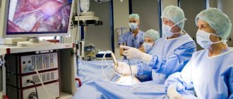
VIDEO
Video description:
the operation is performed without clamping the renal vessels (under conditions of zero ischemia). No safety drain was installed. The duration of the operation is 40 minutes. She was not in the intensive care unit. The postoperative period was uneventful. On the third day the patient was discharged home in satisfactory condition. The sutures were removed on the seventh day on an outpatient basis.
Histological conclusion:
in preparations, clear cell renal cell carcinoma (G1) with invasion into the renal cortex. The surgical margin is negative.
Recovery period
Compared to abdominal interventions, patients tolerate laparoscopy much easier. The duration of hospitalization is 2-4 days. The patient receives drug treatment consisting of antibiotics, anti-inflammatory, and painkillers. Already on the day of surgery you can get up and eat.
Despite its minimal invasiveness and minimal risk of complications, laparoscopy has been and remains a full-fledged surgical operation. This means that after removal of the cyst, you should avoid lifting heavy objects, bending your body, and sudden movements.
What is partial nephrectomy?
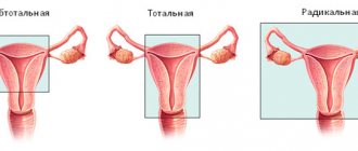
Kidney resection (from the Latin resectio - cutting off) - removal of part of the kidney (other names for this operation: partial nephrectomy, partial nephrectomy) affected by some pathological process (tumor, less often - stones, cysts, injuries, tuberculosis, etc. ), within healthy tissue (i.e. the tumor is removed as much as possible, while the surgeon goes a little into healthy tissue).
Today, classical open resection has faded into the background; its modern minimally invasive alternative, laparoscopic resection, has taken the leading position, which will be discussed further, namely, indications, contraindications, technique, etc.
Cost of laparoscopic kidney resection
The operation is performed under a compulsory medical insurance policy or according to a quota, depending on the volume and type of operation.
Who is indicated for laparoscopic partial nephrectomy?
Kidney resection is most effective for localized cancer (the tumor does not grow into neighboring organs or the kidney capsule), a small tumor size (no more than 4 cm) and a single tumor node. Most often, the operation is performed at stages T1 and T2. With such tumor parameters, the risk of relapse after surgery is extremely small and is less than 10%.
In addition to the above, partial nephrectomy is performed in cases where kidney removal would leave the patient without both kidneys or would require immediate initiation of dialysis.
This situation is possible when:
- bilateral kidney cancer (if both kidneys are affected by the tumor),
- cancer of a single kidney (congenital absence of a kidney or after surgical removal),
- a single functioning kidney (i.e., despite the presence of both kidneys, one of them has lost its function for some reason),
- in case of diseases of the kidney (which is not affected by a tumor) that potentially threaten its function (i.e. if on one side the kidney is affected by a tumor, and on the opposite side there is a disease that subsequently can or will lead to loss of its function).
Indications for cyst removal
If a pathology is detected, the patient should regularly see a doctor and undergo examinations. An ultrasound examination of the organ is mandatory at least once a year. Most often, with a kidney cyst, careful observation is sufficient, but cases when it is necessary to remove a cyst on the kidney cannot be ruled out.
Each patient diagnosed with this pathology must clearly identify the main signs that are an indication for surgical intervention. If you do not start treating a kidney cyst in time, the likelihood of developing dangerous complications increases. First of all, the growing formation compresses the blood vessels supplying the kidneys with blood, as well as the tissues of the organ. As a result, the normal functioning of the kidneys is disrupted and the load on the organ increases. In addition, removal of a kidney cyst is prescribed in the following situations:
- violation of the free passage of urine through the ureters;
- increased blood pressure;
- severe pain in the area of the affected organ;
- rupture or inflammation of the cyst;
- the appearance of blood in the urine;
- symptoms of malignancy of the cyst.
In the latter case, a biopsy is mandatory. If a positive result is obtained, nephrectomy is often prescribed, since removal of part of the kidney often ends in recurrence of the pathology. To treat a kidney cyst correctly, you need to conduct a thorough examination of the patient. In addition to ultrasound examination, magnetic resonance imaging with ascertaining is prescribed.
But the detection of a cyst does not always become the reason for turning to surgeons. In some cases, surgery is not prescribed. These are polycystic disease in the acute phase, asymptomatic kidney cyst, diseases of the cardiovascular and respiratory systems, blood clotting disorders, as well as diabetes mellitus in the decompensation stage.
Our doctors
What are the contraindications to laparoscopic partial nephrectomy?
Like all laparoscopic operations, kidney resection has a general list of contraindications, in which the operation is associated with a high risk of complications such as bleeding, or surgical intervention will be difficult.
Resection is contraindicated in:
- extreme obesity,
- disruption of the blood coagulation system,
- for infectious diseases,
- in late pregnancy.
Acute inflammatory processes due to the high likelihood of infection of the abdominal cavity are also a contraindication to laparoscopic resection. The presence of scars in the peritoneal cavity from previously performed operations causes significant technical difficulties in isolating the kidney and its surrounding tissues, which significantly complicates or makes laparoscopic intervention impossible.
Features of the operation in our clinic
Clinic of Urology named after. Frontshteina is equipped with modern equipment that allows diagnosing kidney cysts at an early stage of development. Experienced specialists will help cure the kidney and reduce the likelihood of recurrence of the pathology to zero. In our clinic you can perform a robot-assisted nephrectomy or remove a kidney using the laparoscopic method. It is also possible to perform an operation to remove part of the kidney (kidney resection) using laparoscopy or a modern robot-assisted method. From the moment you contact us, determine the method of treatment for the cyst, and until complete recovery, you will be under the close attention of doctors and medical personnel, who will create the most comfortable conditions for successful rehabilitation.
How to prepare for surgery?
Before the operation, you must be consulted by specialists of various profiles - an anesthesiologist, therapist, surgeon, and, if necessary, others. After this, your attending physician gives individual recommendations that you must strictly follow - do not eat, do not drink several hours before surgery, etc.
Be sure to disclose any drug allergies or medications you are taking to avoid complications during surgery (especially blood thinners, which can cause serious bleeding both during and after surgery).
Before the operation, a thorough bowel preparation is carried out, and broad-spectrum antimicrobial drugs are prescribed to prevent infectious complications in the postoperative period.
What kind of disease is this?
Kidney tissue has a special structure that has its own functional tasks: it filters blood, retains necessary components and removes toxic metabolic products.
A congenital kidney cyst can form in the prenatal period, which is influenced by genetic factors and adverse external influences. The development of an acquired kidney cyst can be caused by certain provoking factors. The size of the cyst varies from one millimeter to several centimeters. The biggest problem is represented by multiple cysts in the kidneys. Such formations are dangerous because, penetrating the kidney tissue, they disrupt the functioning of a large volume of the filtration system. Due to dysfunction of part of the parenchymal tissue, the activity of the kidneys is disrupted.
How is laparoscopic partial nephrectomy performed?
The operation is performed under general anesthesia (you will be put to sleep throughout the entire operation). The patient is placed in a special position - on the healthy side, while the operating table is extended, thereby bringing the kidney closer to the anterior abdominal wall. Access to the kidney is carried out using 3-4 small holes of about 1 cm on the anterior abdominal wall, through which a special camera (laparoscope) and other necessary surgical instruments are inserted. One of the holes is subsequently extended slightly so that the bag with the tumor can be removed from the abdominal cavity.
The laparoscope increases the size of the surgical field several times, which allows the surgeon to work more accurately and accurately. This is necessary during kidney resection, since resection, unlike kidney removal, is a technically more complex manipulation that requires more precise and careful actions. To increase the working space in the abdominal cavity, carbon dioxide is introduced into it (creating pneumoperitoneum), which is completely removed at the end of the operation.
After creating pneumoperitoneum, a laparoscope is inserted and the abdominal cavity is examined. Next, the surgeon begins to isolate the kidney; to do this, he dissects the tissue and moves the organs to the sides. After isolating and examining the kidney, the stage of isolating the vessels and ureter begins, then the surgeon clamps the renal artery with a special clamp to ensure minimal blood loss during the operation.
After this, the most critical stage begins - the resection itself, which is performed under conditions of kidney ischemia (as mentioned earlier, the renal artery is pinched, therefore, arterial blood does not flow to the kidney, which can lead to its death if the ischemia time is more than 40 minutes) . On average, the ischemia time is about 10–15 minutes, which is absolutely safe for the kidney and does not affect its functions in any way.
After removing the area of the kidney with the tumor, the edges of the kidney are sutured and the clamp is removed from the artery. The abdominal cavity and the bed of the removed tumor are examined for bleeding, the tumor is placed in a special bag and removed from the abdominal cavity. The surgeon then removes the instruments and stitches the tissues together. On average, the duration of the operation varies from 2 to 3 hours.
In recent years, the views of urologists on the treatment of cystic kidney lesions have changed significantly. The choice of treatment method depends on the degree of reliability of the diagnosis, the nature of the cyst, the presence or absence of complications, concomitant diseases, the age and condition of the patient, as well as the personal experience of the doctor.
There are several types of surgical treatment of cysts: open operations, percutaneous x-ray puncture and endovideosurgical methods (transperitoneal or retroperitoneoscopic). There are conflicting opinions about the safety, effectiveness and feasibility of using various methods of treating patients with simple kidney cysts.
Open surgery is currently practically not performed or is performed extremely rarely due to the traumatic nature of the method.
Percutaneous puncture treatment of a simple renal cyst is dominant in domestic urology, however, recurrence of the cyst occurs in 80-90% of cases [1]. Along with puncture of the cyst, the introduction of sclerosing solutions is used, as well as their subsequent drainage with step-by-step sanitation [2, 3]. However, even when using sclerosing solutions, the frequency of cyst recurrence remains high and, according to different authors, ranges from 12.5 to 33% [4-6].
Improvement of equipment and instruments, successes in percutaneous surgical interventions, as well as the use of cystoscopy, have made it possible to propose endoscopic methods for excision of the free wall of the cyst using transperitoneal and retroperitoneal access for the treatment of cystic kidney lesions [7-10].
In 1984, for the first time, urological rigid endoscopes with nephroscope and resectoscope manipulators were used to examine the kidney and excise the walls of a kidney cyst [11-12]. In 1989, a similar operation was carried out in the USSR [13]. However, the first mention of direct visualization of organs and tissues of the retroperitoneal space for diagnostic and therapeutic purposes was published in 1969 when performing lumbar sympathectomy. The scientists examined the retroperitoneum using an endoscope for tumors and also performed a biopsy of suspicious lymph nodes. Currently, laparoscopic resection of simple renal cysts is considered a safe and effective treatment method [14, 15].
Table 1. Characteristics of research methods performed in 84 patients with simple renal cysts
| Research method | Absolute indicators | Relative indicators, % |
| Ultrasonography | 84 | 100 |
| Survey urography | 84 | 100 |
| Excretory urography | 77* | 91,6 |
| X-ray computed tomography with intravenous contrast enhancement | 63 | 75 |
| Dynamic nephroscintigraphy and radioisotope renography | 84 | 100 |
| Note: * In 7 patients, intravenous urography was not performed due to intolerance to iodine-containing radiocontrast agents. | ||
This report presents our own experience of endovideosurgical excision of renal cysts using transperitoneal and retroperitoneal approaches.
Materials and methods
We conducted a retrospective and prospective analysis of the results of laparoscopic excision of simple renal cysts for the period from 1996 to 2008. in 84 patients. The age of the patients ranged from 22 to 78 years (± 28.6 years, mean age was 43.6 years). Of these, there were 49 (58.3%) men and 35 (41.7%) women. In 38 (45.2%) patients, single simple cysts were diagnosed at the preoperative stage, in 19 (22.6%) multiple cysts, and in 22 patients (26.2%) multilocular cysts were identified. It should be noted that in 12 (12.5%) patients the disease was bilateral. In this regard, a total of 92 laparoscopic interventions were performed (in 4 patients with bilateral processes, excision of the cyst walls was performed only on one side, since the cyst of the contralateral kidney was clinically insignificant). The average size of a simple renal cyst was 11.7 cm (range 3.0 to 26 ± 5.4 cm). Endovideosurgical intervention from the transperitoneal approach was performed in 38 patients (45.2%), from the retroperitoneal approach in 46 (56.8%).
In all patients, the preoperative examination was performed according to a standard procedure (Table 1).
Laboratory studies consisted of performing clinical blood and urine tests, biochemical blood tests, bacteriological urine culture to determine the sensitivity of microorganisms to antibiotics.
The indications for surgical treatment of kidney cysts were nephrogenic arterial hypertension, decreased kidney function, deformation of the pyelocaliceal system and impaired passage of urine through the upper urinary tract, a progressive increase in the size of the cyst, pain, and hematuria. The presence of a large renal cyst(s) in the absence of clinical manifestations, but with impaired renal function, was also an indication for surgery.
At the beginning of the introduction of laparoscopic methods, the location and size of the cysts were important for determining access. Transperitoneal access was used when cysts were localized in the upper segment, in the middle segment along the anterior surface. At the same time, visualization of the cysts was carried out without technical difficulties, especially when the latter were large. In other cases, retroperitoneal access was used.
At the beginning of mastering the method of laparoscopic treatment of renal cysts, all interventions were performed by us mainly from the transperitoneal approach. With the patient in the supine position under endotracheal anesthesia, pneumoperitoneum was created and a 10 mm trocar was inserted 1 cm above the navel, after which the patient was transferred to the healthy side and 2 additional trocars were inserted along the mid-axillary line to insert manipulators. After determining the topographic anatomy of the kidney and opening the peritoneum, the perinephric tissue was isolated and the cyst was mobilized. Then the cyst was opened in a small area or punctured and its contents were evacuated. The fluid was aspirated by aquapuncture and sent for cytological examination. Cystoscopy was performed. Endoscissors with coagulation were used to excise the cyst wall. The resected area was removed using a laparoscope and sent for examination. The bed and remaining walls of the cyst were coagulated and treated with alcohol. To stop bleeding, coagulation was used in mono or bipolar mode. A telescope was used to examine the base of the cyst to exclude a tumor. If suspicious areas were present, a biopsy was performed. The cyst cavity was drained. Drainage was drained through the lumbar region. Perirenal tissue was placed into the cyst cavity, and the peritoneal defect was closed with an endosuture or clips were applied.
Subsequently, with the accumulation of experience in performing laparoscopic interventions, we mainly used the retroperitoneal approach. With the patient positioned on the healthy side, a 1 cm long skin incision is made in the lumbar triangle and the retroperitoneal space is reached using an instrument (forceps). Then a trocar with optics is inserted into the retroperitoneal space and a working space is created by CO2 insufflation. The first additional trocar is inserted at the end of the XII rib approximately along the mid-axillary line, the second is slightly above the superior posterior iliac spine. Trocars are inserted into the retroperitoneal space under laparoscope control without damaging the parietal peritoneum. The renal fascia is incised and the lateral surface of the kidney is found. First, it is mobilized, and then the poles of the kidney. At this stage, it is recommended to install a third additional trocar along the anterior axillary line opposite the first one inserted in the lumbar triangle. The third trocar is needed to retract the kidney medially and provide access to its posterior surface and hilum.
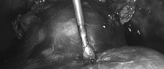
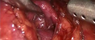
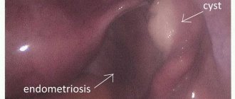
In most cases, a cyst is easily identified by a bulge above the surface of the kidney. After the connective tissue and fatty tissue are carefully removed from the surface of the cyst, the cyst looks like a bluish volumetric formation, clearly delimited from the surrounding renal parenchyma. The cyst wall is carefully excised with an ultrasonic scalpel or electric knife and sent for histological examination. The resulting cavity is carefully inspected. If a malignant tumor is suspected, a biopsy is performed and an urgent histological examination is performed. The retroperitoneal space is drained by installing an emergency drain. Then the trocars are removed and the puncture sites are sutured (some stages of retroperitoneoscopic excision of kidney cysts are shown in Figure 1-4).
| Figure 1. Trocar placement | Figure 2. Cyst mobilization |
| Figure 3. Excision of cyst walls | Figure 4. Final stage of the operation |
In patients with a bilateral process, we used transperitoneal access for the purpose of simultaneous excision of cysts on both sides from the same access.
Safety drains were removed on days 2-4 depending on the amount of discharge. On the evening of the day of surgery, patients were allowed to drink. Patients were activated the next day and discharged the next day after removal of the safety drainage with preliminary ultrasound control.
The average duration of surgery for the transperitoneal and/or retroperitoneal method was 80 minutes. (15-180 min.). With more experience, the operation time was significantly reduced.
Monitoring of patients consisted of their active clinical examination at 1, 3, 6 and 12 months. The follow-up period ranged from 5 months to 10 years: 23 patients from 6 to 12 months, and the remaining 61 for at least 12 months. Recovery time varied from 3 to 12 days.
The average length of stay per day was 4 days.
Results and its discussion
Treatment results were rated as good, satisfactory and unsatisfactory. The criteria for assessing the effectiveness of treatment were based on the results of periodic postoperative examinations. A comprehensive urological examination was carried out for all patients and necessarily included ultrasonography and radioisotope renography (renoscintigraphy); excretory urography was mainly performed in patients who had dilatation of the maxillary tract according to ultrasound data.
Among successfully performed laparoscopic operations, a good treatment result was achieved in 82 (97.6%) cases. Good long-term results of treatment were considered to be the absence of recurrence of the cyst during a control ultrasound examination one year after treatment, the absence of complaints from the patient, the elimination of arterial hypertension, and the improvement or stabilization of renal function in case of defining disorders before surgery.
In 2 (2.4%) cases, a satisfactory treatment result was obtained. Satisfactory results were considered to be the presence of a residual cavity in the projection of the removed cyst, according to ultrasound data, with a diameter of 2-2.5 cm without a tendency to increase and the absence of previously existing clinical symptoms.
In 2 cases, unsatisfactory results were obtained, which were characterized by persistence of pain and arterial hypertension.
Thus, in all cases, the patients were freed from cysts.
Complications that arose during laparoscopic excision of the cyst walls and methods of their treatment are presented in Table 2.
Table 2. Complications that arose during 88 endovideosurgical interventions in 84 patients with simple renal cysts
| Complications | Quantity | Elimination methods |
| Opening the lumen of the pelvis* | 1 | Suturing the walls of the pelvis |
| Perirenal hematoma measuring 4.0 cm | 1 | Drainage of the retroperitoneal space for 10 days |
| Pneumothorax* | 1 | Puncture of the pleural cavity |
| Peritoneal injury | 2 | No additional measures were required |
| Inflammatory complications with* leukocytosis and hyperthermia | 4 | Strengthening antibacterial therapy |
| Large cyst size with dense adhesions to surrounding tissues* | 1 | Conversion |
| | * complications after laparoscopic access | | ||
Pneumothorax noted in one patient resulted from the use of an insufflator with no ability to regulate carbon dioxide pressure.
During a control study 3 months or more after surgery, recurrent cysts measuring up to 3 cm were detected in 2 patients. These cysts did not impair renal function, and therefore, these patients were subsequently kept under dynamic observation.
In 1 patient, due to the large size of the cyst (26 cm), which occupied almost the entire retroperitoneal space and had dense adhesions with the descending colon, pushing it to the medial side, it was necessary to perform a lumbotomy with excision of the cyst wall.
We did not observe active intraoperative bleeding in any of the cases. The volume of blood loss was 58.0 ± 10.5 ml (10100 ml).
It should be noted that in most cases, complications were observed at the stage of mastering the method. By accurately determining the indications and assessing contraindications for the intervention, performing a full preoperative examination, as well as strict adherence to appropriate preventive measures and the correct technique for performing the operation, almost all possible complications with this method of treatment can be avoided.
We did not obtain significant differences in the results of laparoscopic treatment of simple renal cysts depending on the transperioneal and retroperitoneal approach (p > 0.05), which indicates that with sufficient experience, it is possible to remove a cyst of any location using the retroperitoneoscopic method. For bilateral cysts, transperitoneal access is appropriate. Complications occurred more often with laparoscopic access at the stage of implementation of the method.
Based on histological examination, no signs of malignancy were identified in any of the cases.
Conclusion
Laparoscopic resection of a kidney cyst is an effective intervention with a low number of complications and rapid rehabilitation of patients. Emerging intraoperative complications can be eliminated without conversion if the operating surgeon has sufficient skills and appropriate equipment in the operating room. With sufficient experience and skill, the retroperitoneoscopic approach is less invasive and minimizes (although does not eliminate) the risk of internal organ injury. The absence of familiar anatomical landmarks and limited space for manipulation are mitigated by the high professionalism of the operating surgeon and a proven technique for performing transperitoneal endovideosurgical interventions.
Key words: simple renal cyst, laparoscopic resection, complications. Keywords : simple renal cysts, endoscopic excisions, complications.
Literature
- Ignashin N.S. Ultrasonography in the diagnosis and treatment of urological diseases. M. Vidar, 1997. 119 p.
- Li SQ, Li XS, Dong YL, He ZS, Xia TL, Na YQ Ultrasound guided percutaneous puncture and sclerotherapy with alcohol for peripelvic cysts // Zhonghua WaiKe Za Zhi. 2005. Vol. 43. No. 22. P. 1461-1463.
- Choi YD, Ham WS, Kim WT, Cho KS, Lee JH, Cho SY, Seo JW, Jin OH Clinical experience of single-session percutaneous aspiration and OK-432 sclerotherapy for treatment of simple renal cysts: 1-year follow-up / / J Endourol. 2009. Vol. 23. No. 6. P. 1001-1006.
- Nechiporenko N.A., Nechiporenko A.N., Ryazantsev I.V. Evaluation of the effectiveness of treatment of a simple kidney cyst. // Urology and nephrology. 2000. No. 6. P. 9-12.
- De Dominicis C., Ciccariello M., Peris F., Di Crosta G., Sciobica F., Zuccalà A., Iori F. Percutaneous sclerotization of simple renal cysts with 95% ethanol followed by 24-48 h drainage with nephrostomy tube / / Urol Int. 2001. Vol. 66. No. 1. P. 18-21.
- Zenkov S.S., Zakhmatov Yu.M., Trofimov K.S. Percutaneous puncture treatment of simple cysts // Russian Medical Journal. 2003. No. 1. P. 37-40.
- Fryczkowski M., Huk J., Sitko-Saucha A., Kupilas A. Place of laparoscopic cysts decortication (LCD) in the treatment of autosomal dominant polycystic kidney disease (AD PKD) // Prog Urol. 2007. Vol.17. No. 7. P. 1324-1327.
- Micali S., Pini G., Sighinolfi MC, De Stefani S., Annino F., Bianchi G. Laparoscopic simultaneous treatment of peripelvic renal cysts and stones: case series // J Endourol. 2009. Vol. 23. No. 11. P. 1851-1856.
- Castillo OA., DeGiovanni D., Sanchez-Salas R., Foneron A., Vitagliano G., Diaz MA, Fajardo M. Laparoscopic treatment of symptomatic simple renal cysts // Arch Esp Urol. 2008. Vol. 61. No. 3. P. 397-400.
- Porpiglia F., Fiori C., Billia M., Renard J., Di Stasio A., Vaccino D., Bertolo R., Scarpa RM Retroperitoneal decortication of simple renal cysts vs decortication with wadding using perirenal fat tissue: results of a prospective randomized trial // BJU Int. 2009. Vol. 103. No. 11. P. 1532-1536.
- Zilberman M.N., Baev V.A. Direct retroperitoneoscopy. Methodical recommendations. Orenburg. 1978. 61 p.
- Baev V.A., Zilberman M.N. Current issues of constructive and reconstructive surgery. Irkutsk 1989. Part 2. pp. 338-339.
- Ibragimov V.Sh. Percutaneous and endoscopic methods for the diagnosis and treatment of simple kidney cysts: Cand. Thesis. honey. Sci. Tbilisi. 1989. 150 p.
- Lopatkin N.A., Fidarov F.B., Martov A.G. Laparoscopic resection of a simple kidney cyst // Urology and Nephrology. 1999. No. 2. P. 23-25.
- Steg A. Cystic diseases of the kidney in adults // J Urol Nephrol. 1975. Vol. 81.9 Suppl. P. 1-282.
| Attached file | Size |
| Article in PDF format | 157.34 kb |
‹ Pathogenetic treatment of fibroplastic induration of the penis (Peyronie's disease) Up Simultaneous urethroplasty with an increase in the area of the glans penis in the treatment of hypospadias in children ›
Possible complications during and after surgery
- bleeding - usually blood loss during resection is about 500 ml or less, however, more serious blood losses occur that require the infusion of special solutions or blood transfusions;
- infection – to prevent the occurrence of infectious complications, a broad-spectrum antibacterial drug is administered before and after surgery, which reduces the risk of this complication to a minimum;
- damage to neighboring organs is an extremely rare complication; a several-fold increased view of the surgical field helps to avoid it;
- postoperative hernia - as well as organ damage are rare, due to the fact that the holes after laparoscopic operations are small;
- conversion (transition to open surgery) - occurs when it is impossible to perform laparoscopic intervention due to adhesions or bleeding.
Causes of tumors in the kidneys
Despite the fact that kidney cyst is a disease that occurs quite often, experts have not yet figured out the reliable causes of this pathology. As a result of traumatic, hereditary or infectious factors, abnormal tissue growth is possible, which leads to the formation of a kidney cyst. Accordingly, this disease can be classified as either acquired or congenital.
Experts believe that a genetic predisposition to cysts in the kidneys occurs due to a lack of connective tissue in the body. If complications arose during the mother's pregnancy (there were injuries, infections), then the likelihood of the child developing a kidney cyst increases significantly.
Acquired kidney tumors can occur both at the site of a hematoma due to trauma, and when the site of infection is not stopped in a timely manner.
The age factor is also important, playing a role in the formation of kidney cysts. Most often, people over 50 years of age suffer from this disease.
So, the provoking factors for the development of the disease are:
- age-related changes;
- hereditary predisposition;
- pyelonephritis;
- urolithiasis disease;
- kidney tuberculosis;
- malignant tumors;
- injuries,
- prostate adenoma in men.
The risk of developing a kidney cyst increases in people suffering from hypertension and urolithiasis, frequent colds, which are complicated by inflammation of the kidney parenchyma.
Make an appointment
What to expect after surgery?
After laparoscopic resection of the kidney, the patient is transferred to the intensive care unit (ICU) under the supervision of an anesthesiologist-resuscitator to monitor vital functions (blood pressure, heart rate, respiratory function, amount of urine excreted are monitored), in addition, discharge is assessed by drainage ( special tubes inserted into the wound cavity), temperature, urine color and general well-being of the patient.
Most often in the postoperative period there are complaints about:
- pain in the area of the postoperative wound - most patients experience minor pain, which often does not require drug pain relief, which differs from pain during open surgery, when analgesic therapy is required;
- nausea - most often a consequence of the administration of various drugs necessary for anesthesia;
- the presence of a urethral catheter - necessary to control the color and amount of urine; it is removed after a few days.
Types of kidney cysts
The following types of cystic anomalies of the kidneys and upper urinary tract are distinguished:
- Simple renal cyst;
- Multicystic kidney;
- Polycystic kidney disease;
- Multilocular cyst;
- Solitary renal cyst;
- Parapelvic cyst.
The most common are simple renal cysts. They are a single-chamber cavity filled with serous contents. Multiple kidney cysts can be located in both organs.
Multicystic kidney is a congenital anomaly in which the renal parenchyma is replaced by cysts of various sizes. The mechanism of development of a multicystic kidney is based on atresia of the ureteropelvic anastomosis during the embryonic development of the fetus.
Polycystic kidney disease is a genetically determined disease that is manifested by the formation of numerous cysts on both kidneys. They gradually grow, causing atrophy of the organ parenchyma.
Multilocular cyst is a rare, poorly studied developmental anomaly. It has other names: adenoma, cystic lymphangioma, cystic hamartoma, multilocular cystic adenoma, Wilms cystic tumor, cystic nephroma, adenomatous polycystic tumor. Multilocular cyst occurs in patients of different ages - from birth to 80 years. In half of the cases, developmental anomalies are detected in children. Kidney cysts are diagnosed more often in women than in men.
A multilocular cyst is located in the right or left kidney; sometimes doctors detect bilateral and diffuse damage. It is characterized by the following criteria:
- One-sidedness of the lesion;
- Multi-chamber;
- Lack of communication with the pyelocaliceal system;
- Lack of connection between individual cavities;
- The presence of epithelial connective tissue cords;
- Lack of differentiated kidney elements.
A solitary renal cyst is a round-shaped cystic formation that is not associated with the collecting system of the kidney. There are simple solitary and dermoid cysts. A simple cyst does not contain septa or inclusions and is filled with serous contents. Located in the cortical layer (parenchyma) of one of the kidneys.
Parapelvic cysts of both kidneys are located in the region of the pelvis or sinus of the organ. They are a type of simple cyst. They are a hollow, round-shaped formation that has a fibrous membrane, lined with cubic epithelium and filled with fluid.

What to do in the postoperative period?
On average, a patient stays in the hospital for about a week after laparoscopic partial nephrectomy. Drinking and eating are usually allowed the next day after surgery, and walking is allowed in the evening of the same day. After the operation, as before, a broad-spectrum antibacterial drug is administered.
During the postoperative period, you will be advised to:
- drink 1-2 liters of water per day;
- do not lift weights exceeding 5 kg;
- do not undergo heavy physical activity.
You should immediately consult a doctor if you have:
- blood appeared in the urine;
- body temperature increased;
- severe abdominal pain occurred.
After surgery, your doctor will schedule consultations for you to undergo examinations, blood and urine tests, ultrasound examinations, and computed tomography scans to evaluate the effectiveness of treatment. After 5 years, if there is no evidence of tumor progression, the patient is removed from the register.
Make an appointment at the urology clinic of the First Moscow State Medical University named after. Sechenov, you can see a urologist, oncologist, Doctor of Medical Sciences G. N. Akopyan by phone or through the interactive Appointment form on our website.
September 11, 2019
Hakobyan Gagik Nersesovich - urologist, oncologist, MD, doctor of the highest category, professor
All clinical observations...
Diagnostics
- Survey and excretory urography
- Ultrasound, Dopplerography
- CT (computed tomography)
- Diagnostic puncture and cystography
X-ray methods: It is not possible to establish an accurate diagnosis of a simple renal cyst only on the basis of survey and excretory urography. With the help of excretory urography, it is possible to identify the “sickle” symptom or the “open mouth” symptom, which are characterized by the spreading of the kidney calyces without their “amputation”.
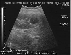
Ultrasound examination (ultrasound) reveals a simple renal cyst as an echo-negative formation of a round or oval shape, with clear, even, continuous contours and thin walls. Ultrasound allows for dynamic observation and use as a screening test. Dopplerography is a method that allows you to study the blood supply to the kidney. This diagnostic method is especially important for the combination of kidney cysts and arterial hypertension.
Computed tomography (CT) in some cases cannot provide 100% confidence in the accuracy of the diagnosis, especially with parapelvic cysts and tumors in the cyst. This somewhat reduces its diagnostic value. The CT scanning criteria for cystic renal lesions are similar to those used in echography: sharp, thin, distinct, smooth walls and edges; round or oval shape; homogeneous content. Density ranges from -10 to +20 HU, similar to that of water, and no enhancement should occur after intravenous injection of contrast medium.
Percutaneous puncture cystography is currently not the main diagnostic method, but can be used in the process of percutaneous puncture treatment of a simple cyst to clarify the location of the cyst and determine its relationship with the pyelocaliceal system.