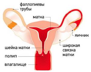
Uterine polyps are local benign neoplasms growing from the endometrial mucosa. They look like tubercles located on a wide base, or round or oval formations on a thin stalk. There may be one or several of them in the uterine cavity; in the latter case they speak of polyposis. The size of polyps varies from a few millimeters to 8 or more centimeters.
- Signs of a polyp in the uterine cavity and their diagnosis
- When is polyp removal necessary?
- Methods for removing endometrial polyps
- What is hysteroscopy of the uterus for polyps
- Types of hysteroscopy
- Indications for hysteroscopy
- Features of the event
- Preparation
- Basic recovery recommendations
- Complications after polyp removal
- Contraindications
Signs of a polyp in the uterine cavity and their diagnosis
Often uterine polyps do not manifest themselves, but some patients have the following symptoms:
- Menorrhagia - heavy menstruation.
- Bleeding outside of menstruation.
- Spotting after sexual intercourse.
- Bleeding during menopause.
- Infertility and miscarriages.
- With large polyps, women may notice copious whitish discharge, discomfort and pain during intercourse, as well as periodic cramping pain in the lower abdomen.
To make a diagnosis, it is necessary to undergo a comprehensive examination, which includes the following procedures:
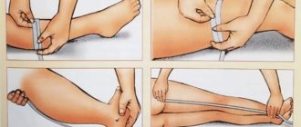
- Gynecological examination - particularly large polyps can be palpated during manual examination; cervical polyps can be detected during examination in the speculum.
- Ultrasound of the pelvis - in the uterus, growth of the endometrium with a local increase in its mucous layer is detected. The structure of this neoplasm is homogeneous.
- Separate diagnostic curettage followed by histological examination of the obtained material.
- Hysteroscopy - allows you to examine the endometrium from the inside, visualize single or multiple polyps and determine their location. The procedure is also therapeutic in nature, since it allows for the simultaneous removal of all identified tumors and their subsequent histological examination. This is the most effective method for diagnosing a polyp in the uterus.
Causes of the disease
No doctor has the right to judge the cause and nature of polyposis until the patient undergoes a histological examination after removal of the formation itself. Only after the procedures can we talk about the cause of the manifestation of the neoplasm and its benign quality.
If a “glandular” component is found in it, then the source of the disease is the entry of a large amount of estrogen into the blood. These hormones are produced by the ovaries. Due to the human papillomavirus (HPV) or herpes, fibrous growths form on the genitals. This pathology does not have pronounced symptoms, so it is diagnosed accidentally during an ultrasound scan.
When is polyp removal necessary?
The choice of patient management tactics and determination of the need to remove the polyp is determined individually. If a woman is of reproductive age and has no symptoms of a polyp, conservative management tactics can be chosen. In this case, the patient should undergo regular monitoring.
In all other cases, removal of polyps is indicated:
- Presence of symptoms of the disease.
- Pre- and postmenopausal periods, even if there are no symptoms.
Types and course of surgical intervention
The essence of the operation
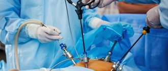
The choice of anesthesia depends on the size of the polyp. For large tumors, general anesthesia is preferred (painkiller is delivered by injection into a vein, the patient is conscious) and hospitalization. Small polyps are removed under local anesthesia; using low-traumatic techniques, the operation can be performed on an outpatient basis.
A woman sits on a gynecological chair. A hysteroscope is inserted into the cervix - this is a tube with a light source and a camera. It allows you to accurately see the location of the polyp. Sometimes a hysteroresoscope equipped with an attachment with a cutting surface is used.
The doctor unscrews the polyp , completely removing it; if necessary, the pedicle is excised, which may be located in the thickness of the epithelial tissue (this should be visible on an ultrasound). Multiple tumors are excised. After this, curettage is performed - complete cleansing of the mucous membrane of the cervical canal and uterus. It is performed using a special instrument - a curette.
Note. A curette is a medical spoon, which is a rod with an attachment resembling a spatula or a loop with a pointed edge.
Some doctors have a negative attitude towards this practice because it is aphysiological, but most are inclined to use it because it reduces the risk of relapse. With low-traumatic removal methods and a small polyp, curettage can be abandoned.
The removed tissue and polyp are examined. It is necessary to confirm the benign nature of the tumor. Tests are prepared within 1 to 10 days.
Types of surgical treatment of polyp
Despite the same essence of the operation, technologies may differ in the method used for removal.
Main types of surgical intervention:
- Polypectomy. The neoplasm is twisted until it is completely detached from the wall of the cervical canal or truncated using a special conchotome instrument. The operation is indicated for the removal of polyps up to 3 cm in size. The bed is cauterized.
- Laser coagulation. The stalk of the polyp is excised using radiation. This method allows the vessels feeding the neoplasm to be coagulated, which minimizes the risk of bleeding. Laser coagulation is effective for removing polyps of any size.
- Cryodestruction. This way you can get rid of small polyps. The stem is frozen with liquid nitrogen, after which the polyp is removed. The method is considered low-traumatic; after its use there are no scars left.
- Diathermoexcision. This method involves destroying the base of the polyp using a loop through which an electric current is passed. There is a risk of formation of adhesions and erosions. The method is used for deformation of the cervix and dysplasia of its walls.
- Radio wave coagulation using the Sugitron apparatus. The doctor touches the leg of the polyp with an electrode; when the wave passes through the cellular structures, the latter heat up and are destroyed. When using a Sugitron generator, thermal damage is reduced by three times compared to the action of a loop with electric current.
Video: polyp of the cervical canal. Radio wave, loop polypectomy
Methods for removing endometrial polyps
Surgical removal of endometrial polyps is called polypectomy. It involves curettage of the endometrium followed by histological examination of the resulting material. Curettage can be performed blindly, under visual control using ultrasound or hysteroscopy. The latter method is the most preferable because it allows you to control the completeness of polyp removal and avoid the development of surgical complications (uterine perforation, excessive curettage with damage to the basal layer of the endometrium, etc.).
If treatment does not help, and the patient is in menopause, she may be offered surgery to remove the uterus in the following cases:
- Persistent relapses of multiple polyposis.
- High risk of malignancy of polyps.
- The presence of severe symptoms that significantly worsen a woman’s quality of life.
Types of neoplasms on the mucosa
They can be single or multiple, have the shape of an oval or a circle ranging in size from several mm to 1-2 cm. They are dense in structure and consist of fibrous tissue that has a blood vessel and a thin stalk - these are fibrous polyps.
Glandular, on the contrary, consists of a delicate spongy structure, similar to the normal endometrium, and only histological examination allows us to establish pathological changes at the cellular level. They look outwardly like chaotic growths. Based on the mixed structure, size and shape, a combined polyp, glandular-fibrous, can be determined.
Types of hysteroscopy
There are diagnostic or surgical hysteroscopy. Diagnostic involves only examining the endometrium and endocervix for the presence of pathologies. The study is performed on an outpatient basis and does not require serious anesthesia (local anesthesia and sedation are sufficient) and a long recovery period. Therefore, it was called office hysteroscopy. Based on its results, the doctor draws up a plan for further management of the patient.
Surgical hysteroscopy allows for diagnosis and immediate treatment of identified pathologies. It is performed in an operating room and requires full anesthesia. The procedure is prescribed either when the diagnosis has already been established, or when the likelihood of detecting a pathology is extremely high.
We specialize in surgical hysteroscopy because we believe that invasive diagnostic techniques can be successfully replaced by safer tests. This will help make the correct diagnosis without the risk of surgical complications.
Preparing for surgery
- Vaginal smear for the presence of pathogenic bacteria - ureaplasma, mycoplasma, chlamydia.
- PCR analysis for the presence of viruses - HIV, hepatitis B and C, papillomavirus, HPV (herpes simplex virus).
- Ultrasound. You may have to do a series of tests, the last one immediately before the operation or a few days before it.
- Some clinics require an X-ray of the lungs and ECG results.
- Consultation with a phlebologist, especially for varicose veins of the lower extremities. If you have this disease or a predisposition to it, your doctor may recommend wearing compression stockings or using elastic bandages during the operation. This is necessary to prevent vein thrombosis.
- Hysteroscopy is an examination of the uterus and cervical canal using an endoscope.
If the decision to undergo surgery is positive, the patient is recommended to:
- Two weeks before the procedure, stop drinking alcohol and smoking (or reduce the number of cigarettes you consume);
- On the eve of the operation, do a cleansing enema, shave the hair around the genitals;
- On the day of the procedure, avoid eating and drinking water.
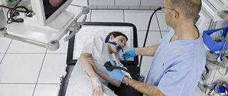
Features of the event
Therapeutic hysteroscopy is carried out on a hospital basis, including a one-day hospital in an operating room. An hour before the start of the intervention, premedication is performed - a sedative injection is given. After being delivered to the operating room, the patient is placed in a gynecological chair and given intravenous anesthesia. Next, proceed to the operation:
- Dilation of the cervical canal.
- A certain amount of sterile liquid is injected into the uterine cavity to “straighten” its walls.
- A hysteroscope is inserted through the cervical canal. As it is introduced, a step-by-step examination of all parts of the uterus is performed. First, the cervical canal is examined, then the uterine cavity and its angles.
- If necessary, surgical procedures are performed - taking a biopsy, removing polyps, dissecting synechiae, etc.
- At the end of the operation, fluid is removed from the uterine cavity.
Atypical endometrial hyperplasia
typical endometrial hyperplasia is a pathological growth of the inner layer of the uterus with the appearance of atypical cells. It is provoked by an excess of estrogen and a lack of progesterone. It is considered as a precancerous disease. It can develop at any age, but is more often detected after 45 years. Accompanied by menstrual irregularities and uterine bleeding (menorrhagia, metrorrhagia). The diagnosis is established on the basis of complaints, anamnesis and additional research data. Treatment is hormone therapy, curettage or ablation of the mucous membrane.
Symptoms of atypical endometrial hyperplasia
Symptoms of atypical endometrial hyperplasia occur in the form of certain manifestations inherent in each specific character and type of development of the pathological process. The main symptom of this disease is uterine bleeding. In most patients, such bleeding occurs due to a delay of menstruation for a period of 1-3 months. Less commonly (as a rule, in the absence of obesity and obvious endocrine pathology), regular cycles are observed with a duration of menorrhagia of more than 7 days. In approximately a quarter of patients with atypical endometrial hyperplasia, anovulatory uterine bleeding is detected. In 5-10% of cases metrorrhagia is diagnosed. Scanty bleeding is possible in the middle of the menstrual cycle or in the absence of menstruation. So, in the glandular form of the disease, which is essentially benign, proliferation of the stroma and endometriotic glands occurs. There is thickening of the mucous membrane, and the glands in the stroma are located incorrectly. The severity of the processes of glandular hyperplasia determines its differentiation into the active, acute stage of the disease and its quiescent, chronic form. The active form is characterized by a large number of cell mitoses in the stroma and epithelium of the glands, which manifests itself as a consequence of prolonged excessively high levels of estrogen. At the stage of chronic disease, mitoses are rarely formed, which is caused by insufficient hormonal stimulation due to the small amount of estrogen. The symptoms of atypical endometrial hyperplasia of the glandular-cystic type are similar to the manifestations of glandular hyperplasia, with the only difference being that they have a slightly greater degree of severity. One of the characteristic signs is cystic enlargement of the glands.
Treatment of atypical endometrial hyperplasia
Treatment of this pathology can be either conservative or surgical, carried out on an outpatient basis or in a hospital setting. Indications for planned hospitalization in reproductive age are bleeding and spotting, in postmenopause - bleeding, prolonged watery or purulent discharge. Emergency hospitalization is indicated for heavy bleeding. Treatment tactics for atypical endometrial hyperplasia are determined taking into account the patient’s age, her desire to have children, the presence of somatic diseases and diseases of the reproductive system (especially adenomyosis or fibroids), the form of atypical endometrial hyperplasia and the number of relapses. The main thing is the timely detection of this disease or the initial stages of cancer, when atypical cells have just appeared in the basal layer of the endometrium. Therefore, all menstrual irregularities should be immediately examined. To do this, women first undergo an ultrasound examination of the uterus, and then, if changes are detected, an endoscopic examination (hysteroscopy). Hysteroscopy can be diagnostic and therapeutic. Most often, diagnostic hysteroscopy, when the doctor examines the endometrium enlarged with optical equipment, turns into a therapeutic one, that is, the endometrium is removed. But this is not always done. In childbearing age, today they try to use mainly hormonal therapy: suppression of estrogen secretion using drugs with antiestrogenic properties, progestogens (synthetic analogues of progesterone) or analogues of Riesling hormones of the hypothalamus (they suppress the secretion of pituitary hormones). If it is not necessary to preserve reproductive function, then ablation of the mucous membrane of the uterine cavity is performed - its complete destruction in various ways along with the basal layer, after which the endometrium is no longer restored. Subsequent hormonal correction is also carried out. To prevent endometrial cancer, any irregularities in a woman’s menstrual cycle should be promptly identified and treated.
Surgical treatment of endometrial hyperplasia
By curettage of the uterine cavity, the doctor uses a curette to remove the hyperplastic endometrium under the visual control of a hysteroscope. Polyps are removed with special scissors or forceps, and under visual control they are “unscrewed” or cut off. The operation to remove a polyp is called a “polypectomy.” Then, after receiving the results of histological examination, depending on the type of hyperplasia, the patient’s age and concomitant diseases, hormonal therapy is selected (except for fibrous polyps that do not require hormonal treatment). The goal of hormone therapy is to suppress further proliferation (overgrowth) of the endometrium and regulate hormonal imbalance.
The following groups of hormones are used to treat endometrial hyperplasia:
- COCs - combined oral contraceptives (Regulon, Zhanin, Yarina) are prescribed for six months according to the contraceptive regimen. The drugs are suitable for women of reproductive age up to 35 years of age, as well as teenage girls with heavy and/or irregular menstruation with glandular and glandular-cystic types of hyperplasia or polyps.
COCs can be used for 'hormonal hemostasis' (taking hormones in large doses) in girls in emergency situations to stop bleeding, so as not to resort to curettage. COCs are prescribed 2-3 tablets per day, then the dose is reduced, bringing it to 1 tablet per day. The course of treatment is 21 days. If hormonal hemostasis is ineffective - if the bleeding continues and threatens the life of the child, they resort to curettage of the uterine cavity.
- gestagens (Duphaston, Utrozhestan) from the 16th to 25th day of the menstrual cycle are prescribed for 3-6 months. Suitable for women of any age with any type of hyperplasia. The gestagen-containing contraceptive intrauterine device Mirena is successfully used, which has a local effect on the endometrium, in contrast to gestagens used orally, which have a systemic effect. The IUD is installed for 5 years. The disadvantage of the IUD is that quite often a side effect occurs in the form of intermenstrual bleeding within 3-6 months after the IUD is installed. In addition, many patients are confused by the spotting nature of menstrual discharge against the background of Mirena and the presence of a foreign body in uterine cavity;
- GnRH gonadotropin releasing hormone agonists (Zoladex, Buserelin) are the most effective group of hormones. Used in women over 35 years of age and during perimenopause from 3 to 6 months for any form of hyperplasia. An unpleasant side effect of drugs in this group is symptoms of early menopause (hot flashes). Gonadotropic releasing hormones are formed in the nerve cells of the anterior and middle hypothalamus and regulate the synthesis and release of gonadotropic hormones of the pituitary gland, indirectly the formation of sex hormones in the ovaries. The mechanism of action of GnRH agonists (as well as natural ones) is to bind to the receptors of pituitary cells that secrete gonadotropic hormones. As a result, a picture develops similar to that observed with hypogonadotropic amenorrhea. This phenomenon is also called 'medical castration'. The process is reversible: after stopping the administration of GnRH agonists, after 14-21 days the function of the entire hypothalamic-pituitary-ovarian system in women of reproductive age is restored. GnRH drugs are widely used in gynecological clinics, primarily for estrogen-dependent pathologies: endometrial hyperplasia, uterine fibroids, endometriosis, and breast cancer. The drugs are administered once every 28 days for 3-6 months, depending on the nature of the pathological process.
Patients with atypical endometrial hyperplasia require special dynamic monitoring by a gynecologist. Control ultrasound should be performed 3, 6 and 12 months after curettage and the start of taking hormones to assess the effectiveness of treatment. If adenomatosis recurs, removal of the uterus is indicated.
In case of recurrence of endometrial polyps, glandular and glandular-cystic forms of hyperplasia, if hormone therapy is ineffective - if the patient is not interested in childbearing - ablation (resection) of the endometrium is indicated - complete destruction of the endometrium. For this purpose, electrosurgical (with a cutting loop) and laser ablation methods are used under the control of a hysteroscope. The operation is performed under general intravenous anesthesia.
After curettage of the uterine cavity and/or resection of the endometrium, the patient can be discharged home on the day of surgery or the next day. Within 3-10 days after the manipulation, there may be light bleeding from the genital tract. After ablation, the remains of resected tissue usually come out along with the discharge. Such discharge is normal and should not be embarrassing. In parallel with hormone therapy, for a quick recovery, taking vitamins is indicated: ascorbic acid, B vitamins, iron supplements for anemia (Sorbifer, Maltofer). Sedative therapy (tincture of valerian or motherwort) is prescribed. Physiotherapeutic procedures (electrophoresis) and acupuncture are useful. Nutrition must be nutritious, it is necessary to observe a regime of work and rest. Sexual abstinence is also recommended for 2 weeks after curettage.
« Back to previous page
Preparation
Preparation for hysteroscopy is similar to other surgical interventions in gynecology. First, an examination and, if necessary, certain treatment are carried out. The following tests are performed:
- General blood and urine analysis.
- Coagulogram.
- Blood type and Rh factor.
- Vaginal smear for flora.
- ECG.
- Test for STIs.
- Tests for HIV and parenteral hepatitis.
Hysteroscopy is planned at the beginning of the menstrual cycle (before day 10), since endometrial tumors are clearly visible during this period.

Immediately before surgery, it is recommended to follow these rules:
- Two days before hysteroscopy, avoid sexual contact.
- On the day of the operation, take a shower and then put on underwear made from natural fabrics.
- Remove jewelry and contact lenses.
- Immediately before entering the operating room, empty your bladder.
- Do not eat for at least 6 hours before anesthesia.
Menstruation after hysteroscopy
It should be noted that after hysteroscopy, discharge may last for three days. They are bloody, but there is no need to be afraid of them, because this is a natural cleansing of the uterine cavity after external intervention. Therefore, after the diagnostic procedure, the discharge will be minimal, and after the operation it will be more significant. You should consult a doctor only if blood clots appear on the pad and the discharge lasts longer than three days. As for menstruation after hysteroscopy, in most cases they occur according to the usual schedule for a particular woman. But sometimes it is the procedure that becomes the catalyst for the start of a new menstrual cycle. When menstruation begins after hysteroscopy, you should monitor the color and composition of the discharge. If they are strikingly different from those of previous menstruation, then you should consult a gynecologist for additional advice.
It should be borne in mind that pregnancy after hysteroscopy is possible only six months after the end of the procedure, although medical practice has described cases of successful conception earlier than the stated period. Do not forget that this procedure serves for a detailed examination and removal of certain pathologies. It cannot be a guarantee of pregnancy, because the causes of infertility may lie elsewhere.
In our clinic in Arkhangelsk you can always count on the help of experienced gynecologists. After all the examinations carried out on high-quality equipment, you can count on making the correct diagnosis. In addition, with us you can get rid of, for example, polyps after hysteroscopy and many other pathologies. Come to us and never get sick.
Basic recovery recommendations
- If an antibiotic is prescribed, take it for as long as your doctor recommends.
- To relieve pain after hysteroscopy, you can take antispasmodics or analgesics.
- Avoid sexual intercourse and heavy physical activity until the bleeding stops, but not less than 10 days.
- Perform hygiene measures in the shower. Taking a bath is not recommended.
- Toilet your genitals as needed, but at least 2 times a day.
- During the recovery period, it is not recommended to use tampons and menstrual cups; discharge should come out of the vagina freely.
Most women generally feel satisfactory after therapeutic hysteroscopy. The main complaints are nagging pain in the lower abdomen and weakness. The full recovery period takes about two weeks. However, if complications develop - fever, heavy bleeding, foul-smelling discharge, you should immediately consult a doctor.
Frequently asked questions about the disease
Why remove or treat a polyp?
99% of polyps removed using hysteroscopy are benign. Cancer cells in such formations are rare, but removal of any tumors is mandatory for several reasons.
Neoplasms disrupt the menstrual cycle. If they are not removed, the likelihood of cancerous or precancerous cells forming increases. A woman with this diagnosis increases the likelihood of miscarriage, frozen fetus, and even infertility.
What difficulties exist in treatment and diagnosis?
Today, doctors are trying to find new alternative methods for treating endometrial polyposis, but their efforts do not bring the desired results. It should be noted that almost all formations that a doctor sees on an ultrasound are called polyps, but in medical reports they are written with “?” This means that the pathology has been identified, but its nature remains unknown.
Any structure in the uterus, at first glance, has a similar structure, but may contain not only benign, but also malignant tissue. For this reason, in addition to finding the best non-surgical interventions, doctors are trying to improve surgical techniques in order to perform operations as safely as possible for patients.
In advanced clinics, conventional uterine curettage is replaced by hysteroscopy, as a more gentle and effective procedure. This method is both a diagnostic method and an operation to remove various formations. Non-surgical methods for determining pathology sometimes include office hysteroscopy, but in essence this is not true, since this is a surgical intervention, but the diagnostic value of MRI is not questioned.
Endometrial polyposis and cancer. Is there a connection?
Typically, a polypous neoplasm is benign and is not associated with cancer, but there is a small probability of its development. According to static data, it is known that 0.5 percent of such formations are uterine cancer.
This is the main reason why the disease should be treated: removed, examined at the cellular level, and histologically examined. After this, you can calm down and not think about the “bad things.”
How is a benign polyp harmful and dangerous?
The disease causes various cycle disorders, including heavy bleeding and, albeit rarely, gives rise to pain in the lower abdomen.
Plus, it increases the miscarriage rate, which is especially tragic for women who became pregnant through IVF. If the tumors are located near the place where the fallopian tubes flow, then such a diagnosis can generally cause infertility.
Who is susceptible to the disease?
This is the age group of women from 35 years to 50. Risk factors include being overweight, high blood pressure and weak immunity. The onset of the disease can be affected by any hormonal therapy and estrogen intake.
Is the diagnosis related to infertility?
The fact that a polyp leads to infertility is confirmed by scientific data and facts:
- They occupy a large area, can disrupt the normal process of fertilization and implantation, and those formed in the tubes have a negative effect on the patency of sperm.
- Few scientific works have been written about their nature and development, but this is associated with the absence of the need to conduct serious research - in any case, the formations need to be removed in order to exclude cancer.
- Often the tumor is found in women undergoing examination due to infertility. It has been scientifically proven that its elimination increases a woman's chances of becoming pregnant.
- After removal of the tumor, in 65% of cases, women were able to conceive a child, and if hysteroscopy was not performed, then this figure was 25%.
- Most often they form on the back wall of the uterus, where implantation of the embryo occurs.
- If the formations are localized at the confluence of the fallopian tubes and are eliminated through hysteroscopy, the patency of the tubes increases several times.
- There is an opinion that the disease provokes irritation of the uterine mucosa, in which the fetus develops. Due to this irritation, the fertilized egg cannot be implanted.
Complications after polyp removal
As after any gynecological operation, the following complications may develop after hysteroscopy:
- Damage to the wall or cervix - treatment may require additional surgery to close the perforation.
- Damage to the cervix - may require stitches.
- Bleeding.
- Infectious complications.
It should be noted that complications after hysteroscopy are much less common than after other gynecological interventions performed “blindly”. But the possibility of their development cannot be completely excluded.
Conservative treatment
Treatment of polyps is carried out not only surgically, but also conservatively. Drug treatment can be suggested by a doctor in the absence of painful symptoms and large size of the formation. But this method does not exclude constant gynecological monitoring and regular additional diagnostic measures.
Treatment of uterine polyp without surgery also includes undergoing a number of physiotherapeutic procedures:
- Magnetotherapy;
- Ultrasound treatment;
- Electrophoresis with the use of medications;
- Electrical stimulation technique;
- Laser therapy.
Indications for one or another type of conservative complex therapy depend on many reasons, among which are:
- No complications;
- Mild symptomatic picture;
- Slight and slow growth of benign growth in the uterine cavity;
- The presence of contraindications for a woman to undergo surgical removal of a polyp.
Drug treatment
The main stage of treatment for polypous growths of the endometrium is the use of hormonal drugs (COCs and gestagens).
The formation of a pathological process in the uterine cavity in 70% of cases is associated with a hormonal imbalance in a woman’s body, which leads to excessive production of estrogen. Hormonal contraceptives eliminate pain in the lower abdomen and quickly stop uterine bleeding. Antibacterial and anti-inflammatory drugs are used in the treatment of endometrial polyps if the formation of a benign process occurred against the background of the presence of a sexually transmitted disease, chronic inflammation (adnexitis, cervicitis, oophoritis). The combination of these two classes of medications gives the most effective and high-quality results.
Vaginal suppositories for polyps in the uterus are also one of the methods of drug therapy for this disease. Suppositories have a number of advantages over other types of therapy. The active substance exerts its therapeutic effect directly on the pathological focus, quickly penetrating through the mucous membrane of the vaginal wall, bypassing the liver and kidneys.
Treatment of uterine polyps also includes the use of a vitamin complex and drugs with a high iron content to prevent anemia.
Contraindications
- The presence of decompensated chronic diseases that may interfere with anesthesia, for example, cardiovascular pathology.
- Presence of STIs and vulvovaginitis.
- The presence of diseases accompanied by a tendency to bleeding.
Euroonco specialists closely monitor the condition of patients not only in the postoperative period, but also at the initial stage. In this regard, we perform the procedure only after we are convinced that the patient has no contraindications. You can make an appointment for a consultation about uterine polyps by calling:
Book a consultation 24 hours a day
+7+7+78
Discharge after polyp removal
The nature, volume and longevity depend on various factors, and therefore are individual for each patient.
What influences?
How much and how it will be is given by:
- Removal method. As a result of curettage, bleeding may take longer, since a large area of the endometrium is affected. The volume of discharge after hysteroscopy is significantly less. Laser removal promotes very rapid tissue restoration;
- Polyp size. Large formations or a wide base contain larger vessels that may not heal for a long time;
- Quantity. Areas of polyposis are carefully cleaned; they cannot be treated with current or laser, so many damaged vessels bleed;
- Localization. Removal of a tumor from the cervical canal is much easier to tolerate than manipulations inside the uterus;
- Depth of the polyp root;
- Blood clotting. This parameter determines the speed of wound healing. If the woman is normal, then 3 days after the operation, the discharge will darken and become brown and insignificant;
- Concomitant therapy. Hormonal drugs that are prescribed before a crust appears on the wound interfere with cell regeneration and can therefore cause bleeding;
- Compliance with recommendations. In most cases, heavy discharge occurs due to violation of the prohibitions on lifting weights, sex and playing sports.
Carefully! A hot bath or warm compresses on the abdomen can cause bleeding.
Volume
In the first 1-3 days there is quite noticeable red discharge, but it is inferior in quantity to menstrual discharge. Further, a daub is noted. A non-decreasing abundance for more than 3 days should be alarming.
Duration
Normally, discharge is present for 3 to 10 days. Moreover, long ones should be meager in quantity.
Color
In the first days it is fresh bright red blood, then a pink mucous substance, and then brown. At the initial stage, clots of a dark shade may be present.