Indications
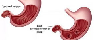
- suspicion of an ulcerative process;
- detection of a neoplasm;
- protrusions or other deformations of the gastric walls;
- inflammatory processes in the stomach;
- state of dysphagia (functional impairment of swallowing);
- abdominal pain;
- severe constant heartburn;
- sudden involuntary release of gas into the oral cavity from the stomach or esophagus with a sour odor;
- the appearance of scarlet blood in the stool;
- decrease in red blood cells;
- sudden weight loss without objective reasons.
- pain in the epigastric region;
- detection of mucous and purulent discharge, as well as blood impurities, in feces;
- chronic increase in intervals between acts of defecation, hardening of stools, feeling of incomplete bowel movement;
- frequent diarrhea with changes in stool color (black, tar-like);
- rapid weight loss not due to dieting.
X-rays of the large and small intestines are necessarily indicated for suspected congenital malformations, oncopathology, polyposis, saccular protrusions of the intestinal wall, granulomatous inflammation with segmental damage to different parts of the digestive tract, chronic colitis and enterocolitis.
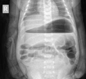
Indications for abdominal x-ray
A review of an x-ray of the abdominal cavity is carried out for pain in the abdomen (acute abdominal syndrome) and lower back, bloating, abdominal trauma, retroperitoneal abscesses, acute intestinal obstruction (obstruction of the lumen of the cyst, polyps, tumor, etc.). X-rays are also taken for intussusception (intestinal obstruction due to the introduction of one part of the intestine into the lumen of another); diverticulitis (inflammation of protrusions in the intestinal wall - diverticulum).
It is also performed to diagnose inflammation of the pancreas (pancreatitis) and gallbladder (cholecystitis), with kidney or bladder stones.
After the review, an X-ray will be taken - if there is no damage to the mucous membranes of the intestinal wall - radiography or fluoroscopy with a contrast agent.
Contraindications
X-ray of the stomach with barium is contraindicated in the following cases: disturbances in the functioning of the hematopoietic system, a pathological condition associated with clouding of the lens of the eye, affecting visual acuity, oncopathology of the bronchopulmonary system, carrying a child at any stage, endocrine pathologies of the thyroid gland.
There are the following contraindications for performing an X-ray examination of the intestine:
- intestinal perforation;
- unconsciousness of the patient;
- general serious condition of the patient;
- nonspecific ulcerative colitis;
- a combination of segmental or total dilatation of the colon against the background of signs of systemic toxicity;
- severe diseases of the cardiovascular system;
- complete disruption of the passage of contents through the intestines;
- internal bleeding;
- severe pain in the epigastric region;
- pregnancy.
Contraindications for X-rays
In fact, there are no contraindications for performing abdominal x-rays for diagnostic purposes. This examination is prescribed by a doctor - in the appropriate direction, where a preliminary diagnosis of the patient that requires clarification may be indicated.
However, an abdominal x-ray is not recommended for a child under 14 years of age, as well as a pregnant woman at all stages of pregnancy; an ultrasound should be done.
Contraindications to this behavior include perforation of any part of the gastrointestinal tract; obstruction of the colon; acute diverticulitis; dehydration during vomiting and diarrhea; ulcerative colitis in the acute stage; bronchial asthma; allergies; intestinal or mixed form of cystic fibrosis of the pancreas (cystic fibrosis).
Preparation
In order for the procedure to be successful and give reliable results, it is necessary to properly prepare for an X-ray of the stomach with barium. This is a mandatory condition, if not met, the radiologist may refuse to perform the diagnosis. Preparation for an x-ray of the stomach should begin 3 days before the scheduled examination and include the following.
It is necessary to adhere to a certain diet, which involves excluding from the diet foods that cause increased gas formation (legumes, sauerkraut, black bread, fresh fruits and vegetables, whole milk). On these days you need to eat low-fat foods, steamed or boiled/baked.
12 hours before the scheduled x-ray examination, you must completely stop eating. The procedure is performed exclusively on an empty stomach. Some patients are advised to cleanse the intestines with an enema and gastric lavage.
Drinking alcohol and smoking before the examination is strictly prohibited. Immediately before the procedure in the X-ray room, the patient takes off clothes with metal fittings, jewelry, removable dentures, etc., which can negatively affect the quality of the images.
An X-ray of the small intestine will also be most effective if the patient prepares for it correctly. The subject must adhere to a strict diet three days before the procedure, which will prevent flatulence and fermentation in the intestines. The doctor who sends the patient for such an examination, as a rule, explains what needs to be excluded from the diet and gives the patient a reminder.
If the patient suffers from chronic constipation, then he will need to use laxatives and try to drink more clean water without gas. Colon cleansing must be done in the evening the day before the scheduled x-ray. This is done in the classical way using a series of enemas, which are also repeated on the day of the examination, or using special pharmaceutical preparations - microenemas.
The day before the test, you should completely give up your usual food. Allowed to drink are broths, herbal teas, clear fruit juices. Before the procedure, it is recommended to refrain from smoking and alcohol for at least 7 days.
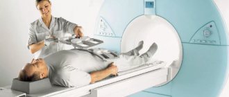
How is an abdominal x-ray performed?
An X-ray of the abdominal cavity is taken while wearing clothes (but without jewelry) in a standing or lying position. The patient must stand in front of the X-ray machine or lie on a special table and stand (or lie) absolutely still for several minutes. After irradiation, the device turns off and the radiologist reports that the procedure is complete. Often the test is carried out simultaneously in two positions: first standing and then lying down.
For contrast radiography of the abdominal cavity, the patient must drink a barium sulfate suspension before the diagnostic procedure begins.
Carrying out
An X-ray of the esophagus and stomach begins with a general image of the abdominal cavity. After this, the patient is asked to drink prepared barium sulfate. The initial targeted photograph is taken after two sips of the drug. At this moment, the relief of the walls of the esophagus with barium is determined. Then the patient is allowed to finish the rest of the drug. During the examination, the doctor may press on the patient's abdomen to promote better distribution of the contrast.
During the process, various pictures are taken in different positions - lying on your back as standard or with the pelvis raised at an angle of 45°, lying on your side, standing. At the same time, at the command of the radiologist, the patient must hold his breath. Modern X-ray rooms are equipped with a special table that rotates while taking pictures.
As a rule, to examine the upper part of the gastrointestinal tract, it is enough for an adult to take 250-300 ml of dissolved barium sulfate (in some cases the dose may be increased). If radiography is prescribed for a child, then the required amount of suspension is calculated based on the age category. For children, as a rule, 100 ml of barium paste is sufficient.
Bowel examination
X-ray of the small intestine with barium is performed in stages. The procedure begins with the patient drinking 0.5 liters of barium suspension. If the study is carried out with double contrast, the drug enters the body through a special tube that is inserted into the patient’s mouth. Along with the contrast, air or inert gas is supplied.
After this, wait at least 2 hours - during this time the barium has time to reach the small intestine. As contrast fills the small intestine, the radiologist takes a series of pictures, asking the patient to assume different body positions. And after relieving themselves, they take the last control shot. After filling them with contrast, the diagnostician examines the various segments of the small intestine in detail on the monitor for half an hour.
The contrast procedure allows you to evaluate intestinal motility and its mucous membrane. While the contrast agent is still present in small quantities, the relief of the inner wall of the intestines is examined, and when there is a lot of contrast, the shape, size, contours and functionality of the intestines are assessed. With maximum barium filling, it is possible to identify inflamed segments, ulcerative processes and identify neoplasms.
If the passage of barium is disrupted, the radiologist gently presses on the anterior wall of the peritoneum to distribute it evenly. During a procedure with contrast, as the substance is distributed in the intestinal lumen, an experienced radiologist can draw conclusions about the presence of pathological processes. If the barium suspension is distributed in the form of flakes, then this is a clear sign of impaired absorption. And if the contrast fills the lumen unevenly, this may indicate oncopathology.
X-ray of the stomach with barium - reviews
[email protected]
https://irecommend.ru/content/rentgen-zheludka-konechno-ne-zamenit-gastroskopiyu-no-mozhet-vyyavit-koe-chto-drugoe
I became acquainted with stomach x-rays quite by accident. In general, I have IBS (irritable bowel syndrome) with a predominance of diarrhea, and then suddenly my stomach began to hurt in the navel area and constipation began (sorry for the details). I went to a gastroenterologist. When I went to the doctor, I explained that I had this situation. He examined me, felt my stomach and wanted me to do a gastroscopy, but I immediately warned that I had asthma and panic attacks. The issue was immediately removed from the agenda because literally a month ago, a man had a gastroscopy and he began to panic, and it all ended in death. Of course, she gave me the phone number of a professor who could deal with such a problem, but I said that I would agree only with medicated sleep. Yes, I forgot to write, I came straight to the appointment with tests, general, renal-liver and ultrasound of the abdominal organs, and they did it for me twice and twice there were different results. One diagnosed me with cholecystitis, the second that I was completely healthy. Since I did the second one with an ultrasound doctor, whom the gastroenterologist knew, she took it as a basis. In general, since such a kitchen, they sent me for an x-ray of the stomach and prescribed a test for Helicobacter.
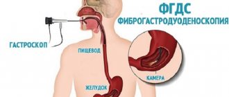
What is barium fluoroscopy of the stomach?
When diagnosing diseases of the stomach, intestines and esophagus, X-ray with barium is the most highly accurate and informative method, since without introducing a contrast agent into the body it is impossible to examine the shape and structure of organs and determine the presence of some darkening in the lumen of these organs.
When performing fluoroscopy of the stomach and intestines, the doctor can examine internal organs with a hollow structure on the monitor, study their functional abilities, and also study the relief of the intestinal mucosa. During the x-ray, the specialist also takes a series of x-ray photographs, which represent a documentary record of the examination.
So I came for an x-ray. They told me not to drink or eat anything in the morning. The doctor placed me on a pedestal that resembled a table with a footrest and handles. Then he gave me a glass with a white, tasteless liquid (barium) and told me to drink. At first I drank a little, then at his command I drank it all. After this, the table with the footrest began to lower horizontally and rotate, and the doctor, coming out of his closet, told me how to lie down. The procedure took about 15 minutes and then after another 15 minutes they gave me the result with which I went to the doctor.
What does a barium stomach x-ray show?
X-ray of the stomach with barium helps to detect most pathologies and changes in the organs of the digestive system in the early stages. Among them:
- ulcers, gastritis, decreased peristaltic function of the organ being diagnosed;
- tumors of benign and malignant nature;
- inflammatory processes;
- diverticular manifestations;
- gastric obstruction;
- displacement or prolapse of organs;
- other pathologies and changes.
In general, my x-ray showed that I have prolapsed stomach and duodenogastric reflux.
Then I began to remember what could lead to a prolapse of the stomach and I remembered. I lost a lot of weight due to stress and at this time I had to carry heavy things. In addition, I was diagnosed with thoracic osteochondrosis.
I am 172 cm tall and at the moment when I came to the doctor I weighed about 57 kg. For me, this is the weight at which I feel comfortable. to which the doctor told me that I needed to get better. But at the moment when everything happened (stress) I weighed 52 kg. Let’s just say I’ve gotten better.
In general, she gave me an appointment:
- Mosid MT;
- Ursonost.
THANK YOU DEAR DOCTOR!!!!
After 3 weeks I had such diarrhea...!!!!!
Her prescription caused an exacerbation of IBS and I lost weight again to 54 kg. But that is another story. About IBS HERE.
There is nothing scary about an X-ray of the stomach other than radiation. I did not feel any consequences after the barium. The procedure was paid (we don’t have anything free now, they hide behind charitable foundations) I gave 130 hryvnia.
ps I tested for Helicobacter - it was not detected.
Be healthy.
Sane4ka74
https://irecommend.ru/content/ne-alternativa-fgds-no-v-nekotorykh-sluchayakh-ee-zamenyaet-k-schastyu
So.
I suffered from stomach problems for about six months before I stopped sleeping and, having gotten so fed up, finally decided to go to the doctor. The worst thing for me was the referral to the FGS (FGDS). I do not agree to swallow a pipe under any circumstances (well, except under general anesthesia, which in our city costs about 13k). I told the doctor about this. She said that in my case she saw that it was possible to get by with an x-ray for now, and sent me for one. It cost me 1000 rubles.
About the procedure itself.
I was warned that the night before dinner should be light (I ate around 6 pm, just in case), and in the morning not to eat or drink anything. They told me to take a barium mug with me.
Immediately there they asked me to take off my chain and bra, allowing me to remain in a T-shirt, they placed me on a stand on the device, and gave me half a mug of barium. They told me to drink on command.
I took one sip at a time, thank God. No matter how much I read that barium is tasteless, I didn’t think so. It reminded me of the taste of skim milk with sourness and spice; it burned my throat a little. Although, perhaps this is a consequence of the fact that it already hurt me.
So, I drank one sip at a time, turned around, then the device “lay down” - it turned into a horizontal plane, and I went with it (they took the mug). She lay there, swallowing the barium previously stored in her mouth. She turned over onto her stomach and back. Then again vertically, a couple more shots and that’s it.
It all lasted about 3 minutes.
I had, one might say, a light version, without eating porridge and drinking barium saturated with air, perhaps that’s why it came out quickly)
You can eat and drink right away. I apologize to the impressionable, but for the sake of completeness I will say that the barium left my body the very next morning) in the evening I felt a slight twist in my stomach, but you can’t guess whether it was because of it or because of the food.
I received a relatively large dose - 4.5 mSv. In my opinion, this corresponds to the dose of radiation we receive over three years.
Overall, I am pleased that I managed to avoid FGS, because... The x-ray revealed a pathology in me that entailed the symptoms I had.
But I want to say right away that the indications for FGS and X-ray are different, their information content is different. Where one study helps, another will not, so they are not interchangeable. I was lucky, although it is possible that I will also have FGS sometime in the distant future.
Among the disadvantages of the procedure, I can only mention radiation, which is still harmful, as well as a number of contraindications for this procedure. At a minimum, it is strictly forbidden for pregnant women.
Among the advantages - it’s definitely less disgusting)
And also fast.
There is no need to be afraid of the procedure, and the taste of barium is quite tolerable.
By the way, I drank nothing at all - less than a quarter of an ordinary mug.
So good luck and health!
Mo-skal-Lad888
https://otzovik.com/review_9623833.html
Advantages:
Painless, comfortable
Flaws:
does not replace FGSD
Before the X-ray procedure of the esophagus with barium, I was very nervous, and there were reasons for this: firstly, I was afraid that the taste of barium would cause gag reflexes (I can’t even drink bitter pills); secondly, due to the fact that I suffer from poor passage of food and liquid through the esophagus, I was worried that the barium would rise somewhere in the middle and I would not be able to drink the required amount. So, all my fears turned out to be in vain - the barium solution is absolutely tasteless (for example, smecta is much nastier, it has a taste), I drank it calmly, the consistency was like liquid sour cream. First, the doctor suggests taking a small sip and taking a photo, after a while, take a larger sip or two (as many as you can) and take another photo. Then I sit in the corridor for about 10 minutes, and so the whole procedure lasts for about 40 minutes. I think if you just do an X-ray of the stomach without observing the patency of the esophagus, then it will take much less time. That's all! There is absolutely nothing to be afraid or worry about, FGSD is much worse, but these procedures do not replace each other, unfortunately.
kotenok0883
https://otzovik.com/review_3084369.html
Advantages:
stomach check
Flaws:
taste of barium
managed to end up in the hospital with my stomach. Before the hospital I had an FGS - not the most pleasant procedure. Therefore, the hospital ordered an x-ray of my stomach. Phew, I thought - x-ray is nonsense, get up... don't breathe... get dressed. But it was not there. I came into the office, they told me to undress from the waist up, I pressed my back against this machine and they gave me a glass, 200 ml. So the white substance is barium. First they told me to take a sip, it tasted like regular chalk. Didn't give me any difficulty swallowing. Then another sip... and immediately gulped down the entire glass. Of course, it’s a little hard to swallow after half a glass, but for the sake of your health you can endure it. The barium left me for more than a week. The procedure is painless, but if you don’t like chalk, it will simply not be pleasant.
Yayuya
https://irecommend.ru/content/rentgen-s-bariem-dlya-isklyucheniya-gryzhi-pishchevoda-nashli-opushchenie-zheludka-i-ezofagi
I was bothered by pain in the middle of my chest. Such constant pressing pains that appeared after eating. Based on these complaints, I was prescribed an x-ray of the esophagus with barium to rule out a hiatal hernia.
X-rays of the esophagus and stomach with barium are done strictly on an empty stomach. Therefore, it is better to sign up for the morning time. I had an X-ray with barium at a regular clinic at my place of residence. There was no need to bring anything with you.
To undergo an x-ray, you had to undress to the waist and remove all metal objects. The barium was brought out to me in a faceted Soviet glass - it was filled to the brim with a boiling-white viscous liquid. Although it’s difficult to call it a liquid. This is a very thick and dense mass. The spoon was practically standing in the glass (the solution was mixed with this spoon).
————————/How the X-ray with barium was done/————————
I was told to put barium in my mouth, but not to swallow. You had to swallow on command while the picture was being taken.
Barium tastes nasty. It immediately takes up literally all the space in the mouth. Unpleasant sensations. And it is quite difficult to swallow; you need to make an effort to push it inside. He even tried to jump back out, but I finally overcame him.
And there were several such sets of barium in the mouth - a sip on command. We took a lot of pictures:
- just straight
- the body is slightly turned to the left,
- left side,
- the body is slightly turned to the right,
- right side.
Then it was necessary to drink all the barium to the end. They took the picture again.
At the end, the picture was taken in a very interesting way: you had to lie on your stomach, and a small stool was placed under the pelvic area. It turns out you’re lying with your butt up, well, the position is so-so
As I read the reviews, I noticed that everyone takes pictures differently. Apparently it depends on the equipment that the institution has, and on the doctors themselves, of course.
Taking photos of X-rays is a thankless task, but that’s how it is:
--------/Results/--------
The result was ready the next day. No hiatal hernia was detected (URA). But they found gastritis, reflux esophagitis and prolapse of the stomach.
The research is specific. And if it is prescribed, then be sure to do it. You can get quite a lot of information. Based on the results, I was prescribed treatment for esophagitis. Esophagitis (inflammation of the esophagus) can cause pain.
X-ray of the esophagus and stomach does not replace FGDS. I had an FGDS a year ago (relatively recent). Maybe that’s why FGDS was not prescribed to me.
————————/After X-ray/————————
If you think that you've had an X-ray and that's it, you're wrong. Barium remains in your body. And this barium needs to come out. He is very heavy. I felt like there were stones in my stomach. The stomach was rocky and heavy. It was as if he wasn't moving at all. I even felt a little scared. It’s good that I didn’t read then about possible complications after an X-ray with barium. And don’t read)) All my barium came out successfully after 1-2 days. If you are prone to constipation, it is better to discuss taking laxatives with your doctor or focus on nutrition. To remove barium easily and freely.
Good health to everyone
_____________________________________
You might also be interested in the “intestinal” topic
- Moviprep or Fortrans? On GW. My tricks
- Quick preparation for an appointment with a proctologist. Preparation for sigmoidoscopy. I experienced all the side effects of Enema Klin myself!
- When is sigmoidoscopy needed? In what position is sigmoidoscopy performed? What does this have to do with a gynecological chair?
- Colonoscopy for breastfeeding. Without anesthesia. Twice. Paid and free. +Third colonoscopy after surgery
- Alpha normix. Quickly relieves symptoms of inflammation (bloating, cramps, pain, mucus). But if it helped you, this does not mean that the problem is completely solved! Examine your intestines completely!
Katyundrik
https://irecommend.ru/content/neobkhodimaya-protsedura-dlya-diagnostirovaniya-zabolevanii-bez-diskomforta-i-boli
Good day.
It just so happened that I needed to undergo several different examinations, among which was an x-ray of the stomach with barium.
About the procedure itself:
X-ray of the stomach is a diagnostic method that allows you to identify a wide range of disorders of the gastrointestinal tract (esophagus, stomach and duodenum): peptic ulcers, neoplasms of various types, protrusion of the wall of the esophagus, stomach and duodenum. This procedure provides an opportunity to evaluate several important parameters: the condition of the circular muscles (sphincters), the shape of the stomach, its size, etc.
X-ray of the stomach with a series of radiographs - during the study, the patient takes a contrast agent (water-barium suspension 200 ml), and the doctor evaluates morphological and functional changes in the esophagus, stomach, duodenum in real time on the monitor and performs a series of radiographs, in order to take a closer look at the morphological changes.
Indications for x-ray of the stomach.
- Suspicion of a peptic ulcer of the stomach or duodenum.
- Suspicion of a tumor process in the stomach.
- The study is part of a comprehensive diagnostic program for gastritis and gastroduodenitis.
- Suspicion of gastroesophageal and duodenogastric reflux.
- Malformations of the stomach, diverticula (protrusions of the wall).
- Control after gastric surgery.
- Symptoms characteristic of stomach diseases: belching, heartburn, pain around the navel, indigestion, bloating, pain under the left rib.
X-rays of the stomach were performed by appointment.
There was no preparation for the procedure.
You had to bring with you: barwist, a glass with a spoon, water (for diluting barium). It was necessary to arrive on an empty stomach.
Barium Toxicity:
Barium sulfate, due to its low solubility in water, is not toxic substance to the body, unlike all soluble barium salts, and therefore it can be used as a radiopaque substance.
And then that day came.
The office in which the procedure took place was not lit, even the windows were closed. The X-ray itself was similar to the one used for fluorography, but the monitor was placed at the level of the abdomen.
I was asked to undress to the waist. Then I was placed near the X-ray monitor and the doctor gave me an almost full glass of barium solution.
First I was told to take one sip of barium. The taste of barium is simply no, tasteless. Did not cause a gag reflex.
Next, the doctor ordered me to take a few more sips of barium, and then gave me some water to drink. Repeatedly, the doctor came up to me and adjusted my position to the monitor. All this happened while standing.
And only then, the doctor told me to drink all the rest in one gulp. There was approximately 1/2 cup left. A little more time passed and I was released.
I didn't feel any pain or discomfort. The whole procedure lasted about 30 minutes. I was given the result immediately after the procedure (conclusion and pictures).
I'm glad I went through this procedure. X-ray of the stomach with barium helps to detect most pathologies and changes in the organs of the digestive system in the early stages.
After the results were given, I at least calmed down that no serious pathologies were identified.
For those who need to undergo this procedure, it is better to take wet wipes with them, as barium dries out when it gets on your lips. I didn’t take a napkin with me, so I wiped off the barium with my palm. After that, the whole palm was white.
If you have been prescribed an x-ray of the stomach, there is nothing wrong with this procedure. Feel free to come in. Although everyone’s body is individual, the procedure is still necessary for the timely diagnosis of diseases.
Reviews about other procedures:
1. Curettage (gynecology) - https://irecommend.ru/content/ved-strakhov-bylo-bo...
2. Puncture of the posterior fornix - https://irecommend.ru/content/inogda-neobkhodima-d...
Thanks everyone for your attention.
results
- abnormalities in the structure of the digestive tract;
- acute expansion or narrowing of the lumens of the stomach/esophagus;
- malformations of certain organs of the gastrointestinal tract;
- hypertonicity/hypotonicity of the muscular wall of the stomach;
- tumors, papillomas, foreign bodies;
- reduction or radial arrangement of shell folding;
- cicatricial changes at the site of tumors, ulcers, chemical burns.
- pathological narrowing;
- saccular protrusions and elasticity of the walls;
- the introduction of one section of the intestine into another with the possible development of gastrointestinal obstruction;
- intestinal motor function;
- the presence of ulcerative and inflammatory processes;
- tumors, polyposis.
An examination can show how the bauhinium valve functions. This is the structure that separates the small and large intestines and is responsible for passing food between them. If it has pathological changes, then the food gets back access, and this poses a danger to the patient’s life.
Where can I get an abdominal x-ray?
An X-ray of the abdominal organs is carried out in accordance with the prescription of the attending physician (gastroenterologist, urologist, nephrologist, endocrinologist) in a medical institution. The price depends on factors such as the category of medical institution, equipment model and qualifications of the radiologist. After all, it is the doctor of this specialty who describes in detail everything that the image displays. So the cost of x-rays is also the interpretation of the x-ray.
Other studies may be required to establish the correct diagnosis, since abdominal x-rays cannot provide comprehensive information about existing pathologies.
You can sign up for a consultation and find out more from the administrator by calling +7 (495) 356-30-03.
Preparation for irrigoscopy
There are various ways to prepare for irrigoscopy, but radiologists recommend adhering to the following scheme:
- three days before the test, switch to a low-fiber diet: minimize the consumption of vegetables, fruits, bran, and wholemeal bread;
- on the day before the procedure, you must switch to liquid food and take Picolax twice, following the instructions (at 8:00 and 18:00).
Preparing a patient for irrigoscopy involves following other recommendations. For example, women of childbearing age should schedule the procedure within the first 10 days after their period. Patients who have undergone heart valve replacement with a history of endocarditis or systemic-pulmonary anastomosis require a/b prophylaxis. The doctor may also prescribe a cleansing enema before irrigoscopy.
You can undergo irrigoscopy in Kharkov at a medical office.
What is intestinal irrigoscopy?
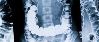
Irrigoscopy is a diagnostic procedure aimed at studying the structure and functions of the large intestine. The technique involves filling the examined part of the gastrointestinal tract with a contrast agent, after which, using an X-ray machine, intestinal function, elasticity of the walls are analyzed and a series of survey and targeted images (irrigograms) are taken.
Rectal irrigoscopy has a number of advantages:
- painlessness;
- high information content;
- minimum number of contraindications.
Absolute contraindications include:
- toxic megacolon (enlargement of the colon caused by toxic exposure or chronic pathology);
- pseudomembranous colitis (infectious bowel disease);
- performing a rectal biopsy: with a flexible endoscope in the previous day, with a rigid endoscope in the previous 5 days.
The procedure is prescribed with caution in the presence of relative contraindications, including poor preparation of the gastrointestinal tract, general weakness of the patient, and recent studies with barium. In this case, the gastroenterologist may refer you for other examinations of the rectum: for example, colonoscopy or sigmoidoscopy.